Articles in PresS. Am J Physiol Renal Physiol (March 29, 2005). doi:10.1152/ajprenal.00389.2004
WT1-Interacting Protein and ZO-1 Translocate into Podocyte Nuclei after Puromycin Aminonucleoside Treatment Maribel Rico 1, Amitava Mukherjee 2, Martha Konieczkowski 2, Leslie A. Bruggeman 2, R. Tyler Miller 1, 2, 3, Shenaz Khan 2, Jeffrey R. Schelling 2, and John R. Sedor 1, 2 Departments of Physiology and Biophysics1 and Department of Medicine2, School of Medicine, Case Western Reserve University, Louis Stokes Veterans Administration Medical Center3 and Kidney Disease Research Center, Rammelkamp Center for Research and Education, MetroHealth System Campus, Cleveland, OH. Running Title: WTIP nuclear translocation changes WT1-induced gene expression
Corresponding author: John R. Sedor, M.D. Department of Medicine, R415 MetroHealth System 2500 MetroHealth Drive Cleveland, Ohio 44109-1998 Phone: (216) 778-4993 FAX: (216) 778-8248 E-mail:
[email protected]
1 Copyright © 2005 by the American Physiological Society.
Abstract Podocyte differentiation is required for normal glomerular filtration barrier function and is regulated by the transcription factor WT1. We identified WT1 Interacting Protein (WTIP) and hypothesized it functions both as a scaffold for slit diaphragm proteins and a co-repressor of WT1 transcriptional activity by shuttling from cell-cell junctions to nucleus after injury. Endogenous WTIP co-localizes with ZO-1 in cultured mouse podocyte adherens junctions. To model podocyte injury in vitro, differentiated podocytes were incubated with puromycin aminonucleoside (PAN, 100 µg/ml) for 24 h, which disassembled cell-cell contacts, rearranged actin cytoskeleton, and caused process retraction. Podocyte synaptopodin expression diminished after PAN treatment, consistent with podocyte dedifferentiation in some human glomerular diseases. To assess podocyte function, we measured albumin flux across differentiated podocytes cultured on collagen-coated Transwell filters. Albumin transit across PAN-treated cells increased to levels observed with undifferentiated podocytes. Consistent with our hypothesis, WTIP, as well as ZO-1, translocated from podocyte adherens junctions to nuclei in PAN-treated cells. Since WTIP is a transcriptional co-repressor for WT1, we examined the effect of PAN on expression of Rbbp7 (also known as RbAp46), a WT1 target gene expressed in S-shape bodies during nephrogenesis. Rbbp7 expression in PAN-treated podocytes was reduced compared to untreated cells. In conclusion, WTIP translocates from cell-cell junctions to the nucleus in PANtreated podocytes. We suggest WTIP monitors slit diaphragm protein assembly and shuttles into the nucleus after podocyte injury, translating changes in slit diaphragm structure into altered gene expression and a less differentiated phenotype. Keywords: nucleo-cytoplasmic translocation, glomerulosclerosis, LIM domain, slit diaphragm, cell-cell contacts
2
Introduction The glomerular filtration barrier is composed of a highly fenestrated endothelium, the glomerular basement membrane (GBM) and the podocyte. After a tightly orchestrated differentiation program, podocytes develop foot processes and assemble a specialized adherens junction, the slit diaphragm, which mediates contact between adjacent cells (44). In proteinuric diseases, regardless of the etiology, podocytes undergo marked morphologic change. The actin cytoskeleton rearranges into a mat below the plasma membrane opposed to the GBM, slit diaphragm structures are lost and the podocyte assumes a cuboidal shape. At a molecular level in both human biopsies and experimental models, this stereotypical morphological response is associated with changes in cytoplasmic, plasma membrane and nuclear podocyte differentiation marker expression (5, 6). In addition, podocytes in some glomerular diseases also revert to a less differentiated phenotype, more characteristic of the developing, rather than the fully differentiated glomerulus (5). Appropriate treatment can restore normal podocyte structure and filtration barrier function, suggesting that regulation of foot process architecture is plastic and dynamic. Given its unique microenvironment with exposure to hemodynamic forces and high ultrafiltrate flow, the podocyte must rapidly respond to changes in physical forces or soluble signals that occur with injury. We have hypothesized that slit diaphragm-associated proteins monitor the podocyte microenvironment and may trigger signaling cascades that alter in podocyte differentiation state (52). Cell-cell junction molecules can transmit extracellular cues by shuttling into the nucleus to regulate gene expression (9; 12; 14). Two podocyte molecules, WTIP and ZO-1, are candidates for this important adaptive role. The Wilm’s tumor gene 1 (WT1) is a zinc finger transcription factor, whose function is required for normal nephrogenesis (18; 28) and podocyte
3
differentiation (18; 28). We previously reported that a WT1 co-regulator, the LIM domain protein WTIP, localized to nascent cell-cell contacts and interacted with the cell junction scaffold proteins Mena and CD2AP (52). A truncated WTIP, containing only its LIM domains, colocalized with WT1 in nuclei, co-precipitated with WT1, and inhibited WT1-dependent transcriptional activation of the amphiregulin promoter. Although full-length WTIP was excluded from cell nuclei, it accumulated in the nucleus and co-precipitated with WT1 after the addition of an inhibitor of Crm1-mediated nuclear export, leptomycin B. These data suggest that WTIP may function to transfer information from the slit diaphragm microenvironment into the nucleus. After wounding, the MAGUK family member zonula occludens-1 (ZO-1) translocates into epithelial cell nuclei from tight junctions (15) with its cognate Y-box transcription factor ZONAB to regulate proliferation and to promote epithelial cell dedifferentiation (4; 16; 29). An analogous ZO-1 function has not been described in the podocyte, where ZO-1 is normally colocalized with P-cadherin and Neph-1 (20). In this study, we test the hypothesis that WTIP and ZO-1, components of podocyte cell-cell junctions, translocate into the nucleus after injury. As an in vitro model of damage, podocytes were treated with puromycin aminonucleoside (PAN), and injury was assessed by morphological characteristics and functional response by an albumin diffusion assay. Both WTIP and ZO-1 translocate to the nucleus after PAN treatment, a finding associated with downregulated expression of a WT1 target gene, Rbbp7 (also known as RbAp46).
4
Materials and Methods Cell lines. The conditionally immortalized podocyte cell line MPC was a generous gift of Dr. Peter Mundel (Albert Einstein Medical College, NY, NY) (35). Cells were maintained in RPMI-1640 medium (Cambrex, Walkersville, MD) supplemented with 10% fetal bovine serum (FBS), 100 U/ml penicillin, and 100 mg/ml streptomycin. As published (35; 52), podocytes were cultivated at 5% CO2 and 33°C (permissive conditions), and culture medium was supplemented with 10 U/ml mouse recombinant γ-interferon (Sigma Chemical, St. Louis, MO) to enhance expression of the SV40 Large T-antigen. To induce differentiation, podocytes were maintained on type I collagen at 5% CO2 and 37°C without γ-interferon (non-permissive conditions) for at least 10 days. A detailed characterization of these cells has been published previously (35). All batches of podocytes (between passages 10 and 25) used in these studies expressed the podocyteselective transcription factor WT1. Prior to use in experiments, expression of the podocyte differentiation marker, synaptopodin, was confirmed by immunofluorescence analysis in a parallel well of podocytes. MDCK cells (clone 8) were a generous gift of Dr. Bingcheng Wang (Case School of Medicine) and were maintained in Dulbecco’s Modified Eagle’s Medium (DMEM) supplemented with 10% FBS, 100 U/ml penicillin, and 100 mg/ml streptomycin at 5% CO2 and 37oC. COS7 cells (ATCC CRL-1651; American Type Culture Collection, Manassas, VA) were cultured at 5% CO2 and 37°C in DMEM containing 10% FBS, 100 U/ml penicillin and 100 mg/ml streptomycin. Phylogenetic analysis of zyxin family LIM domain proteins. The ClustalW (slow, accurate) algorithm from the MegAlign module of Lasergene (DNAstar) was used to determine unrooted phylogenetic relationships between LIM domain family proteins. To generate the multiple alignment analysis, the Gap penalty was 10.0, the gap length penalty was 0.2, the DNA
5
transition weight was 0.5 and the Gonnet protein weight matrix was used. The length of each pair of branches represents the distance between sequence pairs, while the units at the bottom of the tree indicate the number of substitution events. Protein sequence sources are provided in the figure legend. Generation of Expression Vectors and Transient Transfections—Expression vectors for full-length WTIP, WTIP with deletion of the pre-LIM domain (N∆WTIP), ajuba and zyxin were generated by cloning the coding regions into pCMV-Tag (Stratagene, La Jolla, CA) with fusion to an N-terminal FLAG or Myc tag to generate pCMV-Tag-FLAG-ajuba, pCMV-Tag-myczyxin, pCMV-Tag-mycN∆WTIP, and pCMV-Tag-myc-WTIP. COS7 cells were transiently transfected with the indicated expression vector using Superfect reagent (Qiagen, Valencia, CA) as we have published (52). Briefly, cells were incubated for 2.5 h with Superfect-DNA complex at a ratio of 1:5 in serum-free DMEM, after which medium was changed to Dulbecco's modified Eagle's medium containing 10% fetal bovine serum. Cells were either lysed 48 h after transfection for biochemical analyses or fixed in 4% paraformaldehyde in PBS for immunocytochemical studies (see below). WTIP antibody generation. A WTIP antibody was raised in rabbit immunized with purified glutathione S-transferase (GST)-N∆WTIP fusion protein (52), which contained the LIM domain region of WTIP (N∆WTIP, aa 186-430). The crude anti-serum was purified against GST-N∆WTIP after pre-clearing on a GST affinity column. To eliminate residual anti-GST antibodies, the antibody was further affinity-purified using a 6 x His-WTIP (His-WTIP) fusion protein conjugated to cyanogen bromide (CNBr)-activated beads (Amersham Biosciences, Piscataway, NJ). Antibody was eluted from the CNBr column by using 0.1M glycine (pH 2.4) followed immediately by neutralization with 1M Tris base (pH 9) and subsequently dialyzed
6
against PBS. In some immunoblots, we assessed anti-WTIP specificity by incubating the affinity purified anti-WTIP antibody with the His-WTIP fusion protein. Immunofluorescense microscopy. Differentiated podocytes grown on collagen-coated slides were analyzed by immunofluorescence, as we have published (52). Briefly, cells were fixed with 4% paraformaldehyde for 30 minutes at room temperature and then permeabilized with PBS containing 0.2% Triton X-100 and 2% BSA (permeabilization buffer) for additional 30 minutes at room temperature. After blocking, cells were incubated with primary antibodies at 1:50 dilution in permeabilization buffer for 1h at 37oC or overnight at 4oC. Primary antibodies included mouse monoclonal anti-myc (Santa Cruz Biotechnology, Santa Cruz, CA), rabbit polyclonal anti-P-cadherin (Zymed Laboratories, San Francisco, CA), rat monoclonal anti-ZO-1 (Chemicon International, Temecula, CA), mouse monoclonal anti-synaptopodin (Maine Biotechnology, Portland, MA), mouse monoclonal anti-TFIIβ, both rabbit polyclonal and mouse monoclonal anti-WT1 (Santa Cruz Biotechnology, Santa Cruz, CA) and anti-WTIP (0.2 mg/ml, see above). Antibody binding was detected with secondary anti-rabbit (Molecular Probes, Eugene, OR), anti-mouse (Molecular Probes), or anti-rat (Jackson ImmunoResearch Laboratories, West Grove, PA) antibodies conjugated with either FITC or rhodamine at a concentration of 1/200. Antibody staining was visualized using a Nikon epifluorescence E600 microscope, and photographs were taken with a SPOT Digital System camera model 2.3.0. Confocal images were obtained with a Leica TCS SP2 Confocal system (Leica Microsystems, Wetzlar, Germany). Digital images were processed and grouped using Adobe Photoshop v7.0.1 (Adobe Systems Inc., San Jose, CA). Podocyte bovine serum albumin (BSA) filter assay. Transwell-Col PTFE filters (3 µM pore, Corning Inc., New York, NY) were seeded with 3x10⁵ podocytes/filter and cultured under
7
differentiating or permissive conditions. Cell density after plating was monitored by fluorescence microscopy of parallel filters stained with Cell Tracker (Molecular Probes). After 10-18 days, podocyte differentiation was confirmed by synaptopodin expression by immunofluorescence analysis of cells on parallel filters. Upon confirmation of differentiation, cells were washed twice with PBS supplemented with 1 mM MgCl2 and 1 mM CaCl2 in order to preserve the cadherinbased junctions. The upper compartment was then refilled with 0.5 mL 1640 RPMI and the lower compartment with 1 mL BSA-media (1640 RPMI supplemented with 40 mg/ml BSA) and incubated for two hours or as indicated at 37oC. Total protein concentration in the upper compartment was determined using a Bio-Rad protein assay (Bio-Rad Laboratories, Inc., Hercules, CA). Coomassie-stained gels demonstrated that virtually all of the protein in the upper chamber is derived from the DMEM containing BSA and that the contribution of cellular proteins is negligible (not shown). In some experiments, podocytes were treated with PAN, as described below. Induction of podocyte injury by PAN treatment. Podocytes differentiated for 10 to 18 days on coverslips or Transwell filters were incubated with 100 µg/mL PAN (Sigma Chemical. St. Louis, MO). After 24h at 37oC, cells were analyzed by immunofluorescence microscopy or by the BSA filter assay described above. Apoptosis assays. Podocytes were cultured to confluence, treated with PAN or vehicle as described above, fixed in paraformaldehyde (4%, 30 min, room temperature) and mounted in DAPI-containing media. Cells were assayed for apoptosis by examiners blinded to experimental conditions and indicated by DAPI-labeled pyknotic nuclei or apoptotic bodies from greater than 300 cells, as previously described (23).
8
Quantification of Nuclear Fluorescence. Differentiated podocytes were fixed on coverslips and stained with the nuclear dye TOPO (Molecular Probes) and either anti-WTIP, anti-ZO-1 or anti-TFIIβ before and after treatment with PAN. Stacks of non-overlapping 3 µmthick sections of 20 to 50 cells were generated and projected into a single photograph with the Leica TCS SP2 confocal system. The fluorescence intensity of the nuclear area, as determined by a region of interest that corresponded to the TOPO nuclear dye, was quantified for WTIP, ZO-1 and TFIIβ with the Leica TCS SP2 confocal system software (Leica Mycrosystems. Wetzlar, Germany). Isolation of Nuclear Extracts. Podocytes (5x10⁴) were treated with 100 µg/mL PAN or vehicle for 24 h at 37oC, washed with PBS and scraped into 1 ml PBS containing Protease Inhibitor Cocktail (Sigma-Aldrich, St. Louis, MO). Nuclear protein extracts were obtained as described (49). Briefly, cells were pelleted at 250 x g at 4oC for 2 min and immediately resuspended in 400 µL chilled Buffer A (10 mM HEPES, pH 7.9, 10 mM KCl, 0.1 mM EDTA, 0.1 mM EGTA, 1 mM DTT, 0.5 mM PMSF). Resuspended cells were incubated on ice for 15 minutes without vortexing and then 25 µL 10% NP-40 was added. This mixture was vortexed vigorously for 10 sec and centrifuged at 1500 x g for 1 min at 4oC. The remaining nuclear pellet was resuspended in 50 µL ice-cold Buffer B (20 mM HEPES, pH 7.9, 400 mM NaCl, 1 mM EDTA, 1 mM EGTA, 1 mM DTT, 0.5 mM PMSF), rocked vigorously at 4oC for 15 min and then centrifuged at 14,000 x g for 10 min at 4oC. Nuclear proteins were separated by 4–20% SDS-PAGE, transferred to Immobilon membranes (Millipore Corp., Billerica, MA) and analyzed by Western blotting. Western Blot. Primary antibodies used in western blots were rabbit polyclonal antiRbbp7 (rabbit polyclonal anti-RbAp46, Affinity Bioreagents, Golden, CO), mouse monoclonal
9
anti-GFP (gift of Dr. Jeffrey Simske, Case School of Medicine), rabbit polyclonal anti-myc (Santa Cruz Biotechnology, Santa Cruz, CA) and mouse monoclonal anti-FLAG (Sigma) and rabbit anti-WTIP (described above). Immobilon membranes were blocked with 5% milk for 30 min at room temperature then incubated with primary antibody (1:2000 dilution in 5% milk for antibodies except anti-WTIP, which was used at 1:500 dilution) overnight at 4oC. Membranes were subsequently washed in TBST buffer (10 mM Tris, pH 7.5, 150 NaCl and 0.05% Tween). Membranes were incubated with protein A conjugated to horseradish peroxidase (Sigma) diluted 1:5000 in 5% milk for 1 h at room temperature and then washed in TBST. Bound antibody was detected by chemiluminescence (Western Lightning, PerkinElmer Life Sciences, Boston, MA). His-WTIP pull down experiments. Cells were scraped in chilled PBS containing Protease Inhibitor Cocktail (Sigma), pelleted by centrifugation and resuspended in RIPA buffer for lysis. Lysates were cleared by centrifugation and then incubated overnight at 4°C with an anti-WTIP antibody (0.03µg/µl) and His-WTIP fusion protein. Antibody-bound WTIP was precipitated from the lysates using protein A-conjugated sepharose beads. After washing, ZO-1 was detected by immunoblotting using monoclonal anti-ZO-1 antibody (1:1000 dilution). Generation of adenoviral expression vector containing GFP-WTIP fusion protein. WTIP was cloned in frame with GFP in pEGFP-C2 (BD Biosciences Clontech Laboratories, Palo Alto, CA) and then the GFP-WTIP fusion protein gene was amplified by PCR using specific primers. The PCR product was subcloned into pShuttle-CMV, an AdEasyTM transfer plasmid for recombinant adenovirus construction. Recombinant transfer vector was linearized and co-transformed with pAdEasy-1 DNA into BJ5183 according to the manufacturer’s instructions. Bacteria were selected on LB plates containing kanamycin. Plasmids were amplified, purified (Qiagen, Valencia, CA), linearized and transfected into 293 cells (American
10
Type Culture Collection, Manassas, VA) for viral particle generation. Recombinant viral particles were then amplified and purified using Adeno-X virus purification kit (BD Biosciences) and titered. Infecting podocytes with 200-300 pfu/cell was sufficient to achieve uniform WTIP expression. Statistics. Data are presented as the mean + standard error (S.E.M.) of at least three experiments unless otherwise specified. Statistical comparisons between two groups used either a Mann-Whitney rank sum test or a T-test for samples with skewed or normal distributions, respectively. For experiments with more than two groups, differences between median values were analyzed for statistical significance using Kruskal-Wallis ANOVA. Dunn’s method was used to identify significant differences between groups.
11
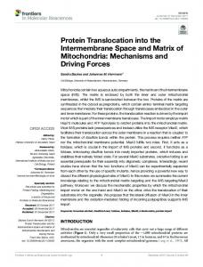
Results WTIP phylogeny. Sequence alignments suggest WTIP is a member of the zyxin family of LIM domain-containing proteins [(52) and figure 1A]. As indicated by analysis of nucleotide substitutions using an unrooted method (ClustalW, see methods), WTIP appears to belong to a zyxin subfamily, which is related to the Drosophila zyxin paralogue CG11063 (45), and is most similar to ajuba, which is contained in cadherin-based cell-cell contacts and translocates into nucleus to regulate mitotic commitment. WTIP LIM domains share significant sequence homology with other zyxin family paralogues (52), although ajuba and LIMD1 mRNA transcripts are not detected by RT-PCR in isolated glomeruli (A. Padiyar and J.R. Sedor, unpublished data). WTIP antibody specificity. We generated a rabbit anti-WTIP antibody against a GST-N∆WTIP fusion protein. To test specificity of the affinity purified WTIP antibody, we immunoblotted lysates from COS7 cells transiently expressing either the immunogen (myc epitope-tagged N∆WTIP), a myc epitope-tagged full length WTIP, FLAG-epitope tagged ajuba or myc epitopetagged zyxin (figure 1B). The WTIP antibody detected a single band of the appropriate size for N∆WTIP and full length WTIP and did not cross-react with ajuba or zyxin, other LIM domain proteins that also translocate from sites of cell adhesion into the cell nucleus (22; 37; 53). No proteins were recognized in control COS7 cells transfected with empty vector (pCMV-Tag). Western blot analysis of podocyte cell lysates revealed that both differentiated and undifferentiated podocytes expressed equivalent amounts of WTIP (figure 1C), suggesting that WTIP function would be regulated by its subcellular localization rather than by differences in abundance. Antibody specificity was verified by competition of anti-WTIP binding with the 6 x His-WTIP fusion protein (figure 1C). The same single band was detected in rat glomerular
12
lysates (not shown). To further characterize the antibody for immunocytochemistry, we transiently transfected COS7 cells with myc-WTIP. Both the confocal image (figure 1D, middle panel) and quantification of signal intensity (figure 1D, right panel) demonstrate complete overlap of the signal from affinity purified anti-WTIP (green channel,) and anti-myc antibodies (red channel). Untransfected COS7 cells were also fixed and incubated with both commercial anti-myc and affinity-purified anti-WTIP antibodies. A low power magnification shows only DAPI-stained nuclei (figure 1D, left panel). No signal could be detected from either channel in untransfected cells indicating that the affinity purified anti-WTIP antibody specifically recognizes WTIP and does not cross-react with other cellular proteins Localization of endogenous WTIP. Since the anti-WTIP antibody detected endogenous podocyte WTIP by Western blotting (figure 1C), we next localized WTIP in cultured podocytes. In undifferentiated podocytes, WTIP was detected in the nucleus as well as at the plasma membranes (figure 1E). In differentiated podocytes, nuclear localization of WTIP is reduced and WTIP localization is relatively more prominent in cytoplasms and plasma membrane (figure 1E). Significant perinuclear WTIP expression was observed in both differentiated and undifferentiated podocytes. Co-localization of ZO-1 and WTIP at podocyte adherens junctions. Since endogenous WTIP is localized in the cell periphery and we have hypothesized that it would be localized to the specialized cell junction of the differentiated podocytes (52), we next characterized the cell-cell contacts of cultured differentiated podocytes. Differentiated podocytes form cell-cell contacts, which can be visualized with Cell Tracker (figure 2A) and contain both ZO-1 and a cadherin identified by an anti-pan-cadherin antibody. As previously reported by Mundel and co-workers, P-cadherin is the major cadherin isoform expressed in cultured, immortalized podocytes. E-
13
cadherin does not localize at cell-cell junctions but is distributed diffusely throughout cytoplasm (44). A specific anti-P-cadherin antibody confirmed that the cadherin expressed at cell-cell contacts of cultured podocytes was P-cadherin (not shown). High-resolution immunofluorescence micrographs revealed that P-cadherin and ZO-1 are juxtaposed at most cellcell junctions in differentiated podocytes (figure 2B). ZO-1 localizes to the cytoplasmic side of filtration slits, where it has been shown to co-localize with P-cadherin (21) and to interact with Neph1 (20), a slit diaphragm protein of the immunoglobulin superfamily with a PDZ binding site. As reported, WTIP contains a PDZ binding domain, suggesting it may associate with ZO1 at sites of cell-cell contact. To test this hypothesis, cells were seeded on collagen-coated permeable Transwell filters to maximize epithelial polarity, differentiated for 10-18 days until synaptopodin was expressed and then assessed for co-localization of WTIP with ZO-1 by confocal microscopy. WTIP and ZO-1 co-localize precisely (figure 2C), suggesting that ZO-1 and WTIP may be components of a multi-protein complex of slit diaphragm proteins. MDCK cells abundantly express ZO-1, and immunoprecipitation assays, using a 6 x His-WTIP fusion protein, confirmed WTIP physically associated with ZO-1 (figure 2D). Podocyte injury with puromycin aminonucleoside (PAN) causes cytoskeletal reorganization and loss of synaptopodin expression. In vivo, podocyte differentiation state is critical for establishment and maintenance of the slit diaphragm, which functions to exclude proteins from the ultrafiltrate. In order to have a functional correlate to assess podocytes injury, we first developed a method to measure vectorial BSA diffusion across podocytes, which were maintained on permeable Transwell supports, as an assay of filtration barrier function. Using this system, confluent but undifferentiated podocytes permitted much greater albumin flux over time
14
compared to confluent and differentiated podocytes (figure 2E, left panel). At 2 hr, albumin diffusion across filters on which undifferentiated podocytes were cultured was not different compared to Transwell filters on which no cells were cultured (figure 2E, right panel). In contrast, albumin transit was significantly less across Transwell filters that contained differentiated podocytes. While this in vitro assay does not fully model the complexity of the podocyte slit diaphragm, it does provide information about barrier function of cell-cell contacts. Consistent with this premise, MDCK cells, which develop tight junctions, do not permit significant albumin diffusion as expected (not shown). We next treated cultured podocytes with PAN, which causes podocyte injury and proteinuria in animal models (25; 54) and determined its effects on podocyte morphology, cytoskeleton and cell-cell junction assembly . We detected podocyte morphology changes with 100 µg/ml PAN, which has been used before to simulate injury in cultured mouse podocytes (13; 47). Figure 3A shows that differentiated podocytes robustly express filamentous actin, whereas podocytes treated with PAN displayed remarkable rearrangement of actin filaments. These data are consistent with a prior report that PAN promotes cytoskeletal changes in podocytes (47). Upon closer examination of adherens junctions, actin filaments were highly organized in control, differentiated podocytes (figures 3A and 3B, upper left panels). Conversely, after treatment with PAN, actin filaments were distributed in a less organized, actin mat (figures 3A and 3B, lower left panels), consistent with observations of podocytes in situ from animal models of glomerular injury (51). Tubulin arrangement paralleled actin patterns in control cells, but was not as dramatically affected by treatment with PAN (figure 3B, right panels). Withdrawal of PAN permitted podocytes to revert to a normal morphology (not shown), suggesting that PAN did not stimulate irreversible cell death after the 24-hour treatment. Moreover, morphological analysis of
15
DAPI-stained nuclei revealed no significant difference in frequency of apoptosis between PANtreated (0.3%) and untreated (0%) podocytes (n > 300 cells for each treatment [not shown]). Differentiated podocytes assemble extensive P-cadherin-based adhesive contacts between cells (figure 3C, left panel), which disassemble after PAN treatment and permit cells to retract from one another (Figure 3C). Consistent with this morphological change, PAN also led to enhanced intercellular albumin diffusion (figure 3D, right panel). In contrast to untreated differentiated podocytes, albumin easily diffused across Transwell filters containing equivalent number of PAN-treated podocytes. Phalloidin staining of parallel filters demonstrated similar numbers of attached treated and untreated cells (figure 3D, left panels), suggesting that increased diffusion of albumin in these conditions is not due to cell loss. Cell counts also confirmed that PAN treatment did not cause significant cell detachment. The actin-binding protein, synaptopodin, was highly expressed in cultured, differentiated podocytes (figures 3E and 3F), in agreement with previous reports (33). After PAN treatment, synaptopodin distribution became punctate (figure 3E), and expression was reduced (figure 3E and 3F), consistent the dysregulated phenotype that characterizes some human glomerular diseases (5). As expected, undifferentiated podocytes did not express synaptopodin (figure 3F). PAN induces translocation of WTIP and ZO-1 from cell junctions to the nucleus. Slit diaphragm injury causes disassembly of ZO-1 from cell junctions (32; 40), and injury to nonpodocyte epithelial cells causes ZO-1 to traffic from cell junctions to the nucleus (15; 46). Similarly, PAN treatment caused ZO-1 to disassemble from the podocyte cell-cell junctions and relocate to the nucleus (figure 4A, upper panels). We previously demonstrated that WTIP contains a nuclear export signal and shuttled between the nucleus and cytosol, a process that was disrupted by treatment with the nuclear export inhibitor leptomycin B (52). Consistent with data
16
from Figures 1 and 2, WTIP localized in perinuclear regions and at cell-cell contacts in differentiated podocytes. However, upon PAN treatment, WTIP moved to the nucleus (figure 4A, lower panels), similar to the re-distribution pattern for ZO-1. Quantification of the nuclear fluorescence showed that nuclear WTIP content increased significantly after PAN treatment, as did ZO-1. In contrast, nuclear content of the general transcription factor TFIIβ was unchanged with PAN treatment (Figure 4 B). Immunoblot analysis of nuclear lysates also showed increased levels of endogenous ZO-1 and WTIP in PAN-treated podocytes compared to untreated controls (figure 4C) in contrast to the unchanged expression of WT1. To confirm nuclear translocation of WTIP, we transfected podocytes with an adenoviral construct that encoded a GFP-tagged full length WTIP. Compared to the untreated cells, nuclear lysates from PAN-treated podocytes revealed a robust increase in the amount of WTIP fusion protein as detected by an anti-GFP antibody (Figure 4C, bottom panel). Our prior work in WTIP-expressing 3T3 and HeLa cells showed that WTIP inhibited WT1-dependent transcription using an amphiregulin promoter-luciferase reporter assay (52). In the current studies, we examined the effect of PAN on expression of the retinoblastoma binding protein Rbbp7 (also called RbAp46), an endogenous kidney protein expressed during nephrogenesis and a known WT1 target gene (17). Translocation of WTIP in PAN-treated cells was associated with reduced expression of Rbbp7 protein (figure 4D), a finding consistent with our previous work but that does not exclude WTIP effects on post-transcriptional regulatory mechanisms. The data suggest that WTIP may play a pathophysiological role in the podocyte, by translocating from cell junctions to the nucleus, where it regulates WT1-dependent podocyte differentiation.
17
Discussion Podocyte differentiation is critical for the filtration function of the glomerulus (44). The tumor suppressor gene WT1 regulates podocyte differentiation (34; 43) and is mutated in syndromes of familial glomerulosclerosis (5; 6; 18) We have demonstrated that WT1 SNPs are associated with focal segmental glomerulosclerosis in African Americans, suggesting dysfunction of WT1 pathways may contribute to more common causes of nephropathy (39). We have identified WT1 Interacting Protein (WTIP) and hypothesized it functions both as a scaffold for slit diaphragm proteins and a co-repressor of WT1 transcriptional activity by shuttling from cell-cell adhesions to nucleus after injury (52). Consistent with our hypothesis, WTIP, as well as ZO-1, translocated from podocyte adherens junctions to nuclei in PAN-treated cells, findings associated with loss of the podocyte marker differentiation marker, synaptopodin, and increased albumin transit across podocytes cultured on Transwell filters. Taken together, this study and our published data suggest WTIP monitors podocyte cell-cell junction assembly and shuttles into the nucleus after podocyte injury, translating changes in junctional structure into altered gene expression and a less differentiated phenotype. In vivo, podocyte differentiation is required for normal filtration barrier function, and podocyte injury results in proteinuria (5; 6). Differentiated, cultured podocytes develop cell-cell junctions that are modified adherens junctions, containing P-cadherin and ZO-1 (44). WTIP colocalizes and physically interacts with ZO-1 at these cell junctions. PAN, an agent that causes proteinuria in animal models (31), has been proposed to cause changes in podocyte morphology by the same mechanisms as fibroblast growth factor-2 (48). In our studies, PAN caused podocyte actin filaments at cell-cell contacts to reorganize into a subcortical ring of F-actin, similar to the diffuse cytoplasmic sheet along the glomerular basement membrane described in experimental
18
glomerulopathies (51). We used an albumin diffusion assay as a functional correlate to corroborate our immunocytochemical findings in vehicle- and PAN-treated podocytes. PANtreated, differentiated podocytes permitted greater albumin diffusion compared to untreated, differentiated podocytes. Total protein levels in upper compartments from filters containing PAN-treated, differentiated podocytes was similar to that measured in filters with undifferentiated podocytes and significantly greater than that of filters seeded with differentiated, but untreated, podocytes. Of course, this in vitro assay incompletely models the barrier of slit diaphragm, since cultured podocytes only express some of the protein components (35; 44). We believe that loss of mature, cell-cell contacts after PAN treatment is the proximate cause of increased albumin diffusion since undifferentiated, but confluent, podocytes without mature cellcell contacts permit albumin diffusion similar to that observed across filters without cells. Coupled with the immunocytochemical changes that demonstrate disassembly of cadherin-based cell-cell contacts and nuclear translocation of both WTIP and ZO-1 with PAN treatment, the data suggest that injured podocytes use these molecules to signal changes in the nucleus concomitant with albumin leak. Of course, these data do not directly demonstrate that WTIP is necessary for either assembly of cell-cell contacts in vitro or filtration barrier function in vivo. Further experiments are needed to demonstrate the functional importance of WTIP and its translocation into nuclei after podocyte injury, experiments that are ongoing in our laboratory. The correct temporal and spatial expression of the Wilm’s tumor suppressor gene (WT1) is critical for the induction and maintenance of a differentiated podocyte phenotype. Although persistent expression of WT1 protein in podocyte nuclei suggests that podocyte differentiation requires ongoing transcription of WT1-dependent genes, none of the previously known WT1 protein partners appear to regulate podocyte WT1. Another WT1 interacting protein (WTAP)
19
was identified through yeast two-hybrid screening and localizes in nuclear spliceosomes but its function in kidney is unknown (28). Given the increasing number of molecules known to shuttle between cytoplasm and nucleus to regulate gene expression, we speculated that specific molecules may transmit information from the filtration barrier to the nucleus to allow the podocyte to regulate its differentiation state in response to environmental signals. A yeast two hybrid assay screen identified WTIP as a candidate molecule for this function (52). WTIP LIM domains are similar to LIM domains in zyxin, the prototype protein for a family of molecules that localize to cell adhesion junctions (37; 38). The LIM domain is a conserved zinc finger protein-interaction motif, and proteins containing LIM domains mediate cytoskeletal organization, cell lineage specification, organogenesis and oncogenesis (1; 2; 10; 24; 27). A number of LIM domain-containing zyxin paralogues shuttle from sites of cell-cell contacts to the nucleus and can regulate cell differentiation state (22; 41; 42; 50; 53). Nuclear accumulation of ajuba, which is phylogenetically closest of the zyxin paralogues to WTIP, regulates endodermal differentiation (22) and mitotic commitment (19). Other junctional proteins also translocate to the nuclei of epithelial or endothelial cells and bind to transcription factors, including ZO-1, PECAM-1 and β-catenin (4; 7; 8; 14; 15; 46). ZO-1, a MAGUK family member, tethers the Y-box transcription factor ZONAB (ZO-1associated nucleic acid binding protein) at cell-cell contacts but translocates into nuclei with disruption of these junctional complexes to influence cellular proliferation and expression of the EGF receptor Erb2b and E-cadherin (3; 4; 15). In this study, we demonstrated that PAN-induced podocyte injury stimulates ZO-1 shuttling to the nucleus. These data suggest that cell junctions, including the highly specialized podocyte adherens junction, contain proteins that transmit information to the nucleus and regulate cell responses to extracellular stresses and function as
20
molecular switching stations. We suggest that molecules such as WTIP and ZO-1, either independently or in concert, signal the podocyte to simplify its differentiation state in response to environmental stress, a potentially adaptive response that would promote cell survival until the resolution of the injury. The response of human and experimental glomerular diseases to therapy suggests that podocytes can reestablish their differentiated phenotype. Altered subcellular compartmentalization of signaling molecules has been implicated in human disease pathogenesis. IGF-1-activated Akt phosphorylated of p27Kip1 in human breast cancers and prevented its import into the nucleus (30), showing that an extracellular stimulus can change cell phenotype by redirecting an intracellular signaling molecule to a different subcellular location. While the in vitro data contained in this paper support our hypothesis, translocation of junctional molecules like WTIP and ZO-1 from sites of cell-cell contact into the nucleus needs to be demonstrated in animal models of podocyte injury. When WTIP is retained in the nucleus, our prior data suggest that it functions as a transcriptional repressor of WT1 activity (52). In the current study, we determined that translocation of WTIP into nuclei of PAN-treated podocytes was associated decreased expression of the WT1 target gene Rbbp7 (also known as RbAp46). While this data is consistent with WTIP function as a transcriptional repressor, our studies do not exclude regulation at the level of RNA processing or translation. WT1 has been shown to bind to RNA and associate with proteins comprising the splicing machinery (11). WT1 also can shuttle into cytoplasm, where it is associated with actively processing polysomes suggesting a role in regulation of translation (36). Further experiments are needed to specifically determine if WTIP interferes with both WT1-dependent transcriptional and post-transcriptional regulation of gene expression.
21
Rbbp7 is a retinoblastoma (Rb)-associated protein, which was discovered to be upregulated by WT1 using suppression subtractive hybridization PCR and co-expressed with WT1 in E13 developing kidney (17). Reduction in Rbbp7 protein expression might have important consequences for podocyte development, since Rbbp7 is a member of the NuRD and Sin3 transcriptional co-repressor complexes and has been shown to repress the c-FOS transactivation domain (55). In addition, its C. elegans orthologs have been shown to inhibit Rasdependent signaling during worm development (56). Although podocytes do not express Ras isoforms in normal glomeruli, however, expression of some isoforms is observed in biopsies from a variety of glomerular diseases (26). Downregulation of Rbbp7 promotes Ras signaling either directly by increasing Ras expression or indirectly by disinhibiting Ras-dependent signaling pathways. The evidence presented in this study for WTIP-dependent repression of WT1 transcriptional activity is indirect. Chromatin immunoprecipitation assays are necessary to prove that WTIP functions as a transcriptional repressor but require that actual WT1 target genes are known. WT1 has been shown to both transcriptionally activate and repress a number of target genes in vitro using promoter-reporter assays, but none of these target genes have been confirmed in vivo either in cells or in animals. Based upon the data in this report, our model of WTIP function in the podocyte continues to evolve. In normal glomeruli WTIP is part of a multi-protein complex in podocyte foot process at cell-cell contacts. After injury, WTIP translocates into the nucleus, where it represses WT1dependent gene expression and dysregulates podocyte phenotype. Loss of WTIP from its location as cell-cell junctions may promote concomitant redistribution of slit diaphragm proteins, which leads to filtration barrier dysfunction. We suggest WTIP regulates podocyte phenotype by
22
monitoring slit diaphragm protein integrity, ultimately translating changes in slit diaphragm structure or function into altered expression of podocyte differentiation genes.
23
Acknowledgements
Support for this project was provided by National Institutes of Health grants DK07470, P50 DK054178, DK064719 and the Kidney Foundation of Ohio.
24
References 1. Akazawa H, Kudoh S, Mochizuki N, Takekoshi N, Takano H, Nagai T and Komuro I. A novel LIM protein Cal promotes cardiac differentiation by association with CSX/NKX2-5. J Cell Biol 164: 395-405, 2004.
2. Bach I. The LIM domain: regulation by association. Mech Dev 91: 5-17, 2000.
3. Balda MS, Garrett MD and Matter K. The ZO-1-associated Y-box factor ZONAB regulates epithelial cell proliferation and cell density. J Cell Biol 160: 423-432, 2003.
4. Balda MS and Matter K. The tight junction protein ZO-1 and an interacting transcription factor regulate ErbB-2 expression. EMBO J 19: 2024-2033, 2000.
5. Barisoni L, Kriz W, Mundel P and D'Agati V. The dysregulated podocyte phenotype: a novel concept in the pathogenesis of collapsing idiopathic focal segmental glomerulosclerosis and HIV-associated nephropathy. J Am Soc Nephrol 10: 51-61, 1999.
6. Barisoni L, Mokrzycki M, Sablay L, Nagata M, Yamase H and Mundel P. Podocyte cell cycle regulation and proliferation in collapsing glomerulopathies. Kidney Int 58: 137143, 2000.
7. Benmerah A, Scott M, Poupon V and Marullo S. Nuclear functions for plasma membrane-associated proteins? Traffic 4: 503-511, 2003.
25
8. Conacci-Sorrell M, Simcha I, Ben Yedidia T, Blechman J, Savagner P and Ben Ze'ev A. Autoregulation of E-cadherin expression by cadherin-cadherin interactions: the roles of beta-catenin signaling, Slug, and MAPK. J Cell Biol 163: 847-857, 2003.
9. Conti E and Izaurralde E. Nucleocytoplasmic transport enters the atomic age. Curr Opin Cell Biol 13: 310-319, 2001.
10. Dawid IB, Breen JJ and Toyama R. LIM domains: multiple roles as adapters and functional modifiers in protein interactions. Trends Genet 14: 156-162, 1998.
11. Discenza MT and Pelletier J. Insights into the physiological role of WT1 from studies of genetically modified mice. Physiol Genomics 16: 287-300, 2004.
12. Gama-Carvalho M and Carmo-Fonseca M. The rules and roles of nucleocytoplasmic shuttling proteins. FEBS Lett 498: 157-163, 2001.
13. Gloy J, Reitinger S, Fischer KG, Schreiber R, Boucherot A, Kunzelmann K, Mundel P and Pavenstadt H. Amino acid transport in podocytes. Am J Physiol Renal Physiol 278: F999-F1005, 2000.
14. Gorlich D and Kutay U. Transport between the cell nucleus and the cytoplasm. Annu Rev Cell Dev Biol 15: 607-660, 1999.
26
15. Gottardi CJ, Arpin M, Fanning AS and Louvard D. The junction-associated protein, zonula occludens-1, localizes to the nucleus before the maturation and during the remodeling of cell-cell contacts. Proc Natl Acad Sci USA 93: 10779-10784, 1996.
16. Gross SS and Levi R. Tetrahydrobiopterin synthesis. An absolute requirement for cytokine-induced nitric oxide generation by vascular smooth muscle. J Biol Chem 267: 25722-25729, 1992.
17. Guan LS, Rauchman M and Wang ZY. Induction of Rb-associated protein (RbAp46) by Wilms' tumor suppressor WT1 mediates growth inhibition. J Biol Chem 273: 2704727050, 1998.
18. Hastie ND. Dominant negative mutations in the Wilms tumour (WT1) gene cause Denys-Drash syndrome--proof that a tumour-suppressor gene plays a crucial role in normal genitourinary development. Hum Mol Genet 1: 293-295, 1992.
19. Hirota T, Kunitoku N, Sasayama T, Marumoto T, Zhang D, Nitta M, Hatakeyama K and Saya H. Aurora-A and an interacting activator, the LIM protein Ajuba, are required for mitotic commitment in human cells. Cell 114: 585-598, 2003.
20. Huber TB, Schmidts M, Gerke P, Schermer B, Zahn A, Hartleben B, Sellin L, Walz G and Benzing T. The carboxyl terminus of Neph family members binds to the PDZ domain protein zonula occludens-1. J Biol Chem 278: 13417-13421, 2003.
27
21. Johnson KR, Lu S, Murtha MT, Ruddle FH and Davisson MT. Genetic mapping of a new homeobox gene to mouse chromosome 7. Genomics 14: 1107-1109, 1992.
22. Kanungo J, Pratt SJ, Marie H and Longmore GD. Ajuba, a cytosolic LIM protein, shuttles into the nucleus and affects embryonal cell proliferation and fate decisions. Mol Biol Cell 11: 3299-3313, 2000.
23. Khan S, Cleveland RP, Koch CJ and Schelling JR. Hypoxia induces renal tubular epithelial cell apoptosis in chronic renal disease. Lab Invest 79: 1089-1099, 1999.
24. Khurana T, Khurana B and Noegel AA. LIM proteins: association with the actin cytoskeleton. Protoplasma 219: 1-12, 2002.
25. Kim YH, Goyal M, Kurnit D, Wharram B, Wiggins J, Holzman L, Kershaw D and Wiggins R. Podocyte depletion and glomerulosclerosis have a direct relationship in the PAN-treated rat. Kidney Int 60: 957-968, 2001.
26. Kocher HM, Moorhead J, Sharpe CC, Dockrell ME, Al Nawab M and Hendry BM. Expression of Ras GTPases in normal kidney and in glomerulonephritis. Nephrol Dial Transplant 18: 2284-2292, 2003.
27. Krause A, Zacharias W, Camarata T, Linkhart B, Law E, Lischke A, Miljan E and Simon HG. Tbx5 and Tbx4 transcription factors interact with a new chicken PDZ-LIM protein in limb and heart development. Dev Biol 273: 106-120, 2004.
28
28. Lee SB and Haber DA. Wilms tumor and the WT1 gene. Exp Cell Res 264: 74-99, 2001.
29. Li L, Backer J, Wong AS, Schwanke EL, Stewart BG and Pasdar M. Bcl-2 expression decreases cadherin-mediated cell-cell adhesion. J Cell Sci 116: 3687-3700, 2003.
30. Liang J, Zubovitz J, Petrocelli T, Kotchetkov R, Connor MK, Han K, Lee JH, Ciarallo S, Catzavelos C, Beniston R, Franssen E and Slingerland JM. PKB/Akt phosphorylates p27, impairs nuclear import of p27 and opposes p27-mediated G1 arrest. Nat Med 8: 1153-1160, 2002.
31. Lowenborg EK, Jaremko G and Berg UB. Glomerular function and morphology in puromycin aminonucleoside nephropathy in rats. Nephrol Dial Transplant 15: 15471555, 2000.
32. Macconi D, Ghilardi M, Bonassi ME, Mohamed EI, Abbate M, Colombi F, Remuzzi G and Remuzzi A. Effect of angiotensin-converting enzyme inhibition on glomerular basement membrane permeability and distribution of zonula occludens-1 in MWF rats. J Am Soc Nephrol 11: 477-489, 2000.
33. Mundel P, Gilbert P and Kriz W. Podocytes in glomerulus of rat kidney express a characteristic 44 KD protein. J Histochem Cytochem 39: 1047-1056, 1991.
29
34. Mundel P, Reiser J and Kriz W. Induction of differentiation in cultured rat and human podocytes. J Am Soc Nephrol 8: 697-705, 1997.
35. Mundel P, Reiser J, Zuniga Mejia BA, Pavenstadt H, Davidson GR, Kriz W and Zeller R. Rearrangements of the cytoskeleton and cell contacts induce process formation during differentiation of conditionally immortalized mouse podocyte cell lines. Exp Cell Res 236: 248-258, 1997.
36. Niksic M, Slight J, Sanford JR, Caceres JF and Hastie ND. The Wilms' tumour protein (WT1) shuttles between nucleus and cytoplasm and is present in functional polysomes. Hum Mol Genet 13: 463-471, 2004.
37. Nix DA and Beckerle MC. Nuclear-cytoplasmic shuttling of the focal contact protein, zyxin: a potential mechanism for communication between sites of cell adhesion and the nucleus. J Cell Biol 138: 1139-1147, 1997.
38. Nix DA, Fradelizi J, Bockholt S, Menichi B, Louvard D, Friederich E and Beckerle MC. Targeting of zyxin to sites of actin membrane interaction and to the nucleus. J Biol Chem 276: 34759-34767, 2001.
39. Orloff MS, Iyengar SK, Winkler CA, Goddard KA, Dart RA, Ahuja TS, Mokryzcki M, Briggs WA, Korbet SM, Kimmel PL, Simon EE, Trachtman H, Vlahov D, Michel DM, Berns JS, Smith MC, Schelling JR, Sedor JR and Kopp JB. Variants In
30
The Wilms Tumor Gene Are Associated With Focal Segmental Glomerulosclerosis In The African American Population. Physiol Genomics, in press, 2005 (ePUB February 1).
40. Patek CE, Fleming S, Miles CG, Bellamy CO, Ladomery M, Spraggon L, Mullins J, Hastie ND and Hooper ML. Murine Denys-Drash syndrome: evidence of podocyte dedifferentiation and systemic mediation of glomerulosclerosis. Hum Mol Genet 12: 23792394, 2003.
41. Petit MM, Fradelizi J, Golsteyn RM, Ayoubi TA, Menichi B, Louvard D, Van de Ven WJ and Friederich E. LPP, an actin cytoskeleton protein related to zyxin, harbors a nuclear export signal and transcriptional activation capacity. Mol Biol Cell 11: 117-129, 2000.
42. Petit MM, Meulemans SM, Alen P, Ayoubi TA, Jansen E and Van de Ven WJ. The tumor suppressor Scrib interacts with the zyxin-related protein LPP, which shuttles between cell adhesion sites and the nucleus. BMC Cell Biol 6: 1, 2005.
43. Quaggin SE. Transcriptional regulation of podocyte specification and differentiation. Microsc Res Tech 57: 208-211, 2002.
44. Reiser J, Kriz W, Kretzler M and Mundel P. The glomerular slit diaphragm is a modified adherens junction. J Am Soc Nephrol 11: 1-8, 2000.
31
45. Renfranz PJ, Siegrist SE, Stronach BE, Macalma T and Beckerle MC. Molecular and phylogenetic characterization of Zyx102, a Drosophila orthologue of the zyxin family that interacts with Drosophila Enabled. Gene 305: 13-26, 2003.
46. Riesen FK, Rothen-Rutishauser B and Wunderli-Allenspach H. A ZO1-GFP fusion protein to study the dynamics of tight junctions in living cells. Histochem Cell Biol 117: 307-315, 2002.
47. Saleem MA, Ni L, Witherden I, Tryggvason K, Ruotsalainen V, Mundel P and Mathieson PW. Co-localization of nephrin, podocin, and the actin cytoskeleton: evidence for a role in podocyte foot process formation. Am J Pathol 161: 1459-1466, 2002.
48. Sasaki T, Hatta H and Osawa G. Cytokines and podocyte injury: the mechanism of fibroblast growth factor 2-induced podocyte injury. Nephrol Dial Transplant 14 Suppl 1: 33-34, 1999.
49. Schreiber E, Matthias P, Muller MM and Schaffner W. Rapid detection of octamer binding proteins with 'mini-extracts', prepared from a small number of cells. Nucleic Acids Res 17: 6419, 1989.
50. Sharp TV, Munoz F, Bourboulia D, Presneau N, Darai E, Wang HW, Cannon M, Butcher DN, Nicholson AG, Klein G, Imreh S and Boshoff C. LIM domainscontaining protein 1 (LIMD1), a tumor suppressor encoded at chromosome 3p21.3, binds
32
pRB and represses E2F-driven transcription. Proc Natl Acad Sci USA 101: 16531-16536, 2004.
51. Shirato I. Podocyte process effacement in vivo. Microsc Res Tech 57: 241-246, 2002.
52. Srichai MB, Konieczkowski M, Padiyar A, Konieczkowski DJ, Mukherjee A, Hayden PS, Kamat S, El Meanawy MA, Khan S, Mundel P, Lee SB, Bruggeman LA, Schelling JR and Sedor JR. A WT1 co-regulator controls podocyte phenotype by shuttling between adhesion structures and nucleus. J Biol Chem 279: 14398-14408, 2004.
53. Wang Y and Gilmore TD. Zyxin and paxillin proteins: focal adhesion plaque LIM domain proteins go nuclear. Biochim Biophys Acta 1593: 115-120, 2003.
54. Yamazaki T. Podocytic degeneration and regeneration in puromycin aminonucleoside nephropathy in the rat. Pathol Int 45: 465-472, 1995.
55. Yang J, Kiefer S and Rauchman M. Characterization of the gene encoding mouse retinoblastoma binding protein-7, a component of chromatin-remodeling complexes. Genomics 80: 407-415, 2002.
56. Zhang Y, Ng HH, Erdjument-Bromage H, Tempst P, Bird A and Reinberg D. Analysis of the NuRD subunits reveals a histone deacetylase core complex and a connection with DNA methylation. Genes Dev 13: 1924-1935, 1999.
33
Figure Legends Figure 1. A. Predicted phylogenetic relationship between the indicated zyxin family, LIM domain-containing proteins. Protein sequences were analyzed using the ClustalW algorithm with the PAM 250 residue weight table within the Lasergene MegAlign module (DNASTAR). Human and mouse WTIP sequences have been reported (52). GenBankTM sequences include NP 116265.1 (human ajuba), AAF48328.2 (CG11063), NP 055055.1 (human LIM domains containing protein 1 [LIMD1]), NP 005569.1 (human lipoma partner protein (LPP)), NP 003293.1 (human thyroid receptor-interacting protein 6 [TRIP6]), and Q15942 (human zyxin). The units at the bottom of the tree indicate the number of substitution events. B. Cell lysates from COS7 cells transiently expressing a myc-tagged full length WTIP (myc-WTIP), myctagged N∆WTIP (myc-N∆WTIP), FLAG-tagged ajuba (FLAG-Ajuba) or myc-tagged zyxin (myc-zyxin) were separated by 4–20% SDS-PAGE and incubated with either anti-myc, antiFLAG or affinity purified, anti-WTIP antibodies as indicated. C. Equal amounts of cell lysates protein from undifferentiated and differentiated podocytes were separated by SDS-PAGE and transferred to Immobilon membrane. Western blots with the anti-WTIP antibody a single protein in lysates from both undifferentiated (1) and differentiated (2) podocytes that is competed by His-WTIP, a recombinant protein different from the immunogen (GST-N∆WTIP). D. Left panel, Low power field of DAPI-stained, untransfected COS7 cells were fixed and incubated with monoclonal anti-myc antibody (red channel) and affinity purified, anti-WTIP antibody (green channel). Absence of non-specific staining is demonstrated by the image, which shows on DAPIstained nuclei. Bar, 50 µm. Middle panel, Confocal image of a COS7 cell line transiently expressing myc-WTIP. Cells on slides were fixed and incubated with both monoclonal anti-myc antibody (red) and affinity purified, anti-WTIP antibody (green). Bar, 10 µm. Left panel, Signal
34
intensity was quantified using Leica TCS SP2 confocal system software. E. Immunolocalization of endogenous WTIP in of both undifferentiated and differentiated podocytes using affinity purified anti-WTIP antibody. In the undifferentiated podocytes, WTIP localizes in the nucleus (arrow head) as well as in the incipient cell-cell junctions (arrows) whereas in the differentiated podocyte nuclear localization is reduced and WTIP localizes slightly more to the cell-cell junctions. Both micrographs reveal significant perinuclear localization. Podocyte cell size increase significantly with differentiation (34). Bar, 50 µm Figure 2A. Confocal microscopy images showing two podocytes differentiated for 15 days and stained Cell Tracker (upper panels) to show cell morphology, anti-ZO-1 antibody (middle panels) and anti-cadherin antibody (lower panels) that recognizes E- and P-cadherin. Whole cells are shown on the left (Bar, 50 µm) and magnified images of cell-cell contacts on the right of each panel (Bar, 10 µm). B. Confocal zoom image of cell-cell contact between two podocytes differentiated for 15 days demonstrating that ZO-1 and P-cadherin are in close spatial association but do not necessarily overlap. Bar, 10 µm. C. Podocytes were seeded on collagen-coated Transwell filters and then analyzed by confocal microscopy for localization of WTIP (green, panel on left) and ZO-1 (red, middle panel). Panel on left shows merged images and demonstrates close association between both molecules. Bar, 50 µm. D. His-WTIP and endogenous MDCK ZO-1 co-precipitate (see Methods). E, Left panel. A representative graph of time course BSA-diffusion across collagen-coated Transwell filters alone or seeded with podocytes. F (
), collagen-coated filter only; UND (
5x10³ undifferentiated podocytes; (
), collagen-coated filter seeded with
) collagen-coated filter seeded with 5x10³ podocytes and
differentiated for 8-12 days. Right panel. After 2 hr, BSA diffusion was quantified in Transwell assays using collagen coated filters (F), collagen-coated filters seeded with 5x10³
35
undifferentiated podocytes (UND), collagen filter seeded with 5x10³ (8-12)-days-differentiated podocytes. N=5 independent experiments. Data presented as mean + S.E. *p < 0.01 by KrusalWallis ANOVA followed by individual comparisons of medians using Dunn’s method. Figure 3 A. Differentiated podocytes treated with vehicle (upper panels) or with 100 µg/mL PAN for 24h (lower panels), which promoted dramatic changes in actin rearrangements and in overall cell morphology. Bar, 50 µm. B. Confocal, zoom image of a cell-cell contact between two podocytes differentiated for 15 d showing actin filament (left panel) and tubulin filament (right panels) arrangement in untreated (top panels) and PAN (100 µg/mL, 24h)-treated cells (bottom panels). Bar, 10 µm. C. Upper panels, Confocal images showing P-cadherin staining of differentiated podocytes treated with either vehicle or with 100 µg/mL PAN for 24h. Bar, 50 µm Lower panels show a zoom image of cell-cell contacts. Bar, 50 µm D, Left panel. Fluorescence photograph of Phalloidin stained collagen-coated Transwell filters with control podocytes (upper) or podocytes treated with 100 µg/mL PAN for 24 h showing similar cell density for both conditions. Right panel. Albumin-diffusion assay collagen-coated filters alone (F), collagencoated filters coated seeded with 5 x 10³ podocytes and differentiated for 10-days in the absence (D) or presence of PAN (P) for 24 h. Albumin diffusion was allowed to proceed for 2 hr and protein in the upper quantified as described in the Methods. *p < 0.05 using the Krusal-Wallis ANOVA followed by between group comparisons of medians using Dunn’s method. E. Confocal image of an untreated or PAN-treated, differentiated podocyte stained for the differentiation marker synaptopodin. Bar, 50 µm. F. Western blot for synaptopodin demonstrating reduced expression of the protein levels after treatment with PAN (100 µg/mL, 24h). Figure 4A. Confocal images (representative of n = 4) of podocytes (seeded at 5x10³/filter) and differentiated for 10-days. Top panels, ZO-1 distribution in control (left) and PAN (100 µg/mL,
36
24h, right)-treated cells. Bottom panels, WTIP distribution in control (left) and PAN (100 µg/mL, 24h, right)-treated cells. After PAN treatment, both proteins show translocation into nuclei and diminished localization at cell contacts. Bar, 50 µm. Each panel shows an image from a different filter. Colocalization of WTIP and ZO-1 in podocytes from the same filter was not evaluated in this experiment. B. Quantification of nuclear fluorescence intensity of nuclei as defined by TOPO nuclear dye from each condition (see methods for details). After PAN treatment, both WTIP and ZO-1 reveal a significant increase in fluorescence intensity but not the nuclear transcription factor TFIIβ. Data presented as mean + S.E. *p < 0.05 using the Student’s t test or Mann-Whitney rank sum test. C, control cells, P, PAN-treated cells. C. Western blot of nuclear extracts from equal numbers of control and PAN-treated, differentiated podocytes. Both ZO-1 and WTIP protein are increased. In contrast, WT1 expression is constant. Nuclear extracts from podocytes expressing GFP-tagged WTIP confirmed the same result. D. Western blot analysis demonstrating reduced expression of the WT1 target gene Rbbp7 (RbAp46) in total lysates of control and PAN-treated podocytes. In contrast, WT1 levels remain constant, suggesting that reduced Rbbp7 expression is specific and not a reflection of cytotoxicity.
37
mouse WTIP human WTIP human ajuba human LIMD1 Drosophila CG11063 human LPP human Trip6 human zyxin Drosophila zyxin (Zyx102)
A
87.4 80
70
60
50
40
30
20
10
0
A ju c-W ba TIP my c-N ∆W my TI c-Z yxi P n my c-W TIP my c-N ∆W my TI c-Z y xi P n
G
C
G-
MV -FL FL A A
1
2
1
2
kDa 250
my
pC
my
c-W
TIP
yc
ba
MV -m
pC
-Aj u
MV -FL
FL AG
pC
TIP
c-W
my
pC
MV -m
yc
B
AG
Nucleotide Substitutions (x100)
140
kDa 250
80
140
WTIP >
80 40 30
40 30
18 α-WTIP
18
α -myc
α-Flag
α-WTIP
D
α-myc
α-WTIP
α-WTIP + HIS-WTIP
Intensity 250 200 150 10 µm
100 50 0 0
Ch1-T2
E
Undifferentiated
2
4
6
8
10 Distance (µM)
12
14
16
18
Ch1-T3
Differentiated
Rico et al.
Figure 1
A
B Cell tracker
Cell 1
ZO-1 Pan-cadherin
ZO-1
m 50 µ
Cell 2
Pan-Cadherin -
IP: WTIP WB: ZO-1
220 kDa
C
50µm
WTIP
35 f
30
undiff
25
diff
20 15 10 5 0 0
Merged
ZO 1
Protein (mg/ml)
E Protein (mg/ml)
m 50 µ
To tal L Be ysat e ad s Hi s-W TIP
D
1h
3h
6h
10 9 8 7 6 5 4 3 2 1 0
* F
UND
D
Time
Rico et al.
Figure 2
A
β-Tubulin
PAN-treated
Control PAN-treated
Cell 1
Cell 1
Cell 2
Cell 2
Cell 1
Cell 1
Cell 2
Cell 2
Cell 2
Control
Protein (mg/ml)
6 4
*
2
F
D
P
110 kDa55 kDa-
Un
PA N
ff
F
Di
PAN-treated
dif f
Cell 2
7
Differentiated
8
0
s-
Cell 2
Cell 1
Co
Cell 1
10
PAN-treated
PAN-treated
P-Cadherin
Control
Synaptopodin
β-Tubulin
F−Actin
D
C
E
B Control
F−Actin
Synaptopodin β-Tubulin
Rico et al.
Figure 3
N
C
PAN
PA
Control
Co nt ro l
A
ZO-1
ZO-1
-
WTIP WT-1
Co
N
nt
ro
l
D
PA
WTIP
GFP-WTIP
Rbbp7 WT-1
Fluorescence Intensity
B
**
*
**
*
C
P WTIP
C
P ZO-1
C
P TFIIβ
Rico et al.
Figure 4