Jan 22, 2013 - METHODS: To study MMP-2 activity, primary cultures of human periodontal fibroblasts were stimulated with the addition of TLRs 2.
10 – ORIGINAL ARTICLE MODELS, BIOLOGICAL
Zimography is an effective method for detection of matrix metalloproteinase 2 (MMP-2) activity in cultured human fibroblasts1 Rodolfo Assis LisboaI, Marcus Vinícius AndradeII, José Renan Cunha-MeloIII PhD, Department of Pathology, School of Medicine, UFMG, Belo Horizonte-MG, Brazil, Acquisition, analysis and interpretation of data, statistical analysis and manuscript writing. II PhD, Associate Professor, Department of Internal Medicine, School of Medicine, UFMG, Belo Horizonte-MG, Brazil. Interpretation of data, manuscript writing, critical revision, final approval of the version to be published. III PhD, Full Professor, Department of Surgery, School of Medicine, UFMG, Belo Horizonte-MG, Brazil. Substantive scientific and intellectual contributions to the study, responsible for conception and design, interpretation of data, manuscript writing, critical revision, final approval of the version to be published. I
ABSTRACT PURPOSE: To describe a method to characterize the gelatinase activity of cultured human periodontal fibroblasts stimulated with Pam3Cys and E. coli LPS, ligands of TLR2 and TLR4 respectively, and by centrifugation of the cultures, simulating an orthodontic force. METHODS: To study MMP-2 activity, primary cultures of human periodontal fibroblasts were stimulated with the addition of TLRs 2 and 4 ligands and the application of mechanical force by centrifugation at 141 x g for 30 min. Supernatant media was collected 24 hours later to perform protein quantification and zymography. RESULTS: MMP-2 activity suffered an increase in cultures co-stimulated with TLRs 2 and 4 ligands alone or with the presence of mechanical force application compared to basal levels. CONCLUSION: Zymography, one of the several methods to study MMPs activities, is a simple, qualitative and efficient method based on electrophoresis of bis-acrylamide gels copolymerized with a protein substrate. Key words: Zymography. Matrix Metalloproteinase 2. Fibroblasts. Periodontal Ligament. Toll-Like Receptors.
216 - Acta Cirúrgica Brasileira - Vol. 28 (3) 2013
Zimography is an effective method for detection of matrix metalloproteinase 2 (MMP-2) activity in cultured human fibroblasts Introduction Metalloproteinases
orthodontic force, as previously described1. (MMP)
are
Zn2+
dependent endopeptidases responsible for the degradation of extracellular matrix on physiological and pathological conditions such as inflammation, neoplasms, wound healing, angiogenesis and matrix remodeling. Collectively, they are capable of degrading all kinds of extracellular matrix proteins and also process a number of bioactive molecules1. These enzymes are produced and released from cells as a latent form (Pro-MMP) and are activated by the removal of the NH2-terminal pro-peptide of approximately 10 kDa. Their family are composed by 25 members divided on subfamilies: collagenases, gelatinases, matrilisyns, stromelisyns and membrane type MMPs2,3. The gelatinase sub-family is composed by gelatinase A (MMP-2) and gelatinase B (MMP-9) both capable of metabolizing native and denatured collagen, gelatin, elastin, laminin, fibronectin and the basement membrane1,2. Stromelysin (MMP-3) and matrilysin (MMP-7) also demonstrate broader substrate specificity like proteoglycans, gelatins, elastin, and glycoproteins4. Several methods have been reported on literature to study the expressions and activities of MMPs, as an example: ELISA, Western Blotting, Immunohistochemistry, fluorometric oligopeptide substrates, radiolabeled collagen and zymography1-5. Zymography is a simple and sensitive qualitative method based on the electrophoresis of SDS-PAGE gels copolymerized with a protein substrate. Gelatin is used as a protein substrate to evaluate the proteolytic activity of MMP-2 and MMP-9, casein for MMP-3, MMP-7 and MMP-10, and collagen for MMP-16,7. In a complex mixture as conditioned cultured cells media, biological fluids, or purified preparations, the zymography allows the identification of MMPs in their active and latent forms, based on molecular weight4-6,8. Toll-like receptors (TLRs) are a family of patternrecognition receptors responsible for triggering inflammatory responses to microbial invasion. They recognize and distinguish the pathogen-associated molecular patterns (PAMPs), which are highly conserved structures of microorganisms9. TLRs can initiate the intracellular signaling cascade leading to inflammatory and immune gene transcriptions like cytokines and matrix metalloproteinases (MMPs)10. The aim of the study is to demonstrate that the zymography can be used to evaluate the gelatinase activity of cultured human periodontal fibroblasts. The cells were stimulated with Pam3Cys and E. coli LPS, ligands of TLR2 and TLR4 respectively, or by centrifugation of the cultures at 141 x g for 30 min, a simulation of
Methods Fibroblast cultures Human periodontal fibroblasts were obtained from primary cultures of periodontal ligament explants. Healthy donors with indication for exodontia of enclosed third molars were selected at the Dental School of the Minas Gerais Federal University (UFMG), Brazil and invited to participate in the study, donating their extracted teeth. The study was approved by the Ethical Committee for Research, UFMG, Brazil (COEP-UFMG, license no. ETIC 0078.0.203.000-11 – 14/06/2011) and informed consent were obtained from all patients. Teeth which had been sectioned during the surgery and those who hadn’t more than half of the root formed were discarded due to difficulty to immobilize the crown for tissue extraction and lack of periodontal tissue. Extracted teeth were immersed in DMEM culture medium (Gibco, Grand Island, NY), with 1% antibiotic–antimycotic (Gibco) and 10% FCS (Sigma, St. Louis, MO) and transported to the Lineu Freire-Maia Laboratory at the Medical School, UFMG, Brazil. Teeth were rinsed with 70% ethanol solution followed by washing out with 0.9% NaCl. Periodontal ligament were removed from the middle third roots and fragmented. The explants were transferred to a 25cm2 culture flask containing the same culture medium used on transportation. Cultures were incubated at 37oC and 5% CO2 until confluence in which the medium was changed every three days. Fibroblasts were trypsinized (0.25% trypsin; trypsin-EDTA, Gibco) and transferred to a 75cm2 culture flask. On the third passage, the cells were seeded on 48 well plates for treatment. Culture stimulation Cells were washed out with 0.9% saline and a serum-free DMEM medium was added prior to stimulation. Experimental and control groups were planned. On the experimental groups, cultures were stimulated with 1 µg/mL of Pam3Cys or E. coli LPS or by centrifugation at 141 x g for 30 min or combination of the stimuli as follows: Control group (CT), Centrifugation group (CTc), TLR4 ligand (LPS), TLR2 ligand (P3C), TLR4 + centrifugation (LPSc), TLR2 + centrifugation (P3Cc), TLR2 + TLR4 (PL), TLR2 + TLR4 + centrifugation (PLc). The cells were stimulated for 24 hours in a 5% CO2 incubator and supernatant collected. Samples were stored at ‑20oC until the gelatin zymography assay.
Acta Cirúrgica Brasileira - Vol. 28 (3) 2013 - 217
Lisboa RA et al.
Cell viability assay After stimulation, cell viability was tested under optical microscopy after the addition of 0.4% Trypan blue (BioWhittaker, Walkersville, MD, cat. 17-942E). The numbers of stained and non stained cells were determined to avoid bias caused by stimulationinduced cell death.
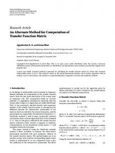
Sample derived from human periodontal fibroblast culture media collected 24 hours after stimulation revealed only activity for 72 kDa MMP-2 on gelatin zymography. MMP-9 was not detected as depicted in Figure 1.
Protein assay A BCA spectrophotometric (490-nm wavelength). Protein Assay Kit (Pierce, Rockford, IL), was used for total protein quantification of culture media samples Zymogram Gelatin zymograms were performed as follows: 10.5% SDS-polyacrylamide separating gels with 2 mg/mL of gelatin (30% bis-acrylamide, 1.5 M tris-HCL pH 8.8, 10% ammonium persulfate, 0.04% TEMED, 10% SDS and distilled water to reach final volume) and 5% SDS-polyacrylamide staking gels (30% bisacrylamide, 1 M tris-HCL pH 6.8, 10% ammonium persulfate, 0.1% TEMED, 10% SDS and distilled water to reach final volume) were prepared. Solutions were cast on Mini Protean (BioRad) with the addition of ammonium persulfate and TEMED to polymerize the gels (1 mm thickness). The gels were loaded with samples diluted in 1:1 non-reducing buffer (12.5% 0.5 M Tris–HCl pH 6.8, 10% glycerol, 4% SDS, and 0.05% bromophenol blue) and the electrophoresis was carried at 4oC using a constant current of 35 mA at 90 V for approximately 5 h. After disassembling the apparatus, gels were washed 2 X in 2.5% triton-x 100 for 15 minutes, incubated on incubation buffer (Tris–HCl 50 mM, CaCl2 10 mM, NaCl 50 mM, pH 7.6) for 18 hours at 37o C, stained by 0.1% Coomassie brilliant blue R-250 solution (with 40% methanol and 10% acetic acid) for 4 h under gentle shaking and destained 2 X for 20 minutes with 25% ethanol and 8% acetic acid solution. Gels were scanned at 600 dpi and analysed for densitometry at Kodak Molecular Imaging Software 4.0.5. The gels were washed out with distilled water and scanned (Genius ColorPage-Vivid Pro II at 600dpi) for densitometry (Kodak Molecular Imaging software v.4.0.1). Results Cell viability assay showed no significant difference between groups (Kruskal Wallis, p>0.05). Non-stained bands were shown at a blue background gel revealing the gelatin digestion by MMPs at its respective molecular weight.
218 - Acta Cirúrgica Brasileira - Vol. 28 (3) 2013
FIGURE 1 - A – Gelatin zymography showing the activity of 72 kDa MMP-2 present in culture medium samples. CT (Control group), CTc (Centrifugation group), LPS (TLR4 ligand group), P3C (TLR2 ligand group), LPSc (TLR4 ligand + centrifugation), P3Cc (TLR2 ligand + centrifugation), PL (TLR4 ligand + TLR2 ligand), PLc (TLR4 ligand + TLR2 ligand + centrifugation). B – Densitometry showing different activities of 72 kDa MMP 2 in medium samples showed in A. The 72 kDa form of MMP-2 showed increase activity in group PL (* Dunn’s posttest, p