CVI Accepts, published online ahead of print on 18 June 2008 Clin. Vaccine Immunol. doi:10.1128/CVI.00157-08 Copyright © 2008, American Society for Microbiology and/or the Listed Authors/Institutions. All Rights Reserved.
Development of a more efficient algorithm for identifying false-positive reactivity in a dengue IgM screening assay
Harry E. Prince* Cindy Yeh, and Mary Lapé-Nixon
D E
Focus Diagnostics Inc., Cypress, CA
*Corresponding author:
T P
5785 Corporate Avenue Cypress, CA 90630
E C
Telephone: 714 503-2047 FAX: 714 821-3364
E-mail:
[email protected]
C A
Running title: Modified dengue IgM testing algorithm
1
Abstract Of 2692 sera screened for dengue IgM using a mu-capture enzyme-linked immunosorbent assay (ELISA), 954 were equivocal (index 0.90-1.10) or positive (index >1.10), and retested using a background subtraction (BS) ELISA that identifies screen false-positives.
D E
No false-positives were found among 427 sera with screen-ELISA
indices >6.00; thus, retesting this specimen subset by BS-ELISA is unnecessary.
T P
E C
C A
2
Dengue viruses are flaviviruses transmitted among humans by Aedes mosquitoes (mainly Aedes aegypti) in tropical and subtropical areas worldwide (5,14).
Dengue virus
infections are associated with significant morbidity, ranging from a nonspecific febrile illness to severe hemorrhagic fever, and in rare cases are fatal (5-7).
D E
A major laboratory tool used in diagnosing dengue infections is measurement of dengue IgM in serum (5,6).
Most dengue IgM tests utilize the mu-capture enzyme-linked
T P
immunosorbent assay (ELISA) format (1,3,4,9,14), which employs capture wells coated
with anti-human IgM, inactivated dengue antigen, and enzyme-conjugated murine anti-
E C
flavivirus monoclonal antibody (reporter reagent).
Because captured IgM with
heterophilic antibody activity may yield false-positive results by directly binding the reporter reagent (8,10), we employ a two-step testing algorithm for dengue IgM
C A
detection. Sera are first screened using a mu-capture ELISA (one well per sample); equivocal or positive samples are then retested using a background subtraction (BS) modification of this same ELISA (two wells per sample), designed to identify false positive screening assay reactivity due to heterophile antibodies (8,12,13). Because the expected screening ELISA reactivity rate is 3.00 in the screening ELISA, 587 (99%) were positive in the BS ELISA. All 427 sera with indices
T P
>6.00 in the screening ELISA were positive in the BS ELISA.
E C
These findings demonstrate that all sera with strong reactivity (index >6.00) in the dengue IgM screening ELISA are also positive in the BS ELISA; thus, our dengue IgM testing algorithm can be modified to eliminate further testing of such sera in the BS
C A
ELISA. Application of this modified algorithm to the current dataset would have reduced the number of samples evaluated using the BS ELISA from 954 to 527, a reduction of 45%. If a laboratory’s quality assurance program allows an overall false positive rate 3.00; application of this algorithm to the current dataset would have reduced the number of samples tested by BS ELISA from 954 to 362 (a reduction of 62%), with only 5 of 2692 sera (0.19%) exhibiting false positive dengue IgM results.
The background subtraction approach for identifying false positive reactivity is routinely applied to other screening mu-capture ELISA systems besides dengue IgM (e.g., West
6
Nile virus IgM) (8,13). Thus, it stands to reason that these other screening assays, like the dengue IgM assay, may also have a characteristic high index cut point above which BS ELISA performance is not necessary. This cut point will undoubtedly vary among different assays, depending on the absorbance value of the screening ELISA calibrator
D E
and the dynamic range of the assay. Each laboratory must therefore define its own reflex testing algorithms for analytes measured by mu-capture ELISA.
T P
References
1. Branch, S. L., and P. N. Levett. 1999. Evaluation of four methods for detection
E C
of Immunoglobulin M antibodies to dengue virus. Clin. Diagn. Lab. Immunol. 6:555-557.
2. Centers for Disease Control and Prevention. 2007. Weekly dengue surveillance
C A
report: CDC Dengue Branch and Puerto Rico Department of Health. http://www.cdc.gov/ncidod/dvbid/dengue/documents/weeklyreport.pdf. Accessed January 30, 2008.
3. Chanama, S., S. Anantapreecha, A. A-nueggonpipat, A. Sa-gnasang, I. Kurane, and P. Sawanpanyalert. 2004. Analysis of specific IgM responses in secondary dengue virus infections: levels and positive rates in comparison with primary infections. J. Clin. Virol. 31:185-189. 4. Falconar, A. K., E. de Plata, and C. M. Romero-Vivas. Altered enzyme-linked immunosorbent assay immunoglobulin M (IgM)/IgG optical density ratios can correctly classify all primary and secondary dengue virus infections 1 day after
7
the onset of symptoms, when all of the viruses can be isolated. Clin. Vaccine Immunol. 13:1044-1051. 5. Gubler, D. J. 1998. Dengue and dengue hemorrhagic fever. Clin. Microbiol. Rev. 11:480-496.
D E
6. Guzman, M. G., and G. Kouri. 1996. Advances in dengue diagnosis. Clin. Diagn. Lab. Immunol. 3:621-627.
7. Hayes, E. B., and D. J. Gubler. 1992. Dengue and dengue hemorrhagic fever.
T P
Pediatr. Infect. Dis. 11:311-317.
8. Hogrefe, W. R., R. Moore, M. Lape-Nixon, M. Wagner, and H. E. Prince. 2004.
E C
Performance of Immunoglobulin G (IgG) and IgM enzyme-linked immunosorbent assays using a West Nile virus recombinant antigen (preM/E) for detection of West Nile virus- and other flavivirus-specific antibodies. J. Clin. Microbiol.
C A 42:4641-4648.
9. Koraka, P., C. Suharti, T. E. Setiati, A. T. A. Mairuhu, E. Van Gorp, C. E. Hack, M. Juffrie, J. Sutaryo, G. M. Van Der Meer, J. Groen, and A. D. M. E. Osterhaus. 2001.
Kinetics of dengue virus-specific serum immunoglobulin classes and
subclasses correlate with clinical outcome of infection.
J. Clin. Microbiol.
39:4332-4338. 10. Levinson, S. S., and J. J. Miller.
2002.
Toward a better understanding of
heterophile (and the like) antibody interferences with modern immunoassays. Clin. Chim. Acta 325:1-15. 11. Pan American Health Organization. 2008. 2007: Number of reported cases of dengue and dengue hemorrhagic fever (DHF), region of the Americas (by country
8
and
subregion).
http://www.paho.org/English/AD/DPC/CD/dengue-cases-
2007.htm. Accessed January 30, 2008. 12. Prince, H. E., and W. R. Hogrefe. 2003. Detection of West Nile virus (WNV)specific Immunoglobulin M in a reference laboratory setting during the 2002
D E
WNV season in the United States. Clin. Diagn. Lab. Immunol. 10:764-768.
13. Rawlins, M. L., E. M. Swenson, H. R. Hill, and C. M. Litwin. 2007. Evaluation
of an enzyme immunoassay for detection of Immunoglobulin M antibodies to
T P
West Nile virus and the importance of background subtraction in detecting nonspecific reactivity. Clin. Vaccine Immunol. 14:665-668.
E C
14. Rigau-Perez, J. G., G. G. Clark, D. J. Gubler, P. Reiter, E. J. Sanders, and A. V. Vorndam. 1998. Dengue and dengue haemorrhagic fever. Lancet 352:971-977.
C A
9
Table 1. Results from a representative dengue IgM screening ELISA run and subsequent BS ELISA run Specimen
Screening ELISA Absorbance
Index
Calibrator
0.254
Negative control
______________BS ELISA______________ Diluent well absorbance 0.107
Corrected absorbance 0.182
Index
NC*
Antigen well absorbance 0.289
0.097
0.38
0.086
0.071
0.015
0.08
Positive control
1.153
4.54
0.887
0.080
0.807
4.43
Screen false positive
0.664
2.61
0.534
0.063
0.35
True positive
0.500
1.97
0.413
0.256
1.41
True positive
2.745
2.750
15.11
T P
E C
*NC = not calculated
10.81
2.839
C A
10
D E
0.471
0.157 0.089
NC*
Table 2. Relationship of dengue IgM BS ELISA results to screening ELISA index values
Screen ELISA
BS ELISA
BS ELISA
BS ELISA positive (%)
index
N
negative (%)
equivocal (%)
0.90-1.10
93
74 (80)
13 (14)
1.11-2.00
180
66 (37)
22 (12)
2.01-3.00
89
9 (10)
2 (2)
3.01-4.00
53
2 (4)
4.01-5.00
54
2 (4)
5.01-6.00
58
1 (2)
6.01-8.00
97
0 (0)
8.01-10.00
103
10.01-14.00
E C
T P
D E
0 (0) 0 (0)
6 (6)
92 (51) 78 (88) 51 (96) 52 (96)
0 (0)
57 (98)
0 (0)
97 (100)
0 (0)
0 (0)
103 (100)
130
0 (0)
0 (0)
130 (100)
>14.00
97
0 (0)
0 (0)
97 (100)
TOTAL
954
154 (16)
37 (4)
763 (80)
C A
11
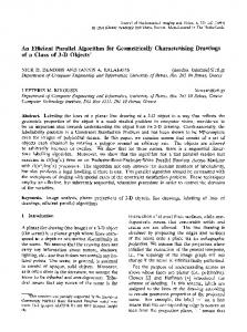
Figure legend
Figure 1. Summary of dengue IgM screening ELISA and BS ELISA results for 2962 consecutive sera submitted for dengue IgM testing. For both assays, indices 1.10 were considered positive.
T P
E C
C A
12
Samples tested for Dengue IgM 2692
Screen ELISA Negative 1738
Screen ELISA Equivocal 93
BS ELISA Negative 74
BS ELISA Equivocal 13
D E
T P
E C
C A
BS ELISA Positive 6
BS ELISA Negative 80
Screen ELISA Positive 861
BS ELISA Equivocal 24
BS ELISA Positive 757