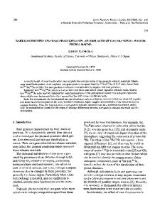
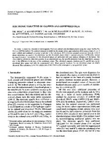
Feb 3, 1984 - the MBRS Program DRR/NIH Grant Number 06168 for financial assistance. We should also like to thank. Professor Michael Weiner (CCNY) for ...
Volume 104, number 2,3
CHEMICAL PHYSICS LETTERS
3 February 1984
THE EFFECT OF MOLECULAR STRUCTURE ON VOLTAGE INDUCED SHIFTS OF CHARGE TRANSFER EXCITATION IN SURFACE ENHANCED RAMAN SCATTERING John R. LOMBARDI, Ronald L. BIRKE, Luis A. SANCHEZ, Irene BERNARD and Song Cheng SUN City University o f New York, City College, Department of Chemistry, New York, New York 10031, USA Received 22 August 1983
The Raman intensity versus electrode voltage behavior of a series of substituted pyridines and saturated nitrogen heterocycUc compounds was studied at two laser excitation energies. The voltage (resonance) maximum shifts to more positive potential with increasing excitation energy for the substituted pyridines (case I) and to more negative potential with increasing excitation energy for the saturated nitrogen heterocyclic compounds (case II). The voltage maxima can be correlated with the Hammett sigma function for substituted pyridines and with pK a for the saturated nitrogen heterocycles. The results are consistent with charge transfer from the metal to molecule (adcluster-molecule complex) in the case I system and from the molecule (adcluster-molecule complex) to the metal in the case II system.
1. Introduction The possibility that a charge-transfer mechanism involving an adsorption induced electronic resonance is at least in part responsible for the phenomenon of surface enhanced Raman spectroscopy (SERS) was first suggested by theoretical work from this lab [1] in 1979. A qualitative discussion of charge transfer mechamisms was also given about the same time [2]. It has since been developed by several groups [ 3 - 7 ] , and stimulated numerous experiments [ 8 - 1 2 ] . The topic has been examined in several recent reviews [13,14]. The basic idea is that proximity to the surface and resulting mixing of molecule-metal energy levels results in a process similar to a resonance Raman effect. If the incident photon energy matches an electronic transition of the combined system, then the polarizabilities of particular vibrational modes of the system are drastically augmented. As in normal resonance Raman spectroscopy, the relative intensities o f the Raman lines differ considerably from those observed at non-resonant frequencies. Since the energies at which SERS is normally observed are in the visible, far below energies required to reach regular excited states of typical molecules involved, it is probable that the resonant state is some type of charge transfer 240
excitation of the molecule-metal complex. Indeed Demuth and Sanda [15] have observed a charge transfer excitation at 1.9 eV in the electron energy loss spectrum (EELS) of pyridine on the (111) surface of crystalline Ag. In addition there is some evidence that metallic adatoms play an important role in the process [14,16,17], since the enhancement factor is greatly diminished on a smooth metal surface. The latter effect could be attributed to the electrodynamic contribution to the mechanism. However, there is evidence that the important species is a molecule-adatom complex [ 1 8 - 2 0 ] most likely associated with an active site [ 8 - 1 2 , 2 1 ]. It is postulated that this complex may have allowed transitions which are absent in the bulk metal since the normal momentum selection rules imposed by highly delocalized electrons would be relaxed for such complexes. The usual picture then is that of photoexcitation of an electron in an adatom cluster causing charge transfer from the metal to the adsorbed molecule, followed by emission of a photon after the electron has returned to the metal while leaving the molecule in a vibrationally excited state. If the hole created by the initial excitation has not migrated, the energy of the scattered photon will be shifted by exactly the energy gain involved in the molecular transition. 0 009-2614/84/$ 0 3 . 0 0 © Elsevier Science Publishers B.V. (North-Holland Physics Publishing Division)
Volume 104, number 2,3
CHEMICALPHYSICSLETTERS
It is possible to scan such resonances by at least two techniques. One is to vary the excitation frequency, or alternatively one can vary the position of the metallic Fermi level by varying the applied potential as in an electrochemical cell [12,22]. In either case a maximum is reached and a resonance energy and its width may be determined [8]. Note that in this model as the excitation energy is increased the voltage at which resonance occurs should shift more positive. This was observed by Furtak and Macomber for pyridine on Ag. We concur with their measurements in this paper and extend them to several other molecules (substituted pyridines). However, there is also another possibility as pointed out originally in 1979 [1 ], and only recently revived [8] which is that charge transfer may also be possible from molecule to metal. In this case, the laser photon excites a molecular electron which has sufficient energy to tunnel into an empty metal band. On return to the molecule it scatters light at a shifted frequency leaving the molecule in an excited vibrational state. In this situation we should expect just the opposite behavior from above, namely that as the excitation energy is increased, the voltage at which resonance occurs should shift more negatively. This was recently observed by Furtak and Roy [10] for SCN-. In this paper we show this effect exists for a series of molecules with saturated nitrogen atoms. Furthermore, we have examined the nature of the shift of voltage at which resonance occurs with the chemical properties of the two series of molecules. These experiments allow us to draw conclusions about the types of energy levels of the adatom-molecule complex important in the enhancement mechanism. It is important to note that whatever the mechanism, it is highly unlikely that any molecular state but the ground state is involved in scattering. This is because, e.g. for pyridine, the observed frequency shifts exactly match those of the (solvated) ground state. For the first excited (A 1B1) state of pyridine the vibrational frequency of the totally symmetric mode is shifted to lower energies by over 20 cm -1 [23]. Similarly, vibrational frequencies of the ground state of the pyridine negative ion [24] displays shifts of up to 30 cm -1 to lower energy. Another important consideration to keep in mind is that the voltage at which resonance occurs varies with vibrational mode. This was first observed by Jeanmaire and van Duyne [25], and of course is con-
3 February 1984
sistent with the observation made above that a resonance Raman spectrum will involve considerably different relative intensities from a normal (non-resonant) Raman spectrum. This led Adrian [7] to include both Franck-Condon and Herzberg-Teller mechanisms in a calculation of charge-transfer effects. Recently [26], we have shown in piperidine that the voltage dependence of the relative intensities of vibrational bands of different symmetry could be explained by a field induced vibronic mixing mechanism similar to Herzberg-Teller types. It is clear then at any complete description of these surface induced charge transfer resonances, must include field effects on vibronic mixing of molecular wavefunctions. In this paper several molecules were studied in two vibrational modes, in order to examine more closely the charge transfer.
2. Experimental The experimental apparatus is similar to that described elsewhere [ 18,26]. Laser excitation was provided at two frequencies by a Spectra Physics model 64 argon ion laser (488.0) nm, and a model 590 dye laser, pumped by the argon laser, operated at 600.7 nm using rhodamine 6G. The laser intensity at the surface of the electrode was between 50 and 120 roW. All samples were dissolved in aqueous KC1 (0.10 M) solution to a concentration of 0.05 M and placed in an electrochemical cell. The pretreatment of the silver electrode was accomplished by applying a double-step oxidation-reduction cycle (ORC) from - 0 . 6 to 40.25 V where the potential was held for 2 s and then returned to -0.2 V. In some cases the anodic potential was +0.35 V. All potentials are versus the saturated calomel electrode (SCE). For each molecule an initial spectrum was obtained in order to select several reasonably intense vibrational modes to study. The spectrometer slits were widened to 2 0 0 - 2 0 0 - 2 0 0 / a m in order to insure that intensity variations do not result from the slight frequency shifts commonly observed in SERS. At the fixed selected scattering frequencies, voltages were scanned usually from 0.0 to - 1 . 2 V. Then the excitation frequency was changed and the process was repeated starting with the ORC.
241
Volume 104, number 2,3
CHEMICAL PHYSICS LETTERS
3. Results and discussion
3 February 1984
(becomes more positive) with excitation energy (table 1) and those for which resonant voltage decreases with excitation energy (table 2). Furtak and Macomber [8] showed that the resonance peak in pyridine changes linearly with excitation in this region. We therefore report in the fifth column o f tables 1 and 2 the slope defined as ( E l - E 2 ) / h ( 6 0 1 - 602) where Elm'2 are the voltages at which maxima are observed at laser frequencies 601 and 602" Thus molecules in tables 1 and 2 are distinguished by whether the slope is positive or negative. We take this as diagnostic as to whether charge transfer is from metal to molecule or the reverse respectively.
Typical results are shown in fig. 1. For 2-methylpyridine (2-picoline) a shift in laser excitation frequency from 488.0 to 600.7 nm causes a shift in the resonance voltage (voltage of maximum intensity) from - 0 . 6 3 tO - 0 . 7 6 V (fig. 1A); whereas, for imidazole the same frequency shift causes this voltage to shift from - 0 . 7 0 to - 0 . 5 0 V, i.e. in the opposite direction. All o f the results are summarized in tables 1 and 2 where we have divided the molecules into two classes, those for which the resonant voltage increases A
-.50
- .73
6 0 0 rlrrl
z
z
4 8 8 nm
'
''.a-.
'"'
8
.
. -.4 . .
.
.
. . 8.
.
E / V vs. SCE
Fig. 1. Relative Raman intensity versus voltage. (A) 0.05 M 2-methylpyridine in 0.1 M KCI for the 1008 cm-1 band. Scan rate: 20 mV/s. (B) 0.05 M imidazole in 0.1 M KCI for the 1163 cm-1 band. Scan rate: 10 mV/s.
Table 1 Molecules for which resonance voltage increases with excitation energy Molecule
Pyridine 2-methylpyridine 3-methylpyridine 4-methylpyridine 4-aminopyridine 3-cyanopyridine 242
Vibration (cm-1)
Voltage maximum (V) 488.0 nm (2.54 eV)
600.7 nm (2.06 eV)
1006 1008 1030 1008 1006 1027
-0.58 -0.63 -0.53 -0.66 -0.77 -0.23
-0.79 -0.76 -0.80 -0.85 -1.05 -0.41
Slope
ax
pKa
ref. [271
+0.44 +0.27 +0.56 +0.40 +0.58 +0.38
0.0 -0.22 -0.07 -0.17 +0.66 +0.56
5.25 5.97 5.68 6.02 9.11 1.5
I.P. ref. [27]
9.6 9.5 9.5 9.2 10.17
Volume 104, number 2,3
3 February 1984
CHEMICAL PHYSICS LETTERS
Table 2 Molecules for which resonance voltage decreases with excitation energy Molecule
Vibration (cm-1)
Voltage maximum (V)
Slope
pKa
ref. [27]
Quinuclidine Imidazole Pyrrolidine Piperidine Piperazine
1324 982 1163 1267 893 1022 1204 1020
488.0 nm (2.54 eV)
600.7 nm (2.06 eV)
-0.52 -0.55 -0.70 -0.71 -0.61 -0.63 -0.50 - 0.64 -0.61
-0.10 -0.17 -0.50 -0.50 -0.20 -0.12 -0.28 -0.32 -0.31
It is worthwhile to see if there are any apparent regularities in the chemical or physical properties o f the molecules in each table. Note firstly that all observed substituted pyridines with an extensive 7r system fall in table 1 whereas all saturated compounds fall in table 2. The only unsaturated compound which appears in table 2 is imidazole which, however, has a saturated nitrogen in its ring. Because o f these observations, it is tempting to implicate the low-lying unfilled 7r* orbitals o f the substituted pyridines in the metal to molecule charge transfer mechanism. That this is a risky course is made clear by several considerations. Firstly, there is no correlation between spectroscopic molecular zr* levels and the observed resonances. Secondly, energetically these orbitals are still considerably higher than can be conveniently reached by visible photon energies. Since charge transfer is involved, the molecule negative ion should be considered. However, these states tend to lie still further up in energy. Nenner and Schultz [24] showed by electron transmission spectroscopy that the electron affinity o f pyridine is 0.62 eV. On the other hand, this molecular negative ion state which is most likely the charge transfer state in metal to molecule charge transfer, could be significantly lowered by hydrogen bonding in solution [20] and the adsorption process. It is clear, then, that we can search the properties o f the molecular ground state for patterns in these results. In fig. 2 we have plotted - E m a x for the substituted pyridine compounds against increasing electron
-0.88 -0.79 -0.42 -0.44 -0.85 -1.06 -0.47 -0.67 -0.63
11.0 6.95 11.27 11.12 9.83
donating power o f the substituent on the pyridine ring, i.e., versus the Hammett o x constant [28]. Correlation coefficients of 0.96 are found with b o t h laser excitation frequencies. It should also be pointed out that extremely good correlations can be made for - E m a x versus t h e p K a ( - 0 . 9 8 for 601 nm and - 0 . 9 6 for 488
)nm
488nm Pyr
-Em
4 -NH 2
3-CH s
i:. •4
3,
.2
L 3,
.I
.z
o
-.z
-.4
-.e
-.e
-~.o
-tz
Sigma Fig. 2. Voltage maxima versus Hammett sigma function. Data in table 1. 243
Volume 104, number 2,3
CHEMICAL PHYSICS LETTERS
3 February 1984
I.O .9
-F m
.8
.7
488nm ~ m i d . 600nm~ . .
~iper.
Pyrr.
.6 -..
.5 .4
Zmid.
•
Pip.
~,,,~iper. APip.
.3 .2
Quin.
~
.I
pKo Fig. 3. Voltage maxima versus p K a. Data in table 2.
nm) and for - E m a x versus gas phase ionization potential (correlation coefficient of 0.997 for 4-NH 2, 4-CH3, 2-CH3, 3-CN, and pyridine itself). All of these correlations must reflect the substituent effect. For the other group of molecules with saturated nitrogens which are implicated in molecule to metal charge transfer, we have plotted - E m a x versus p K a (fig. 3). If the molecules are viewed as weakly bound to a metal adatom-cluster (or possibly to the d orbital of the metal) through the lone-pair of non-bonding electrons on the nitrogen atom, we would expect this interaction to be related to the electron donating strength of the molecule as measured by the p K a in solution [27]. In fig. 3, - E m a x decreases with increasing p K a (correlation coefficients of 0.95 for 601 nm and 0.80 for 488 nm). Note also that in all cases observed the magnitude of the slope is less than one except for pyrrolidine. From simple energetic considerations one might expect the slope to be +1 exactly. However, it is clear that other processes are important. Furtak and Macomber [8] have attributed this to lack of complete screening of the local electrostatic potential by the electrolyte. 244
We have taken this one step further by determining the effect of voltage induced mixing of vibronic states on the relative intensities (and therefore the observed resonance maxima) of different vibrational modes in piperidine [26]. Although these voltages are not sufficient to cause significant vibrational shifting, they can have a drastic effect on their relative intensities. Another contribution to the lowering of the slopes could be the effect of electrode voltage on the energy of the charge transfer state. It is clear that any comprehensive model of the charge transfer contribution to SERS must take into account the following observations: (1) For some molecules the voltage at which resonance occurs shifts more positively with increasing excitation frequency (positive slope) and for others it shifts more negatively (negative slope). (2) For those molecules with a positive slope we find that the resonance maximum becomes more negative with increasing electron donating power of the substituent, while for those with a negative slope the resonance maximum becomes more positive with increasing p K a .
Volume 104, number 2,3
CHEMICAL PItYSICS LETTERS
(3) The slopes observed are typically less than one in magnitude. These observations can be reconciled if we view the molecule as forming a weak complex with either, a single adatom or a small cluster of adatoms on the surface. It is assumed that the molecule binds weakly through a lone pair of electrons on the N atom and that the resulting splitting of the molecular orbitals of the adcluster-molecule complex EHOMO -- ENHOM O is strongly dependent on the electron donating properties ( P K a ) of the molecule. Since the observed Raman frequencies do not appreciably change from those o f the free molecule, it must be that this bond is so weak as to only slightly perturb the vibrations of the molecule. For the case of molecule to metal charge transfer, there must be a low lying charge transfer state, ECT, formed from an excited state of the adcluster-molecule complex and levels of the unf'dled conduction band and this state is strongly dependent on the electron donating power o f the substituent. The adsorption induced resonance energy, EHOMO -- ECT , c a n be tuned by either the electrode voltage, V, or the laser energy, f i w i , since the condition for the resonance transition is EHOMO -- ECT = EHOMO -- [EF(0 ) + e V ] - h¢o i , (1) where EF(0 ) is the Fermi energy o f an electron in the Ag electrode at an arbitrary zero potential and the energies are referenced to the vacuum level. This equation is expressed in terms Of EHoMO to emphasize that a ground state transition is involved. In fact, ECT = EF(0 ) + e V + fi¢o i. This expression has been simplified by omitting the damping term and a term due to the metal-molecule coupling which moves the resonance point to slightly below the Fermi surface [1,29]. When ~ w i is lowered the electrode voltage must be lowered (moved in a negative direction) to reestablish the resonance condition as indicated by the experimental results in fig. 2. Thus the line for 601 nm is always more negative than that for 488 run and the two lines are nearly parallel. Moreover, a change in the substituent on the molecule to a more electron donating group must cause the charge transfer level, ECT, to move to lower energies (with respect to the vacuum level) requiring a shift in the electrode voltage to more negative values to re-establish the resonance condition (fig.
3 February 1984
For the case o f charge transfer from the adclustermolecule complex to the metal, the mechanisms has not been previously considered in any detail. In order to explain our experimental results, we must assume that the lower molecular orbital of the adclustermolecule, ENHOM O, is the ground state of the photoexcited system. Now the adsorption induced resonance condition is ENHOM O -- [EF(0 ) + eV] = h¢o i .
(2)
Ifhcoi is lowered the electrode voltage must be increased (moved in a positive direction) to re-establish the resonance condition as seen in fig. 3. If the molecule is changed so that its p K a is larger and the bond between the adcluster and molecule strengthened, then the energy level ENHOM O is increased with respect to the vacuum level and the electrode voltage must be shifted in a positive direction to re-establish the resonance condition 2. This phenomenon gives the trend in - E m a x versus p K a in fig. 3. Fig. 4 shows the energy level diagrams for the two cases of adsorption induced charge transfer resonance Raman scattering. A tunneling barrier between metal and complex exists at the electrode surface which is not shown in the diagrams. In both cases, the energetics involve photoexcitation o f an electron from the ground state, to an excited state with return of the electron to the ground state and consequent Raman scattering leaving the molecule in the first excited vibrational state. This process is more easy to visualize for case II of complex to metal charge transfer. Chang and Laube [14] have discussed the charge transfer mechanism for case I of metal to complex charge transfer. In this mechanism, in order to give a ground state molecular spectrum, a stationary hole must be produced at an energy Of EHoMO which is destroyed by electron-hole recombination when the electron is returned to the metal. The exact evaluation of energy levels in both mechanisms must await further experimental observations and detailed calculations for the energy levels o f the adsorbed complex. It is clear, however, that the shifts in - E m a x with both molecular properties and ~6oi are consistent with the picture presented in fig. 4.
2). 245
Volume 104, number 2,3
CHEMICAL PttYSICS LETTERS
3 February 1984
h rl ECT
e.~,_ _ E c T
--EcT
E~7~
lhw
//z
eVi?
EF eVi/
eV3//z /
Eg'~ H
--E
H
~ E
N
--E
N
CaseI
EF .
eV'~--'~'~ ""el
-
t~(o,
--EH
--E
(b)
N
(C)
eVz "~/~l~
E
EH
Era,/
--E
(a)
/
t -~
EN
EH
EF ev, :~---I_~-e_-?E.
ETJ
EN ~
(a)
(b)
.....~-E N
(c)
Case IT Fig. 4. Energy level diagrams. Case I: Metal to adcluster-complex charge transfer. (a) Original resonance condition, (b) change in resonance voltage on shift in h w i to/ico 2, (c) change in resonance voltage on shift in ECT. Case II: Adcluster-complex to metal charge transfer. (a) Original resonance condition, (b) change in resonance voltage on shift in h w i to hw 2, (c) change in resonance voltage on shift in ENHOM O.
Acknowledgement
The authors are indebted to the CUNY (PSC-BHE) Faculty Research Award Program (RF-13909) and the MBRS Program DRR/NIH Grant Number 06168 246
for financial assistance.We should also like to thank Professor MichaelWeiner(CCNY) for helpful discussions.
Volume 104, number 2,3
CHEMICAL PHYSICS LETTERS
References [ 1 ] J.I. Gersten, R.L. Bffke and J.R. Lombardi, Phys. Rev. Letters 43 (1979) 147. [2] E. Burstein, Y.J. Chen, S. Lundquist and E. Tosatti, Solid State Commun. 21 (1979) 567. [3] H. Ueba, J. Chem. Phys. 73 (1980) 725. [4] B.N.J. Persson, Chem. Phys. Letters 82 (1981) 561. [51 K. Arya and R. Zeyher, Phys. Rev. B24 (1981) 1852. [6] P.H. Avouris and J.E. Demuth, J. Chem. Phys. 75 (1981)4783. [7] F.J. Adrian, J. Chem. Phys. 77 (1982) 5302. [8] T.E. Furtak and S.H. Macomber, Chem. Phys. Letters 95 (1983) 328. [9] J.R. Campbell and J.A. Creighton, J. Electroanal. Chem. 143 (1983) 353. [10] T.E. Furtak and D. Roy, Phys. Rev. Letters 50 (1983) 1301. [ 11 ] A.D. Ladouceur, D.E. Terault and R.R. Smardzewski, J. Chem. Phys. 78 (1983) 980; C.G. Blatchford, J.R. Campbell and J.A. Creighton, Surface Sci. 120 (1982)435. [12] J. Billman and A. Otto, Solid State Commun. (1983), to be published. [13] T.E. Furtak and J. Reyes, Surface Sci. 93 (1980) 351; I. Pockrand, Springer Tracts in Modern Physics, to be published. [14] R.K. Chang and B.L. Laube, CRC Critical Reviews in Solid State and Material Science, to be published.
3 February 1984
[15] J.E. Demuth and P.N. Sanda, Phys. Rev. Letters 47 (1981) 57. [16] J. Billman, G. Kovacs and A. Otto, Surface Sci. 92 (1980) 153. [17] A. Otto, Appl. Surface Sci. 6 (1980) 309. [18] K.A. Bunding, R.L. Birke and J.R. Lombardi, Chem. Phys. 54 (1980) 115. [19] J.R. Lombardi, A.E. Sheilds-Knight and R.L. Birke, Chem. Phys. Letters 79 (1981) 214. [20] V.V. Marinyuk, R.M. Lazorenko-Manevic and Ya.M. Kolotyrkin, J. Electroanal. Chem. 110 (1980) 111. [21] T. Watanabe, N. Yanagihara, K. Honda and B. Pettinger, Chem. Phys. Letters, to be published. [22] R.L. Birke, J.R. Lombardi and J.I. Gersten, Phys. Rev. Letters 43 (1979) 71. [23] V. Henri and P. Angenot, J. Chim. Phys. 33 (1936) 641. [24] I. Nenner and G.J. Schultz, J. Chem. Phys. 62 (1975) 1747. [25] D. Jeanmaire and R.P. van Duyne, J. Electroanal. Chem. 84 (1977) 1. [26] L. Sanchez, R.L. Birke and J.R. Lombardi, J. Phys. Chem., to be published. [27] B.G. Ramsey and F.A. Walker, J. Am. Chem. Soc. 96 (1974) 3314. [28] P. Zuman, Substituent effects in organic polarography (Plenum Press, New York, 1967). [29] U. Ueba and S. Ichirnura, J. Chem. Phys. 74 (1981) 3070.
247