3D dSTORM SEQUENCE-SPECIFIC VISUALIZATION OF HIGHLY REDUCED MITOCHONDRIAL DNA IN NUCLEOIDS OF PANCREATIC ISLET β-CELLS ISOLATED FROM DIABETIC GOTO KAKIZAKI RATS Nikola Capková1, Tomáš Špaček1, Vojtěch Pavluch1, Lukáš Alán1, Zuzana Berková2, František Saudek2 and Petr Ježek1 1Institute of Physiology, Czech Academy of Sciences v.v.i., Dept.75, Vídeňská 1083, Prague 4, 2Institute for Clinical and Experimental Medicine, Vídeňská 1958, Prague 4, Czech Republic E-mail:
[email protected]
Goto Kakizaki (GK) rats represent a useful model of type 2 diabetes, having hypertrophic pancreatic islets, lower number of β -cells in which highly reduced mitochondrial DNA exists within the fragmented mitochondrial network. Juette et. al., Nature Methods 2008
B
A
ND5 (EdU A647) – RED
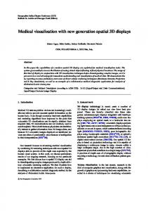
Fig. 4 – A, B images showing reconstructed nucleoid of insulinoma INS1E cells stained with ND5 PCR probe (length 932 bp) colored by EdU click it reaction with Alexa Fluor 647 on TFAM background colored by cy3B.
TFAM ( A647) - RED
Fig. 1 – GK isolated β-Cells stained by primary antiTFAM konjugated by Alexa Fluor 647
B
A
7S (Biotin) - GREEN
7S (Biotin) - GREEN
antiDNA (A647) - RED
Fig.3 - Principe of particle localization
antiDNA (A647) - RED
Fig. 2 - GK isolated β-Cells (A) vs. Wistar (B) stained with 7S PCR probe (250bp) incorporated with Biotin and colored by cy3B on antiDNA background colored by Alexa Fluor 647
D
C
ND6 (EdU A647) – RED
TWINKLE ( cy3b) - GREEN
ND6 (EdU A647) – RED
TWINKLE ( cy3b) - GREEN
Fig. 6 – C, D images showing reconstructed nucleoids of insulinoma INS1E cells stained with ND6 PCR probe colored by EdU click it reaction with Alexa Fluor 647 on TWINKLE background colored by cy3B
G
H
To visualize mtDNA by 3D dSTORM, we immunostained nucleoids of insulin-positive cells by TFAM, Twinkle and antiDNA antibodies. To visualize the specific sequences of mtDNA, we have constructed specific probes (ND5, DLOOP, 7S and ND6) by standard PCR reaction with incorporation of labeled nucleotides either with Alexa Fluor 647, Cy3B, CF568 or biotin. The typical probe had length between 100 and 1500 bp. The employed fluorophores were able to photoblink in reducing media, hence were suitable for dSTORM nanoscopy with ~25 nm x,y-resolution after their hybridization to specific mtDNA sequences. We have combined in situ hybridization [1] technique in the first channel with the dSTORM immunocytochemistry in a second channel, visualizing nucleoids by either anti-DNA, Twinkle or TFAM. 3D dSTORM measurement was performed on a Vutara SR-200 nanoscope. L
ND5 (EdU CF568) – RED
antiDNA (A647)- GREEN
ND5 (EdU CF568) – RED
antiDNA (A647)- GREEN
Fig. 7 – G, H images showing reconstructed nucleoids of insuinoma INS1E cells stained with ND5 PCR probe (length 215 bp) colored by EdU click it reaction with CF568 on antiDNA background colored by Alexa Fluor 647
K
TFAM (A647) - RED
Fig. 9 – Principle of EdU click it reaction for direct coloring of PCR probes
TFAM (cy3B) - GREEN
ND5 (EdU CF568) - GREEN
Fig. 10 – K, L images showing reconstructed nucleoids of insulinoma INS1E cells stained with ND5 PCR probe (length 932 bp) colored by EdU click it reaction with CF568 on antiTFAM background konjugated by Alexa Fluor 647
Fig. 5– Histogram of reconstructed nucleoid upwards
I
TWINKLE (A647) - RED
J
Fig. 8 – I, J images showing reconstructed nucleoids of Wistar cells stained with TWINKLE colored by A647
For nucleoids segmentation and their 3D rendering, Delaunay tessellation and subsequent modeling by principal component analysis will be used [2]. Using such double color 3D imaging, we can characterize positioning on/inside the nucleoid spheroids for socalled D-loop sequences of mtDNA, and we will compare it with the positioning of ND5 gene encoding sequences. We will be also able to resolve a distinct size distribution of nucleoids of the diabetic Goto Kakizaki β-cells.
[1] Alán L, Zelenka J, Ježek J, Dlasková A, Ježek P., Fluorescent in situ hybridization of mitochondrial DNA and RNA. Acta Biochim Pol 57(4):403-8; 2010. [2] Alán L, Špaček T, Ježek P., Delaunay algorithm and principal component analysis for 3D visualization of mitochondrial DNA nucleoids by Biplane FPALM/dSTORM. Eur Biophys J. 45(5):443-61; 2016.
This project was supported by GACR grant No. 17-08565S to A.D.