RESIDENT & FELLOW SECTION Section Editor Mitchell S.V. Elkind, MD, MS
Eric Lee, DO Chafic Karam, MD Nizar Chahin, MD
Correspondence to Dr. Lee:
[email protected]
Clinical Reasoning: A patient with rapidly progressive sensory loss and imbalance SECTION 1
A 52-year-old man presented with sudden onset of acral paresthesia and imbalance. The patient did not have any recent illness, sick contacts, or travel abroad. He denied weakness, pain, bowel or bladder incontinence, dysphagia, dysarthria, or shortness of breath. On neurologic examination, 1 month into his symptoms, he had reduced muscle strength in his finger spread, extension, and flexion on both sides graded on Medical Research Council scale 42/5 and in toe extensors 24/24 and toe flexors 24/4. The rest of his muscle strength was normal. Reflexes were absent throughout. He had reduced sensation to all modalities in a length-dependent
pattern up to his midshin and wrists on both sides. He was severely unsteady when walking and he could not tandem. Romberg was positive. The rest of his neurologic examination was normal apart from high arches and hammertoes. The patient had a family history of Charcot-Marie-Tooth disease type 1A (CMT1A) (PMP22 duplication) and was himself tested, although asymptomatic, and was also found to be carrying the mutation. Questions for consideration:
1. What is your preliminary differential diagnosis? 2. What initial investigations would you propose for this patient?
GO TO SECTION 2
From the Department of Neurology, University of North Carolina at Chapel Hill. Go to Neurology.org for full disclosures. Funding information and disclosures deemed relevant by the authors, if any, are provided at the end of the article. e140
© 2015 American Academy of Neurology
ª 2015 American Academy of Neurology. Unauthorized reproduction of this article is prohibited.
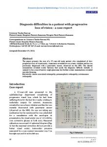
Figure 1
Waveforms generated from the motor nerve conduction studies of the left median and tibial nerves
Nerve conduction study of the left median (A) and tibial (B) nerves reveal markedly prolonged motor distal latencies with prominent temporal dispersion and slowed conduction velocities.
SECTION 2
Despite the genetically confirmed PMP22 mutation, the rapid onset and progression of this patient’s symptoms are concerning. A superimposed process should be considered and ruled out. For example, the patient can have a superimposed inflammatory polyneuropathy such as Guillain-Barre syndrome (GBS) or chronic inflammatory demyelinating polyradiculoneuropathy (CIDP) or a paraneoplastic or neoplastic neuropathy. Certain toxic exposures such as arsenic poisoning can cause subacute neuropathy with a similar presentation. Infectious agents such as Lyme disease, HIV, and hepatitis C can cause subacute GBS-like presentations. Vasculitis can cause sensory disturbance, although it typically presents with painful multifocal neuropathy. Meningeal carcinomatosis can present with rapid motor and sensory disturbance including cranial neuropathies. Finally, a central process such as a cervical cord lesion caused by spinal stenosis or a demyelinating lesion can cause similar bilateral sensory disturbance, although the absence of upper motor neuron signs and a sensory level do not support this. To narrow the diagnosis, the patient underwent additional testing, including nerve conduction study and EMG, MRI of the spine, and CSF Table 1
examination. Nerve conductions studies showed absent sensory responses of the upper and lower limbs with demyelinating features of the motor responses (figure 1, table 1). Table 1 demonstrates markedly prolonged motor distal latencies (DL) (DL in the arms 16–27 ms [normal , 4.2 ms], DL in the legs 14–26 ms [normal , 5.7 ms]). Marked prolonged compound muscle action potential durations as seen in the left median (45 ms at distal site and 50 ms at proximal site) and tibial nerves (47 ms at distal site and 53 ms at proximal site) are consistent with temporal dispersion. On needle EMG, there was abnormal spontaneous activity in the form of fibrillation potentials in the distal legs and longduration, high-amplitude, polyphasic motor unit potentials with reduced recruitment in all muscles tested. A lumbar puncture was performed to rule out an infectious etiology and meningeal carcinomatosis. CSF protein was 70 mg/dL, CSF glucose was normal, cell count was 0. A complete spine MRI revealed mild enhancement of the cervical and lumbar roots. Additional blood tests were performed to rule out other acquired causes of neuropathy. CSF immunoglobulin G index, oligoclonal bands, CSF Venereal Disease Research Laboratory (VDRL),
Values generated from the motor nerve conduction studies of the left median and tibial nerves
Nerve stimulation
Recording site
Latency, ms
Amplitude, mV
Velocity, ms 18.0 . 49
L median
Wrist
27.0 , 4.4
1.5 . 4.2
L median
Elbow
39.7 . 4.0
1.0 . 4.2
L median F response, ms
Wrist
Absent
L tibial
Ankle
14.4 , 5.7
0.9 . 2.8
L tibial
Knee
49.4 . 6.0
0.3 . 2.8
L tibial F response, ms
Ankle
Absent
Neurology 84
Duration, ms 45.0 50.0
11.0 . 41
47.0 53.0
May 12, 2015
e141
ª 2015 American Academy of Neurology. Unauthorized reproduction of this article is prohibited.
angiotensin-converting enzyme, vascular endothelial growth factor, GM1 antibody, cryoglobulin, myelin-associated glycoprotein Western blot, hemoglobin A1c, methylmalonic acid, serum immunofixation, serum free light chains, thyroidstimulating hormone, free T4, complete blood count, B12, sedimentation rate, antinuclear antibodies, extractable nuclear antigen, tissue
transglutaminase immunoglobulin A, hepatitis C antibody, Lyme serology, HIV, and hepatitis B surface antigen were all normal or negative. Question for consideration:
1. What is your diagnosis based on the history, clinical examination, and EMG and laboratory studies?
GO TO SECTION 3
e142
Neurology 84
May 12, 2015
ª 2015 American Academy of Neurology. Unauthorized reproduction of this article is prohibited.
SECTION 3
The uniformly severely slowed conduction velocities are usually consistent with a severe, inherited, demyelinating, sensorimotor peripheral neuropathy such as can be seen in CMT1A. However, the marked temporal dispersion and the reduced recruitment in both distal and proximal muscles are atypical for a patient with asymptomatic CMT1A. Temporal dispersion can sometimes be seen in inherited neuropathies, but the degree of temporal dispersion seen in this patient suggests the presence of a superimposed inflammatory process. The acute onset of paresthesia and imbalance along with the nerve conduction and EMG findings are highly suggestive of a superimposed acquired inflammatory and demyelinating process. The workup ruled out any infectious, paraneoplastic, neoplastic, or metabolic causes. The mildly elevated CSF protein with albuminocytologic dissociation can be seen in either acquired or inherited processes. The patient presented with 4 weeks of progressive symptoms. He did not meet the time course for the diagnosis of CIDP, which is defined as a progression of symptoms for more than 8 weeks. This patient met all the mandatory electrophysiologic criteria1 for acute inflammatory demyelinating polyneuropathy/CIDP, which require only 3 of 4 of the following: 1. Significant reduction in motor nerve conduction velocity in 2 or more motor nerves: a. ,80% of lower limit of normal (LLN) if CMAP .80% of LLN b. ,70% of LLN if CMAP ,80% of LLN 2. Partial conduction block or abnormal temporal dispersion in one or more motor nerves: peroneal
nerve between ankle and below fibular head, median nerve between wrist and elbow, or ulnar nerve between wrist and below elbow a. Criteria suggestive of partial conduction block: ,15% change in duration between proximal and distal sites and .20% drop in negative peak (2p) area or peak-to-peak (p-p) amplitude between proximal and distal sites b. Criteria for abnormal temporal dispersion and possible conduction block: .15% change in duration between proximal and distal sites and .20% drop in 2p area or p-p amplitude between proximal and distal sites 3. Significant prolongation of distal motor latency in 2 or more motor nerves: a. .125% of upper limit of normal (ULN) if CMAP .80% of LLN b. .150% of ULN if CMAP ,80% of LLN 4. Significant prolongation or absence of F-waves in 2 or more motor nerves: a. .120% of ULN if CMAP .80% of LLN b. .150% of ULN if CMAP ,80% of LLN The patient also fulfilled mandatory CSF study findings as he had cytoalbumin dissociation and negative CSF VDRL. Questions for consideration:
1. What treatment would you propose in this situation? 2. What findings would you expect on repeat EMG studies after treatment?
GO TO SECTION 4
Neurology 84
May 12, 2015
e143
ª 2015 American Academy of Neurology. Unauthorized reproduction of this article is prohibited.
Figure 2
Waveforms generated from the motor nerve conduction studies of the left median and tibial nerves after treatment
The follow-up nerve conduction study of the left median (A) and tibial (B) nerves 9 months after the initial study and 1 month after his last treatment with IVIg reveal profound improvement of distal latency, amplitude, and duration, including temporal dispersion and conduction velocity.
SECTION 4
The most widely used treatments for inflammatory neuropathy such as CIDP consist of IV immunoglobulin (IVIg), plasma exchange, and corticosteroids.2 The treatments appear to be equally effective in studies performed over time. The initial dose of IVIg is typically 2 g/kg infused over 4 to 5 days, as was the case with this patient. Pulse IV methylprednisolone (1,000 mg/day) for 3 days can be given with tapering doses given once weekly, then once monthly. The therapy is initiated early in the course of the disease to prevent continuing demyelination and secondary axonal loss leading to permanent disability.2 Our patient was treated with IVIg with complete resolution of his symptoms and normalization of his examination, including deep tendon reflexes. The benefit of treatment is seen in improvement of mobility and strength as well as the return of sensory disturbance to baseline. With treatment of demyelinating peripheral neuropathies, the examiner can see in follow-up studies direct recovery of distal latency, duration, amplitude, and conduction velocity, as noted in figure 2. In consideration of the long-term, sustained improvements in axonal excitability and other measures, clinical improvement seen Table 2
with long-term IVIg therapy may reflect improvements in biophysical properties and possibly even promotion of remyelination and prevention of secondary axonal degeneration (table 2).3 DISCUSSION The
challenge of this case highlights 2 separate pathologic processes that typically do not occur together. The typical findings of a hereditary neuropathy include symmetric distal limb weakness and negative sensory symptoms, isolated absent Achilles or diffusely reduced or absent reflexes, pes cavus, hammertoes, symmetrically and uniformly slowed nerve conduction velocities, and a family history of neuropathy. The stereotypical findings of an acquired neuropathy include symmetric or asymmetric distal or proximal weakness and positive sensory symptoms, diffusely absent reflexes, asymmetric and nonuniformly slowed nerve conduction velocities, and temporal dispersion or conduction block at noncompressive sites. The patient presented herein had both hereditary and acquired neuropathies. An observation such as this case has been described previously, with the authors postulating that patients may have an inflammatory-demyelinating hereditary motor and sensory neuropathy.4
Values generated from the motor nerve conduction studies of the left median and tibial nerves after treatment
Nerve stimulation
Recording site
Latency, ms
Amplitude, mV
Velocity, ms 27 . 49
L median
Wrist
8.8 , 4.4
6.3 . 4.2
L median
Elbow
17.1 . 4.0
5.6 . 4.2
L median F response, ms
Wrist
44 , 31
L tibial
Ankle
7.3 , 5.7
4.2 . 2.8
L tibial
Knee
26.3 . 6.0
2.2 . 2.8
L tibial F response, ms
Ankle
89 , 56
e144
Neurology 84
Duration, ms 7 7
21 . 41
17 17
May 12, 2015
ª 2015 American Academy of Neurology. Unauthorized reproduction of this article is prohibited.
A question has been proposed over the last decade as to whether patients with hereditary neuropathy are more susceptible to inflammatory neuropathy than the general population. It has been suggested and accumulating evidence exists4–9 for a superimposed inflammatory process in a subgroup of patients with CMT disease that is not genotype-specific. It could be related to genetic susceptibility or may relate to a disturbance of the normal function of the protein encoded by the affected gene. The take-home message is that if a patient with CMT disease experiences an acute or subacute deterioration in clinical condition, a search for other causes should be undertaken, and treatment of a coexistent inflammatory neuropathy with immunotherapy should be considered.10 AUTHOR CONTRIBUTIONS Eric Lee: drafting/revising the manuscript, study concept or design, analysis or interpretation of data, accepts responsibility for conduct of research and final approval. Chafic Karam: drafting/revising the manuscript, accepts responsibility for conduct of research and final approval. Nizar Chahin: drafting/revising the manuscript, study concept or design, analysis or interpretation of data, accepts responsibility for conduct of research and final approval, acquisition of data, study supervision.
STUDY FUNDING No targeted funding reported.
DISCLOSURE E. Lee reports no disclosures relevant to the manuscript. C. Karam serves as WriteClick Neurology® deputy editor. N. Chahin reports no disclosures relevant to the manuscript. Go to Neurology.org for full disclosures.
REFERENCES 1. Research criteria for diagnosis of chronic inflammatory demyelinating polyneuropathy (CIDP): report from an Ad Hoc Subcommittee of the American Academy of Neurology AIDS Task Force. Neurology 1991;41:617–618. 2. Köller H, Kieseier BC, Jander S, Hartung H-P. Chronic inflammatory demyelinating polyneuropathy. N Engl J Med 2005;352:1343–1356. 3. Lin CS, Krishnan AV, Park SB, Kiernan MC. Modulatory effects on axonal function after intravenous immunoglobulin therapy in chronic inflammatory demyelinating polyneuropathy. Arch Neurol 2011;68:862–869. 4. Dyck PJ, Swanson CJ, Low PA, Bartleson JD, Lambert EH. Prednisone-responsive hereditary motor and sensory neuropathy. Mayo Clin Proc 1982;57: 239–246. 5. Malandrini A, Villanova M, Dotti MT, Federico A. Acute inflammatory neuropathy in Charcot-Marie-Tooth disease. Neurology 1999;52:859–861. 6. Vital A, Vital C, Lagueny A. Inflammatory demyelination in a patient with CMT1A. Muscle Nerve 2003;28: 373–376. 7. Desurkar A, Lin JP, Mills K, et al. Charcot-Marie-Tooth (CMT) disease 1A with superimposed inflammatory polyneuropathy in children. Neuropediatrics 2009;40:85–88. 8. Donaghy M, Sisodiya SM, Kennett R, McDonald B, Haites N, Bell C. Steroid responsive polyneuropathy in a family with a novel myelin protein zero mutation. J Neurol Neurosurg Psychiatry 2000;69:799–805. 9. Prensky AL, Dodson WE. The steroid treatment of hereditary motor and sensory neuropathy. Neuropediatrics 1984;15:203–207. 10. Ginsberg L, Malik O, Kenton AR, et al. Coexistent hereditary and inflammatory neuropathy. Brain 2004;127: 193–202.
Neurology 84
May 12, 2015
e145
ª 2015 American Academy of Neurology. Unauthorized reproduction of this article is prohibited.
Clinical Reasoning: A patient with rapidly progressive sensory loss and imbalance Eric Lee, Chafic Karam and Nizar Chahin Neurology 2015;84;e140-e145 DOI 10.1212/WNL.0000000000001564 This information is current as of May 11, 2015 Updated Information & Services
including high resolution figures, can be found at: http://www.neurology.org/content/84/19/e140.full.html
References
This article cites 10 articles, 4 of which you can access for free at: http://www.neurology.org/content/84/19/e140.full.html##ref-list-1
Subspecialty Collections
This article, along with others on similar topics, appears in the following collection(s): All Clinical Neurology http://www.neurology.org//cgi/collection/all_clinical_neurology All Neuromuscular Disease http://www.neurology.org//cgi/collection/all_neuromuscular_disease Chronic inflammatory demyelinating polyneuropathy http://www.neurology.org//cgi/collection/chronic_inflammatory_demy elinating_polyneuropathy EMG http://www.neurology.org//cgi/collection/emg Peripheral neuropathy http://www.neurology.org//cgi/collection/peripheral_neuropathy
Permissions & Licensing
Information about reproducing this article in parts (figures,tables) or in its entirety can be found online at: http://www.neurology.org/misc/about.xhtml#permissions
Reprints
Information about ordering reprints can be found online: http://www.neurology.org/misc/addir.xhtml#reprintsus
Neurology ® is the official journal of the American Academy of Neurology. Published continuously since 1951, it is now a weekly with 48 issues per year. Copyright © 2015 American Academy of Neurology. All rights reserved. Print ISSN: 0028-3878. Online ISSN: 1526-632X.