Martin Benning, Pia Heins and Martin Burger. Westfälische Wilhelms-Universität, Institute for Computational and Applied Mathematics,. Einsteinstr. 62, 48149 ...
A Solver for Dynamic PET Reconstructions based on Forward-Backward-Splitting Martin Benning, Pia Heins and Martin Burger Westfälische Wilhelms-Universität, Institute for Computational and Applied Mathematics, Einsteinstr. 62, 48149 Münster Abstract. Dynamic Positron Emission Tomography allows monitoring physiological processes within the body that can be described by kinetic parameters. However, recovery of these parameters often requires the solution of complex and nonlinear operator equations. Advanced operator splitting techniques allow incorporating a-priori knowledge, e.g. sparsity of minimizers with respect to an exponential basis, into the reconstruction process. Keywords: Dynamic Positron Emission Tomography, Inverse Problems, Operator Splitting, Poisson Noise
INTRODUCTION Positron Emission Tomography (PET) is a nuclear medicine imaging technique that - in comparison to other medical imaging techniques like Computerized Tomography (CT) or Magnetic Resonance Imaging (MRI) - allows monitoring of physiological instead of anatomical information. A radioactive tracer is injected into the subject that interacts with the bodies’ molecules in a way the particular tracer is designed for. Subsequently, the tracers’ radioactive emisson is measured via the detectors of a PET scanner. From a mathematical perspective, the aim of computing a PET reconstruction is basically similar to computing a CT reconstruction, since in the easiest setup the reconstruction involves the inversion of the Radon transform K with Z
(K f )(s, θ ) :=
f (x) dx ,
(1)
x·θ =s
if f is two-dimensional (for higher three dimensions the X-ray transform has to be inverted). However, a major difference to CT reconstruction is that the scanned emissions appear randomly and follow a Poisson process. Simplified, the whole process can be described as the solution of the inverse problem ℘(K f ) = g for f , with ℘ describing the Poisson process. A popular approach for solving this inverse problem based on computing the Maximum A-Posteriori (MAP) Likelihood for Poisson distributed data is to minimize � � � Z � g KLg (K f ) := g log + K f − g dθ ds . (2) Kf Σ The standard approach in order to minimize (2) is to use the standard Expectation Maximization (EM) Algorithm [6], i.e. to compute f via the fixed-point iteration � � fk ∗ g fk+1 = ∗ K . (3) K 1 K fk In the following we want to denote for notational simplicity one particular iterate fk+1 with fEM . In order to obtain satisfactory reconstructions, a certain amount of events (the emissions collected with the scanner) needs to be organized into so-called bins (the data g(θ , s)). The goal of dynamic PET reconstruction however is to analyze the tracer interactions over time, e.g. via specific mathematical models, e.g. [3, 9]. Therefore, the data has to be organized into several temporal bins g(θ , s,t). A simple way to compute dynamic PET reconstructions is to compute a standard EM reconstruction for every temporal bin independently. The obvious drawback of this approach is that temporal correlation between the bins is completely neglected. To overcome this problem, recent research has aimed at incorporating mathematical models for the analysis of tracer activity over time into the reconstruction process. The goal of this work is to present a general framework that allows the incorporation of even nonlinear models and the adding of additional smoothness features to the solution.
KINETIC MODELING In order to incorporate the temporal dependency between the bins into the reconstruction process we have to discuss models that incorporate this temporal dependency first. Appropriate operators can be derived on the basis of kinetic modeling of physiological parameters for Emission Tomography. An overview of the basics of these modeling techniques can be found in [13]. We want to present two particular models that have been devolped in previous works for dynamic PET reconstruction.
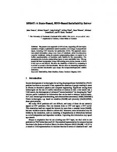
FIGURE 1. Transaxial views of 4D-H215O reconstructions. Each figure shows a particular transaxial slice of a 3D-volume for a certain timestep. From left to right: (1) Independent frames reconstruction, using the Standard EM algorithm only. (2) Independent frames reconstruction, using the Standard EM algorithm with additional Gaussian smoothing after each iterate. (3) Full 4D EM reconstruction, no additional smoothing. (4) Full 4D EM reconstruction, with additional Gaussian smoothing on each frame after each iterate. (5) Proposed full 4D EM reconstruction with TV and `1 regularization on the coefficients, as described in (8).
In [11], Reader et al. have developed the linear operator model Z t
B(h, a) := 0
N
h(τ) ∑ an (x) exp(−bn (t − τ)) dτ
(4)
n=1
for physiological parameters h(t) (input curve) and a(x) = (an (x))n={1,...,N} (coefficients). In [1, 2], we proposed a nonlinear variational model based on standard models for myocardial perfusion quantification developed in [3] and [9]. A similar model to the ones presented in [1, 2] is given by the nonlinear model operator B(F, S, h) := (1 − S(x))ω(x,t) + S(x)h(t) , � � ∂ω ω(x,t) (x,t) = F(x) h(t) − , ∂t λ
(5)
for physiological parameters F (myocardial perfusion), S (arterial spillover) and h (arterial input curve). In the following section we are going to discuss a general forward-backward scheme that allows their computation.
FORWARD-BACKWARD-SPLITTING After having introduced typical modeling operators for dynamic PET reconstruction, the basic aim of this work appears to be the solution of the general scheme (6) ( f , p; µ) = arg min max KLg (K f ) + h f − B(p), µi + αR(p) , f ,p µ {z } | {z } | =:I( f )
=:J( f ,p;µ)
with the operators B : P(Φ) → U(Ω) mapping a vector of physiological parameters p to an image sequence f , and K : U(Ω) → V (Σ) being the X-Ray-transform, mapping an image sequence f to list-mode or sinogram data g. Notice that B does not necessarily has to be linear but can also be nonlinear in at least one of its parameters. Furthermore, KLg represents the Kullback-Leibler functional as presented in (2) and R denotes the regularization functional guaranteeing certain smoothness features of the parameters p, e.g. a bounded total variation as proposed in [8]. In order to solve (6)
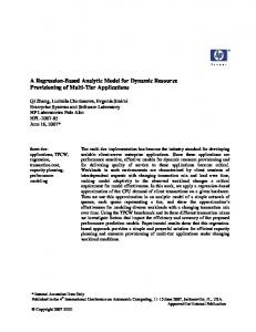
FIGURE 2. Reconstructions of physiological parameters from synthetic 2+1D H215O PET data, computed with the model presented in (9). From left to right: (1) Exact synthetic myocardial perfusion. (2) Reconstructed myocardial perfusion. (3) Exact synthetic spillover. (4) Reconstructed spillover.
we apply a forward-backward-splitting method with variable stepsize, i.e. fk+ 1 = fk − sk I 0 ( fk ) 2
fk+1 = fk+ 1 − sk ∂ f J( fk+1 , p; µ) ,
(7)
2
with sk =
τk f k K ∗ 1 , τk
0
> 0. Inserting I ( fk ) and ∂ f J( fk+1 , p; µ) leads to fk+1
= (1 − τk ) fk + τk fEM −
τk fk (µ + αq) , K∗1
with q ∈ ∂ R(p) being an element of the subgradient of R. Hence, fk+1 satisfies � � �Z T Z � fk+1 2 = arg min max wk ( f − ((1 − τk ) fk + τk fEM )) dx dt + τk (h f − B(p), µi + αR(p)) pk+1 f ,p µ 0 Ω ∗
with the weight wk := Kf 1 . Therefore, the proposed splitting scheme aims in first solving a standard EM step for each k temporal frame, and in a subsequent computation of fk+1 and pk+1 as minimizers of the weighted L2 -problem. The advantage on the one hand is that already developed EM algorithms for standard PET reconstruction can also be used for dynamic PET reconstruction, with all their preferable features as e.g. positivity preservation. On the other hand the weighted L2 -step can usually be solved with fast and efficient numerical schemes, so that the overall process allows easy and efficient implementation schemes.
COMPUTATIONAL RESULTS We want to incorporate the two model operators presented in the Kinetic Modeling Section into the operator splitting scheme developed above. Hence, we consider two specific variational models in order to compute regularized solutions to the inverse problem ℘(KB(p)) = g, such that the temporal correlation between the sinogram-datasets is considered and further regularization of the physiological parameters is allowed.
Linear 4D-EM Based on the linear operator B as described in (4), we want to compute a solution of the variational minimization problem ( ) N N Z γ 2 (h, an ) = arg min KLg (B(h, a)) + α ∑ |∇an | dx + β ∑ |an | + khkH 1 ([0,T ]) (8) 2 h,a n=1 Ω n=1 by using the forward-backward operator splitting method proposed in (7). The minimization of h and an is performed alternatingly; the weighted L2 -minimization for the spatial parameters an is computed via an augmented-Lagrangian implementation. Computational results of real 4D H215O-data can be seen in Figure 1.
Nonlinear 4D-EM Based on the nonlinear model operator as described in (5), we want to solve (6) with B as defined in (5). Figure 2 shows reconstructions of synthetic parameters for the minimization problem n �o α (9) (F, S, h) = arg min KLg (B(F, S, h)) + kFk2H 1 + kSk2H 1 + khk2H 1 2 with B(F, S, h) := (1 − S(x))ω(x,t) + S(x)h(t) , � � ∂ω ω(x,t) , (x,t) = F(x) h(t) − ∂t λ
(10)
computed via the forward-backward-splitting method (7). The weighted L2 -problem is minimized by solving the optimality conditions iteratively, as a mixture of analytically computed parameters and parameters computed via a gradient descent.
CONCLUSIONS We have proposed an efficient scheme for the solution of large scale inverse problems corrupted by Poisson noise as for example Dynamic PET reconstruction. The scheme allows the use of state-of-the-art standard EM algorithms (allowing explicit computation of frames) and efficient algorithms for regularized L2 -schemes, e.g. augmented Lagrangian based or primal dual Newton-type methods.
REFERENCES 1. 2. 3. 4. 5. 6. 7. 8. 9.
10. 11. 12. 13.
M. Benning. A Nonlinear Variational Method for Improved Quantification of Myocardial Blood Flow Using Dynamic H2 15 O PET. Diploma Thesis, 2008. M. Benning, T. Kösters, F. Wübbeling, K. Schäfers, and M. Burger. A Nonlinear Variational Method for Improved Quantification of Myocardial Blood Flow Using Dynamic H215O PET. IEEE Nuclear Science Symposium Conference Record, November 2008. S. R. Bergmann, K. A. Fox, and A. L. Rand. Quantification of Regional Myocardial Blood Flow in Vivo with H2 15 O. Circulation, 70:724–733, 1984. R. E. Carson and K. Lange. A Statistical Model for Positron Emission Tomography. Journal of the American Statistical Association, 80(389):20–22, 1985. K Chen, X Chen, R Renaut, G E Alexander, D Bandy, H Guo, and E M Reiman. Characterization of the image-derived carotid artery input function using independent component analysis for the quantitation of [18f] fluorodeoxyglucose positron emission tomography images. Physics in Medicine and Biology, 52(23):7055, 2007. A. P. Dempster, N. M. Laird, and D. B. Rubin. Maximum Likelihood from Incomplete Data via the EM Algorithm. Journal of the Royal Statistical Society, Series B, 39(1):1–38, 1977. Hongbin Guo, Rosemary A. Renaut, and Kewei Chen. An input function estimation method for fdg-pet human brain studies. Nuclear Medicine and Biology, 34(5):483 – 492, 2007. Hongbin Guo, Rosemary A. Renaut, Kewei Chen, and Eric Reiman. Fdg-pet parametric imaging by total variation minimization. Computerized Medical Imaging and Graphics, 33(4):295 – 303, 2009. H. Iida, I. Kanno, A. Takahashi, S. Miura, M. Murakami, K. Takahashi, Y. Ono, F. Shishido, A. Inugami, and N. Tomura. Measurement of absolute myocardial blood flow with H215O and dynamic positron-emission tomography. Strategy for quantification in relation to the partial-volume effect [published erratum appears in Circulation 1988 Oct;78(4):1078]. Circulation, 78(1):104–115, 1988. M. E. Kamasak, C. A. Bouman, E. D. Morris, and K. Sauer. Direct Reconstruction of Kinetic Parameter Images from Dynamic PET Data. IEEE Transactions on Medical Imaging, 24:636–650, 2005. A. J. Reader, J.C. Matthews, F. C. Sureau, C. Comtat, R. Trébossen, and I. Buvat. Fully 4D image reconstruction by estimation of an input function and spectral coefficients. IEEE Nuclear Science Symposium Conference Record, pages 3260–3267, 2007. A. J. Reader, F. C. Sureau, C. Comtat, R. Trébossen, and I. Buvat. Joint estimation of dynamic PET images and temporal basis functions using fully 4D ML-EM. Physics in Medicine and Biology, 51(21):5455–5474, 2006. M. N. Wernick and J. N. Aarsvold. Emission Tomography: The Fundamentals of PET and SPECT. Elsevier Academic Press, 2004.