G a s t r o i n t e s t i n a l I m a g i n g • Pe r s p e c t i ve Taylor et al. MRI Staging of Rectal Cancer
Downloaded from www.ajronline.org by 54.152.109.166 on 09/13/15 from IP address 54.152.109.166. Copyright ARRS. For personal use only; all rights reserved
Gastrointestinal Imaging Perspective
A Systematic Approach to the Interpretation of Preoperative Staging MRI for Rectal Cancer Fiona G. M. Taylor 1 Robert I. Swift 1 Lennart Blomqvist 2 Gina Brown 3 Taylor FGM, Swift RI, Blomqvist L, Brown G
OBJECTIVE. The purpose of this article is to provide an aid to the systematic evaluation of MRI in staging rectal cancer. CONCLUSION. MRI has been shown to be an effective tool for the accurate preoperative staging of rectal cancer. In the Magnetic Resonance Imaging and Rectal Cancer European Equivalence Study (MERCURY), imaging workshops were held for participating radiologists to ensure standardization of scan acquisition techniques and interpretation of the images. In this article, we report how the information was obtained and give examples of the images and how they are interpreted, with the aim of providing a systematic approach to the reporting process.
O
Keywords: MRI, preoperative staging, rectal cancer DOI:10.2214/AJR.08.1004 Received April 2, 2008; accepted after revision June 25, 2008. The research post held by F. G. M. Taylor is funded by the Croydon Colorectal Cancer Charity. The funding source had no involvement in the study design, collection, analysis, and interpretation of data, or in the writing of the report. The Pelican Cancer Foundation funded the original MERCURY study. 1 Department of Colorectal Surgery, Mayday University Hospital, Croydon, United Kingdom. 2 Department of Diagnostic Radiology, Karolinska University Hospital and Danderyds Hospital, Stockholm, Sweden. 3 Department of Radiology, Royal Marsden Hospital, Downs Rd., Sutton, Surrey SM2 5PT, United Kingdom. Address correspondence to G. Brown (
[email protected]).
CME This article is available for CME credit. See www.arrs.org for more information. AJR 2008; 191:1827–1835 0361–803X/08/1916–1827 © American Roentgen Ray Society
AJR:191, December 2008
ver the past few years, significant progress has been made in the management of rectal cancer. Advances in surgical technique and adjuvant therapies have led to significant improvements in outcome for some patients. The advances in preoperative therapies have led to the need for an accurate preoperative staging technique to select those patients who are most likely to benefit from these interventions without subjecting others to unnecessary treatment. Several studies have been published showing the ability of MRI to accurately stage rectal cancer and to clearly identify the relevant anatomy [1–5]. Although we accept that endoscopic sonography can show comparable accuracy with regard to T and N staging of rectal tumors, sonography has inherent problems. It is operator-dependent, and problems arise when scanning high or stricturing lesions. The limited field of view makes assessment of structures beyond the field of view difficult to interpret. Endoscopic sonography cannot accurately assess the circumferential resection margin or identify other prognostic features such as extramural venous invasion. Similarly, CT has shown poor results for the local staging of rectal lesions. The Magnetic Resonance Imaging and Rectal Cancer European Equivalence Study (MERCURY) study showed that high-resolution MRI can accurately predict involvement of the surgical resection margin (≤ 1
mm) and extramural tumor invasion. The study showed that MRI is reproducible and allows patients to be selected on this basis for preoperative treatment. As a result, this form of preoperative staging is more widespread and is becoming mandatory in certain countries (the United Kingdom, Denmark, Norway, and Sweden) in the management of rectal cancer [6–8]. In this article we describe a systematic approach to the interpretation of MR images that enables all clinically relevant structures to be adequately assessed. Technique We perform this examination for all patients with histologically proven rectal cancer as part of their staging process before initiating treatment. We recommend no bowel preparation, filling of the rectum with contrast agents, or air insufflation. IV or intramuscular antispasmodic agents are also not mandatory but can be helpful in improving image quality. IV contrast enhancement with gadolinium is not recommended for the staging of rectal cancer [9–11]. The technique for acquisition of the scans has previously been described [9], and the parameters are numerated in Table 1. A 1- or 1.5-T system is used with phased-array coils. These coils maintain the high signal required but will obtain greater coverage than endorectal coils. Our experience with 3 T using the present protocol is still limited, but it is likely that there is a benefit from the higher
1827
Taylor et al.
Downloaded from www.ajronline.org by 54.152.109.166 on 09/13/15 from IP address 54.152.109.166. Copyright ARRS. For personal use only; all rights reserved
field strength for the signal to noise required for the high-resolution images. Recommended Sequences Patients must be fully informed about the length of time required for scanning (may be up to 45 minutes) and must be positioned comfortably in the supine position in the scanner. Initial localization images in the coronal and sagittal planes are needed to plan the high-resolution images. The first series is the sagittal, T2-weighted, fast (turbo) spin-echo sequence from one pelvic sidewall to the other, which enables identification of the primary tumor. It is essential that the referring surgeon has accurately indicated the tumor position (low, mid, or high rectal) for proper planning of the sequences. The second series consists of large-field-ofview axial sections of the whole pelvis. The third series consists of the high-resolution images that are T2-weighted thin-section axial images through the rectal cancer and adjacent tissues. These sequences must be performed perpendicular to the long axis of the rectum and at the level of the tumor (3-mm slices); otherwise, the images may be misinterpreted because of partial volume effects. For patients with low rectal cancers, the fourth series consists of high-spatial-resolution coronal imaging that will optimally show the levator muscles, the sphincter complex, the intersphincteric plane, and the relationship to the rectal wall. Avoiding Artifacts To adequately interpret the images to provide the necessary staging information, the scans need to be clear and free of motion artifacts. The potential factors that may impair the quality of the images follow. Coil positioning— It is important that the coil is optimally centered to ensure adequate coverage of the rectum, mesorectum, and anal sphincter complex. It is crucial to cover the lymph node draining territory, which is 5 cm above the tumor [12]; the lower edge of the tumor must lie at least 10 cm below the symphysis pubis and the upper limit of the sacral promontory. The onus is on the referring physician to ensure that accurate information is recorded on the request form. Cross-talk artifact—Cross-talk artifact can cause image degradation and loss of signal. It can be overcome by interleaving slices during the high-resolution acquisition or by increasing the slice gap.
1828
Tumor not visible on initial sagittal sequence—When the tumor is not visible, it may be necessary to obtain high-resolution images along the entire length of the rectum, although adequate clinical information is clearly preferable. Motion artifact—If the patient is unable to lie still for the entire sequence, there may be considerable motion artifact or blurring. Steps to ensure that the patient is comfortable—for example, has an empty bladder and adequate pain relief—should help. Motion artifacts may also be reduced by swapping the phase- and frequency-encoding directions on sagittal and axial images. To achieve this as well as an optimal field of view, no phase-wrap algorithms may be used. If repositioning does not help, the most important
sequence is the oblique high-resolution image through the tumor. The duration of scanning can be shortened by increasing the field of view and decreasing the number of signal acquisitions, or by limiting the sagittal acquisition to include the rectum and mesorectum only. Zand et al. [13] describe in detail the commonly encountered artifacts and pitfalls in pelvic imaging and recommend several remedies. Interpreting the Images Appendix 1 shows the form we use to record our findings. The radiologist should first describe the height of the tumor, ideally from the anal verge, because this is a useful reference point for surgeons. Traditionally, for convenience, surgeons divide the rectum into
TABLE 1: MRI Parameters Fast (Turbo) Spin-Echo Parameter
Standard 5-mm Sagittal and Axial Scans
High-Resolution Oblique Axial and Coronal Scansa
TR Sagittal
5,080
Axial
4,018
5,362
Sagittal
132
100
Axial
80
TE
No. of slices Sagittal
23
Axial
20
16
Thickness/gap (mm) Sagittal Axial
3
3/0.3
5/1
Interleaved
No
Yes
Echo-train length
23
16
Matrix In-phase direction
512
In-phase encoding Phase-encoding direction
256 Anteroposterior
Inferosuperior
Field of view (mm)
250
160
Phase
250
Frequency
250
No. of acquisitions Sagittal Axial Flow compensation Saturation bands
3
6
2 Yes
No
Anterior and superior
None
aFor low tumors.
AJR:191, December 2008
MRI Staging of Rectal Cancer
Downloaded from www.ajronline.org by 54.152.109.166 on 09/13/15 from IP address 54.152.109.166. Copyright ARRS. For personal use only; all rights reserved
TABLE 2: T Staging on MRI Tx
Primary tumor cannot be assessed
T0
No evidence of primary tumor
T1
Tumor invades submucosa: low signal in submucosal layer, replacement of submucosal layer by abnormal signal not extending into circular muscle layer (Fig. 2)
T2
Tumor invades but does not penetrate muscularis propria: intermediate signal intensity (higher signal than muscle, lower signal than submucosa) in muscularis propria; outer muscle coat replaced by tumor of intermediate signal intensity that does not extend beyond outer rectal muscle into rectal fat (Fig. 3)
T3
Tumor invades subserosa through muscularis propria: broad-based bulge or nodular projection (not fine spiculation) of intermediate signal intensity projecting beyond outer muscle coat (Figs. 4 and 5)
T3a
Tumor extends < 1 mm beyond muscularis propria
T3b
Tumor extends 1–5 mm beyond muscularis propria
T3c
Tumor extends > 5–15 mm beyond muscularis propria
T3d
Tumor extends > 15 mm beyond muscularis propria
T4
Tumor invades other organs: extension of abnormal signal into adjacent organ, extension of tumor signal through peritoneal reflection (Fig. 6)
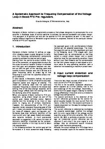
Circular (inner) and longitudinal (outer) muscle layers
Submucosa
Tumor
A
B
C
Fig. 1—62-year-old woman with rectal cancer. A and B, Thin-section T2-weighted axial MR images show rectal cancer (outlined in white, A) Note that invasive margin is at 9-o’clock position, so careful inspection of this area should be performed. Mesorectal fascia is indicated by arrows (B). C, Schematic representation of MR image.
thirds because outcomes appear to relate to the height of the tumor.
will undergo a total mesorectal excision with sphincter-sparing surgery [3].
Upper Third The tumor is in the upper third when its lowest edge is more than 10 cm from the anal verge. The anterior wall of the upper rectum is covered by the peritoneal reflection; the point of attachment occurs at a variable height, particularly in women, and can be as low as 5 cm from the anal verge. Knowledge that the tumor is at this site in relation to the peritoneal reflection must facilitate a careful search for peritoneal perforation of these tumors because of the importance of transcoelomic spread [14, 15].
Lower Third The lower third of the rectum is the area below 5 cm from the anal verge, the area of the rectum and mesorectum below the origin of the levators where the mesorectum tapers sharply. Interpretation at this level is more challenging.
Middle Third The middle third of the rectum is the lowest point between 5 and 10 cm. At this point the rectum is entirely encircled by mesorectal fat; most patients with tumors at this level
AJR:191, December 2008
T Staging the Tumor The process of T staging is shown in Table 2. A morphologic description of the tumor can be helpful in identifying its invasive portion and will indicate the area that causes the most concern. MRI can help in interpreting the relationship of the tumor to the surrounding structures and the bowel wall; these layers can usually be clearly identified (Figs. 1–7). On T2-weighted images, the muscularis mucosal
layer is shown as a fine low-signal-intensity line with the thicker, high-signal submucosal layer seen beneath. The muscularis propria can often be depicted as two distinct layers, the inner circular layer and the outer longitudinal layer. The outer muscle layer has an irregular grooved appearance with interruptions due to vessels entering the rectal wall. The perirectal fat appears as a high signal surrounding the low signal of the muscularis propria and contains signal void vessels (i.e., vessels that do not show any signal). The mes orectal fascia is seen as a fine, low-signal layer enveloping the perirectal fat and rectum; it is this layer that defines the surgical excision plane in anterior total mesorectal resections [3]. MRI diagnosis of a stage T3 lesion is based on the presence of tumor signal extending into the perirectal fat with a broad-based bulging or nodular configuration in continu-
1829
Downloaded from www.ajronline.org by 54.152.109.166 on 09/13/15 from IP address 54.152.109.166. Copyright ARRS. For personal use only; all rights reserved
Taylor et al.
A
B
Fig. 2—T staging in 72-year-old man with rectal cancer. A and B, T2-weighted axial image (A) and corresponding line drawing (B) show stage T1 tumor that has effaced submucosal layer immediately adjacent to tumor (arrow, B). However, muscularis propria comprising inner and outer circular and longitudinal muscle layers is fully preserved, as shown in B.
Fig. 3—62-year-old man with rectal cancer. T2-weighted axial image shows annular tumor with invasion entering outer muscle layer but not extending beyond it (stage T2).
A
B
Fig. 4—73-year-old woman with rectal cancer. A and B, T2-weighted axial image (A) and corresponding line drawing (B) show annular stage T3a tumor with extension less than 1 mm beyond outer muscle coat (arrow).
ity with the intramural portion of the tumor. It is important to note continuity with the intramural component of the tumor because there can be disruption to the outer longitudinal layer as a result of small vessels penetrating the wall that is not necessarily invaded by tumor. Interpreting Stage T4 Tumors and Peritoneal Involvement Stage T4 tumors are defined as those invading an adjacent organ or structure and those that have perforated the peritoneum. MRI is well established in the staging of advanced rectal cancers; common sites for local infiltration of adjacent structures should be sought when examining advanced local tumors. For mid and upper tumors, anterior invasion can often involve the bladder or uterus in addition to peritoneal involvement; lateral exten-
1830
sion can involve the pelvic sidewall; and posterior extension may invade the sacrum. For low tumors, the common sites for stage T4 infiltration are the pelvic floor structures; the anal sphincter; the levator muscles; and the prostate, vagina, seminal vesicles, sacrum, and coccyx. These areas should all be carefully assessed when considering possible T4 lesions. Low rectal tumors deserve special consideration because conventional staging systems are insufficient in these cases. The internal anal sphincter is formed from the lower segment of the circular muscle coat in the distal rectum. At the level of the top of the anal sphincter, fibers from the puborectalis sling join those of the outer muscle coat; together these form the conjoint longitudinal coat, which forms a thin muscular layer between the internal and external sphincters (Fig. 7). We have devised a specific staging system
Fig. 5—51-year-old woman with rectal cancer. T2-weighted image shows stage T4 invasive tumor (arrow) and extension through peritoneal reflection.
that enables identification of tumors that will need a circumferential resection margin and which are at risk if a traditional abdominoperineal excision is performed. This staging is based on the axial and coronal images (see Appendix 1 and Table 3). Extramural Vascular Invasion Extramural vascular invasion is an important and independent prognostic feature that can be readily identified on MRI [16] (Fig. 8). It is defined by the presence of tumor cells beyond the muscularis propria in endothelium-lined vessels. A vessel is defined as a tubular structure containing a signal
AJR:191, December 2008
Downloaded from www.ajronline.org by 54.152.109.166 on 09/13/15 from IP address 54.152.109.166. Copyright ARRS. For personal use only; all rights reserved
MRI Staging of Rectal Cancer
A
B
Fig. 6—56-year-old woman with rectal cancer. A and B, T2-weighted axial image (A) and corresponding line drawing (B) show posterior annular tumor (arrow) with invasive portion at 6-o‘clock position. Note extension < 5 mm beyond outer muscular coat (stage T3b).
TABLE 3: Stages of Low Rectal Cancer as Seen on MRI Stage 1
Tumor confined to bowel wall but does not extend through full thickness; intact outer muscle coat
Stage 2
Tumor replaces muscle coat but does not extend into intersphincteric plane
Stage 3
Tumor invades intersphincteric plane or lies within 1 mm of levator muscle
Stage 4
Tumor invades external anal sphincter and is within 1 mm and beyond levators with or without invading adjacent organs
void on T2-weighted images and shown in continuity on adjacent slices. Extramural vascular invasion is scored on the presence or absence of extramural vascular invasion—that is, positive or negative. If extramural vascular invasion is identified, with tumor growing into or along a recognizable vessel, then it is further classified according to number of vessels and whether they can be identified: for instance, superior or lateral rectal or small and cannot be characterized. If extramural venous invasion is present, a
note is made as to whether the involved veins threaten the mesorectal fascia (i.e., whether they are within 1 mm of the fascia). Mesorectal Lymph Node Morphology The next step is to look in more detail at the mesorectal lymph nodes (Figs. 9 and 10). Nodal staging has traditionally relied on size of the nodes using MRI criteria; however, several studies have indicated the inaccuracy of using this technique alone. Criteria based on the outline of the node and features of sig-
nal intensity have been shown to be more reliable [17, 18]. It may be in the future that identification of involved mesorectal nodes will be augmented by using contrast agents; early results have been quite promising [19]. Lymph nodes are studied using high-resolution images; if nodes are identified they are classified according to their appearance. Uniform nodes having homogeneous signal intensity are not considered to be suspicious. The nodes are judged suspicious if they have irregular borders, mixed signal intensity, or both [4, 17]. If suspicious nodes are present, how many are there? One to three nodes is stage N1 and four or more is stage N2. Finally, record is made whether any node lies within 1 mm of the circumferential resection margin. If that is the case, the node is further identified by suspicious features (see Appendix 1). Margins Measurements are taken of the distance of tumor to the mesorectal fascia, the potential circumferential resection margin. A potentially positive margin is defined as tumor lying within 1 mm (< 1 mm) of the mesorectal fascia [5]. Measurements are also taken of the main tumor, suspicious lymph nodes, extramural vascular invasion, and tumor deposit or satellite. A tumor deposit is classified as a nodule < 3 mm; a nodule > 3 mm is classified as a node [20]. Pelvic Sidewall Lymph Nodes We record the location and size of pelvic sidewall lymph nodes and whether they have any suspicious features according to the morphologic criteria stated previously, which may be a further predictor of survival and local recurrence [21].
Rectal wall
Levators
Puborectalis sling
Internal anal sphincter
Intersphincteric plane
A Fig. 7—58-year-old man with low rectal cancer. A, T2-weighted coronal MR image shows invasion into intrasphincteric plane on left (arrow). B, Diagram shows levels of anorectum.
AJR:191, December 2008
B Fig. 8—57-year-old man with rectal cancer. T2weighted axial image shows extramural venous invasion and extension into lateral rectal vein (arrow).
1831
Taylor et al.
Downloaded from www.ajronline.org by 54.152.109.166 on 09/13/15 from IP address 54.152.109.166. Copyright ARRS. For personal use only; all rights reserved
TABLE 4: Tumor Regression Grade as Seen on MRI Grade 5
No response: intermediate signal intensity, same appearances as original tumor
Grade 4
Slight response: little areas of fibrosis or mucin, but mostly tumor
Grade 3
Moderate response: > 50% fibrosis or mucin and visible intermediate signal
Grade 2
Good response: dense fibrosis; no obvious residual tumor, signifying minimal residual disease; or no tumor
Grade 1
Complete radiologic response: no evidence of ever treated tumor
Note—Modified from grading system of Dworak et al. [22] and altered to represent a radiologic grading system.
Images After Chemoradiotherapy For patients who receive preoperative chemoradiotherapy, we use a tumor regression grade analysis, grades 1–5, modified from Dworak et al. [22] (Table 4). Grade analysis is known to be a better predictor of outcome after treatment than T stage [23]. Discussion The ability of MRI to accurately image the relevant structures of the mesorectum in rectal cancer has become increasingly evident. Advances in imaging technique, together with development of dedicated methods for performing the MRI examination in rectal cancer, have resulted in improved image acquisition. Standardized imaging criteria using thin-section MRI, 3-mm slices, and a small field of view (160–200 mm) allow accurate interpretation of several important prognostic features in rectal cancer. A thorough evaluation of the images can be a daunting task. The aim of this article is to provide an aid to the systematic evaluation of scans to ensure that all clinically relevant structures are adequately assessed. We have also provided some examples to aid in interpretation of these scans. General Description The most common description of a tumor is of an ulcerating lesion with a central indentation and raised, rolled edges. These cancers usually grow circumferentially; most will ultimately cause an annular stenosis of the bowel wall. Polypoid tumors are those that protrude into the lumen; studies have shown that these tend to have a lower grade of malignancy [24–26]. The pattern of invasion by rectal tumors is important. Tumors with a widely infiltrative and ill-defined border will have a worse prognosis (≈ 25% of cases) [20, 27, 28]. However, an inflammatory response at the advancing margin is likely to indicate a more favorable prognosis [20, 29]. It is rare for rectal tumors to show intramural spread, a fact that is noteworthy for the surgeon in planning the distal
1832
resection margin. Current recommendation allows a distal clearance of 1 cm [30–32]. Note should be made whether the tumor is mucinous (10% of large intestinal tumors) because mucinous tumors are associated with a worse prognosis in the rectum. This is thought to be because they infiltrate diffusely and may spread intramurally [31, 33]. Mucinous lesions are identified on T2-weighted images by noting high-signal-intensity mucin pools in the tumors [34]. T Staging and Nodal Status The international TNM staging system [35] is the most widely used pathologic staging system. It is based on the depth of tumor into and beyond the bowel wall, the number of nodal metastases, and the presence of distant metastases. At this time, T staging and margin status give us much of the preoperative information that allows clinical decision making. In the Dutch rectal cancer trial, patients were randomized to preoperative radiation therapy or surgery alone (all underwent total mesorectal excision). That study of mobile operable cancers showed a benefit from radiation therapy, but the degree of benefit depended on the stage of the primary tumor [36]. That study was performed without preoperative staging;
Fig. 9—59-year-old man with rectal cancer. T2weighted axial image shows benign lymph node measuring 7 mm (arrow) that has smooth borders and homogeneous signal intensity. Benign nature of node was confirmed at histopathology.
accurate identification of the preoperative stage can allow greater patient selection. Previous studies have described staging failures due to overstaging of T2 lesions [37, 38], with difficulty in the distinction of spiculation in the perirectal fat caused by fibrosis alone compared with that caused by fibrosis that contains tumor cells. The difficulties of overstaging of T3 tumors by endoscopic sonography have been documented [39]. In that study, 18% of patients with stage T3 or T4 node-positive tumors by endoscopic sonography were shown to be stage T1 or T2 nodenegative tumors at pathology. In our experience, peritumoral fibrosis can be seen as spiculation with lower signal intensity compared with the broad-based or nodular appearance of an advancing tumor margin [40]. The use of preoperative therapy and the selection of patients vary widely and range from routine use of preoperative therapy for all stage T3 and T4 tumors, which form most rectal cancers, to the more selective use of chemoradiotherapy, depending on the risk of both local and distant failure. A more selective approach has been suggested because better patient selection using MRI may be a more effective future strategy that will enable targeted selection of high-risk tumors [41, 42]. This is particularly relevant when considering the morbidity that is associated with chemoradiotherapy [43, 44]. Margin Status Many studies have shown that depth of extramural invasion, nodal involvement, and involvement of the circumferential resection margin are independent markers for poor prognosis [45–48]. Indeed, circumferential
Fig. 10—55-year-old man with rectal cancer. T2weighted axial image shows obviously malignant node (arrow) with irregular border and mixed signal intensity.
AJR:191, December 2008
Downloaded from www.ajronline.org by 54.152.109.166 on 09/13/15 from IP address 54.152.109.166. Copyright ARRS. For personal use only; all rights reserved
MRI Staging of Rectal Cancer resection margin involvement (tumor observed ≤ 1 mm from the resection margin) has been shown to be associated with a higher risk of pelvic recurrence [49, 50] and poor survival. MRI has been shown to accurately identify the depth of extramural invasion and the presence of lymph node metastases, extramural venous invasion, and involvement of the circumferential resection margin [4, 40, 51], and thereby identify those patients who will most benefit from preoperative treatment and those who will not. Despite variation in the use of preoperative treatment, wide agreement exists that all patients with potential margin involvement on MRI should be offered chemoradiotherapy because preoperative discussion of MR images and implementation of this treatment program has led to a decrease in margin positivity rates [6]. The presence of pelvic sidewall nodes is worth noting. In the United Kingdom, pelvic sidewall node dissection is not routinely performed. However, these nodes are probably included in the field when preoperative irradiation is given. In Japan, pelvic wall dissection has been performed since the late 1970s, and a recent publication has suggested that positive lateral lymph nodes are the strongest predictor of both survival and local recurrence [21]. Low Rectal Cancer Low rectal cancers deserve special consideration because the prognosis for tumors at this level is different from that for higher level tumors. This is largely due to anatomic considerations and the fact that the mesorectal envelope tapers downward at this level. We have developed a unique staging system that takes into account the relevant local anatomy, with the aim of providing more information to aid the surgeon in deciding on the most appropriate surgery. The margin positivity rate for abdominoperineal excision has been reportedly as much as 30%, compared with 10% for low anterior resections [36, 50, 52, 53]. Preoperative MRI may be able to predict which patients may be at risk from traditional surgery and enable patients to undergo a more radical excision, thus ensuring adequate resection margins. This is the subject of an ongoing clinical phase II trial that began recruitment in late 2007. Tumor Regression Grade Tumor regression grade is a pathologic grading system based on the degree of tumor regression and fibrosis seen histologically in
AJR:191, December 2008
tumors that have undergone preoperative therapy. The grading system that we use has been adapted to aid in interpreting images of these patients. We have shown that MRI can identify the presence of residual tumor foci, and we have shown good agreement between MRI tumor regression grade and histopathologic tumor regression grade [54]. With some patients showing a complete response to treatment, the interpretation of these images is becoming increasingly important; further work in this area continues. The ability of MRI to accurately image the relevant structures of the mesorectum in rectal cancer has become increasingly evident. Advances in the method and technique for obtaining these scans have resulted in improved image acquisition. Standardized imaging criteria using thin-section MRI, 3-mm slices, and a small field of view allow more accurate interpretation. MRI can now be used to identify several prognostic features that will allow better selection of patients who will benefit from more intensive treatment. In summary, the interpretation of preoperative MR images in patients with rectal cancer allows the identification of several prognostic factors that will enable better selection of patients who may benefit from more intensive treatment without subjecting those who will not to unnecessary treatment. Acknowledgments We thank all the radiologists involved in the MERCURY study—C. D. George, N. Bees, H. Blake, N. Jeyadeven, P. D. Peppercorn, A. Chalmers, A. Guthrie, H. Emblemsvaag, T. Vetrhus, Knut Haakon Hole, H. Massouh, M. Creagh, P. Knuth, R. Bleehen, M. W. Bourne, L. Blomqvist, and M. Torkzad—for all the hard work they put into the project, The Pelican Cancer Foundation for their continued support, and Barbara Bannerman for her ongoing assistance. References 1. Blomqvist L, Rubio C, Holm T, Machado M, Hindmarsh T. Rectal adenocarcinoma: assessment of tumour involvement of the lateral resection margin by MRI of resected specimen. Br J Radiol 1999; 72:18–23 2. Brown G, Davies S, Williams GT, et al. Effectiveness of preoperative staging in rectal cancer: digital rectal examination, endoluminal ultrasound or magnetic resonance imaging? Br J Cancer 2004; 91:23–29 3. Brown G, Kirkham A, Williams GT, et al. Highresolution MRI of the anatomy important in total
mesorectal excision of the rectum. AJR 2004; 182:431–439 4. Brown G, Radcliffe AG, Newcombe RG, Dallimore NS, Bourne MW, Williams GT. Preoperative assessment of prognostic factors in rectal cancer using high-resolution magnetic resonance imaging. Br J Surg 2003; 90:355–364 5. MERCURY Study Group. Diagnostic accuracy of preoperative magnetic resonance imaging in predicting curative resection of rectal cancer: prospective observational study. BMJ 2006; 333:779. Epub 2006 Sep 19 6. Burton S, Brown G, Daniels IR, Norman AR, Mason B, Cunningham D. MRI directed multidisciplinary team preoperative treatment strategy: the way to eliminate positive circumferential margins? Br J Cancer 2006; 94:351–357 7. Glimelius B, Oliveira J. Rectal cancer: ESMO clinical recommendations for diagnosis, treatment and follow-up. Ann Oncol 2008; 9[suppl 2]:ii31–ii32 8. The Association of Coloproctography of Great Britain and Ireland. Guidelines for the management of colorectal cancer. London, UK: ACPGBI, 2007 9. Brown G, Daniels IR, Richardson C, Revell P, Peppercorn D, Bourne M. Techniques and trouble-shooting in high spatial resolution thin slice MRI for rectal cancer. Br J Radiol 2005; 78:245– 251 10. Okizuka H, Sugimura K, Yoshizako T, Kaji Y, Wada A. Rectal carcinoma: prospective comparison of conventional and gadopentetate dimeglumine enhanced fat-suppressed MR imaging. J Magn Reson Imaging 1996; 6:465–471 11. Vliegen RF, Beets GL, von Meyenfeldt MF, et al. Rectal cancer: MR imaging in local staging—is gadolinium-based contrast material helpful? Radiology 2005; 234:179–188 12. Koh DM, Brown G, Temple L, et al. Distribution of mesorectal lymph nodes in rectal cancer: in vivo MR imaging compared with histopathological examination—initial observations. Eur Radiol 2005; 15:1650–1657 13. Zand KR, Reinhold C, Haider MA, Nakai A, Rohoman L, Maheshwari S. Artifacts and pitfalls in MR imaging of the pelvis. J Magn Reson Imaging 2007; 26:480–497 14. Salerno G, Daniels IR, Moran BJ, Wotherspoon A, Brown G. Clarifying margins in the multidisciplinary management of rectal cancer: the MERCURY experience. Clin Radiol 2006; 61:916– 923 15. Shepherd NA, Baxter KJ, Love SB. Influence of local peritoneal involvement on pelvic recurrence and prognosis in rectal cancer. J Clin Pathol 1995; 48:849–855 16. Smith NJ, Barbachano Y, Norman AR, Swift RI,
1833
Downloaded from www.ajronline.org by 54.152.109.166 on 09/13/15 from IP address 54.152.109.166. Copyright ARRS. For personal use only; all rights reserved
Taylor et al. Abulafi AM, Brown G. Prognostic significance of magnetic resonance imaging-detected extramural vascular invasion in rectal cancer. Br J Surg 2008; 95:229–236 17. Brown G, Richards CJ, Bourne MW, et al. Morphologic predictors of lymph node status in rectal cancer with use of high-spatial-resolution MR imaging with histopathologic comparison. Radiology 2003; 227:371–377 18. Kim JH, Beets GL, Kim MJ, Kessels AG, BeetsTan RG. High-resolution MR imaging for nodal staging in rectal cancer: are there any criteria in addition to the size? Eur J Radiol 2004; 52:78– 83 19. Koh DM, Brown G, Temple L, et al. Rectal cancer: mesorectal lymph nodes at MR imaging with USPIO versus histopathologic findings—initial observations. Radiology 2004; 231:91–99 20. Jass JR. Lymphocytic infiltration and survival in rectal cancer. J Clin Pathol 1986; 39:585–589 21. Sugihara K, Kobayashi H, Kato T, et al. Indication and benefit of pelvic sidewall dissection for rectal cancer. Dis Colon Rectum 2006; 49:1663–1672 22. Dworak O, Keilholz L, Hoffmann A. Pathological features of rectal cancer after preoperative radiochemotherapy. Int J Colorectal Dis 1997; 12:19–23 23. Bouzourene H, Bosman FT, Seelentag W, Matter M, Coucke P. Importance of tumor regression assessment in predicting the outcome in patients with locally advanced rectal carcinoma who are treated with preoperative radiotherapy. Cancer 2002; 94:1121–1130 24. Cohen AM, Wood WC, Gunderson LL, Shinnar M. Pathological studies in rectal cancer. Cancer 1980; 45:2965–2968 25. Bjerkeset T, Morild I, Mork S, Soreide O. Tumor characteristics in colorectal cancer and their relationship to treatment and prognosis. Dis Colon Rectum 1987; 30:934–938 26. Michelassi F, Vannucci L, Montag A, et al. Importance of tumor morphology for the long term prognosis of rectal adenocarcinoma. Am Surg 1988; 54:376–379 27. Grinnell RS. The grading and prognosis of carcinoma of the colon and rectum. Ann Surg 1939; 109:500–533 28. Spratt JA, Spjut HJ. Prevalence and prognosis of carcinoma of the colon and rectum. Cancer 1967; 20:1976–1985 29. Halvorsen TB, Seim E. Association between invasiveness, inflammatory reaction, desmoplasia and
survival in colorectal cancer. J Clin Pathol 1989; 42:162–166 30. Hughes TG, Jenevein EP, Poulos E. Intramural spread of colon carcinoma: a pathologic study. Am J Surg 1983; 146:697–699 31. Madsen PM, Christiansen J. Distal intramural spread of rectal carcinomas. Dis Colon Rectum 1986; 29:279–282 32. Andreola S, Leo E, Belli F, et al. Distal intramural spread in adenocarcinoma of the lower third of the rectum treated with total rectal resection and coloanal anastomosis. Dis Colon Rectum 1997; 40:25–29 33. Sidoni A, Bufalari A, Alberti PF. Distal intramural spread in colorectal cancer: a reappraisal of the extent of distal clearance in fifty cases. Tumori 1991; 77:514–517 34. Kim MJ, Park JS, Park SI, et al. Accuracy in differentiation of mucinous and nonmucinous rectal carcinoma on MR imaging. J Comput Assist Tomogr 2003; 27:48–55 35. Sobin L, Wittekind C. TNM classification of malignant tumors, 5th ed. New York, NY: Wiley, 1997:227 36. Kapiteijn E, Marijnen CA, Nagtegaal ID, et al. Preoperative radiotherapy combined with total mesorectal excision for resectable rectal cancer. N Engl J Med 2001; 345:638–646 37. Laghi A, Ferri M, Catalano C, et al. Local staging of rectal cancer with MRI using a phased array body coil. Abdom Imaging 2002; 27:425–431 38. Beets-Tan RG, Beets GL, Vliegen RF, et al. Accuracy of magnetic resonance imaging in prediction of tumour-free resection margin in rectal cancer surgery. Lancet 2001; 357:497–504 39. Sauer R, Becker H, Hohenberger W, et al. Preoperative versus postoperative chemoradiotherapy for rectal cancer. N Engl J Med 2004; 351:1731– 1740 40. Brown G, Richards CJ, Newcombe RG, et al. Rectal carcinoma: thin-section MR imaging for staging in 28 patients. Radiology 1999; 211:215–222 41. Chau I, Brown G, Cunningham D, et al. Neoadjuvant capecitabine and oxaliplatin followed by synchronous chemoradiation and total mesorectal excision in magnetic resonance imaging-defined poor-risk rectal cancer. J Clin Oncol 2006; 24:668–674 42. Koeberle D, Burkhard R, von Moos R, et al. Phase II study of capecitabine and oxaliplatin given prior to and concurrently with preoperative pelvic radiotherapy in patients with locally advanced
rectal cancer. Br J Cancer 2008; 98:1204–1209 43. Dahlberg M, Glimelius B, Graf W, Pahlman L. Preoperative irradiation affects functional results after surgery for rectal cancer: results from a randomized study. Dis Colon Rectum 1998; 41:543– 549; discussion 549–551 44. Wong RK, Tandan V, De Silva S, Figueredo A. Pre-operative radiotherapy and curative surgery for the management of localized rectal carcinoma. Cochrane Database Syst Rev 2007; Apr 18;(2): CD002102 45. Cawthorn SJ, Gibbs NM, Marks CG. Clearance technique for the detection of lymph nodes in colorectal cancer. Br J Surg 1986; 73:58–60 46. Jass JR, Love SB. Prognostic value of direct spread in Dukes’ C cases of rectal cancer. Dis Colon Rectum 1989; 32:477–480 47. Adam IJ, Mohamdee MO, Martin IG, et al. Role of circumferential margin involvement in the local recurrence of rectal cancer. Lancet 1994; 344:707–711 48. Hall NR, Finan PJ, al-Jaberi T, et al. Circumferential margin involvement after mesorectal excision of rectal cancer with curative intent: predictor of survival but not local recurrence? Dis Colon Rectum 1998; 41:979–983 49. Birbeck KF, Macklin CP, Tiffin NJ, et al. Rates of circumferential resection margin involvement vary between surgeons and predict outcomes in rectal cancer surgery. Ann Surg 2002; 235:449– 457 50. Marr R, Birbeck K, Garvican J, et al. The modern abdominoperineal excision: the next challenge after total mesorectal excision. Ann Surg 2005; 242:74–82 51. MERCURY Study Group. Extramural depth of tumor invasion at thin-section MR in patients with rectal cancer: results of the MERCURY study. Radiology 2007; 243:132–139 52. Dehni N, McFadden N, McNamara DA, Guiguet M, Tiret E, Parc R. Oncologic results following abdominoperineal resection for adenocarcinoma of the low rectum. Dis Colon Rectum 2003; 46:867–874; discussion 874 53. Heald RJ, Smedh RK, Kald A, Sexton R, Moran BJ. Abdominoperineal excision of the rectum: an endangered operation. Norman Nigro Lectureship. Dis Colon Rectum 1997; 40:747–751 54. Salerno G, Chau I, Tait D, Cunningham D, Brown G. Interpretation of postchemo-radiation MRI compared with histopathology in low rectal cancer. Colorectal Disease 2007; 9[1suppl]:92–93
F O R YO U R I N F O R M AT I O N
This article is available for CME credit. See www.arrs.org for more information. Appendix I appears on the next page.
1834
AJR:191, December 2008
MRI Staging of Rectal Cancer
Downloaded from www.ajronline.org by 54.152.109.166 on 09/13/15 from IP address 54.152.109.166. Copyright ARRS. For personal use only; all rights reserved
APPENDIX 1: Recording Findings in Rectal Cancer Patient Name
Date:
Date of Birth
Hospital Number
Exam performed elsewhere
Yes
No
Exam technically satisfactory (3 mm)
Yes
No
Image quality
Optimal
Sub-Optimal
Pathology identified
Yes
No
Has the patient received radiotherapy?
Yes
No
Has the patient had a previous rectal MRI?
Yes
No
If yes, where?
If Yes, date of previous examination Morphological description of tumor:
e.g. polypoidal, annular, ulcerating. Site of invasive border, nature of invasive border e.g. smooth, nodular infiltrating. Mucinous tumor? Nodal spread No visible nodes = N0 Homogeneous signal intensity smooth bordered node = N0 1–3: Mixed signal intensity or irregular bordered lymph node or tumor deposit = N1 4 or more: Mixed signal intensity or irregularly bordered node or tumor deposit = N2 T staging Tumor not seen (Tx) Invades submucosa (T1) Invades muscularis propria (T2) Beyond muscularis propria < 1 mm (T3a) Beyond muscularis propria 1–5 mm (T3b) Beyond muscularis propria > 5–15 mm (T3c) Beyond muscularis propria > 15 mm (T3d) Perforation of peritoneal covering (T4b) Tumor invasion into adjacent organ (T4a) Maximum depth of extramural spread beyond muscularis propria______________(mm) Extramural venous invasion No tumor signal in vessels Tumor signal intensity expanding small noncharacterizable veins Tumor signal intensity expanding large anatomical veins (e.g., superior rectal vein) Potential Circumferential Margins (above distal levator insertion) Measure minimum distance of: Main tumor to mesorectal fascia Malignant lymph nodes or tumor deposit EMVI CRM status Distance to mesorectal fascia ≤ 1 mm = potential CRM involved Distance to mesorectal fascia > 1 mm = potential CRM clear Staging tumors at/below the distal levator insertion 1. Tumor on MRI images appears confined to bowel wall but not through full thickness (with intact outer muscle coat) 2. Tumor on MRI replaces the muscle coat but does not extend into the intersphincteric plane 3. Tumor on MRI invading into the intersphincteric plane 4. Tumor invading into external anal sphincter Pelvic sidewall nodes (outside mesorectum, below iliac vessel bifurcation) No visible nodes Homogeneous-signal-intensity smooth bordered node Mixed signal intensity or irregular bordered lymph node or tumor deposit Postchemoradiotherapy assessment Which best describes the tumor regression on MRI? Grade 5: No response (intermediate signal intensity, same appearances as original tumor) Grade 4: Slight response (little areas of fibrosis or mucin but mostly tumor) Grade 3: Moderate response (> 50% fibrosis or mucin and visible intermediate signal) Grade 2: Good response (dense fibrosis; no obvious residual tumor, signifying minimal residual disease; or no tumor) Grade 1: Radiological complete response (rCR) (no evidence of ever treated tumor) Note—EMVI = extramural vascular invasion, CRM = circumferential resection margin. AJR:191, December 2008
1835