A ctive sorting sw itch for biological objects Mojmír Šerý1, Zdeněk Pilát1, Alexander Jonáš1, Jan Ježek1, Petr Jákl1, Pavel Zemánek1, Ota Samek1, Ladislav Nedbal2, Martin Trtílek3 1 Institute of Scientific Instruments of the ASCR, v.v.i., Academy of Sciences of the Czech Republic, Královopolská 147, 612 64 Brno, Czech Republic 2 Institute of Systems Biology and Ecology of the AS CR, v.v.i., Academy of Sciences of the Czech Republic, Zámek 136, 37333 Nové Hrady, Czech Republic 3 Photon Systems Instruments, Drásov 470, 664 24 Drásov, Czech Republic ABSTR A C T Active contactless optical sorting of microobjects represents very useful technique in many areas of biology, chemistry, and medicine. We suggest here a configuration that combines optical sorting, trapping, excitation, and detection paths and provides efficient sorting of biological samples according to their various parameters (fluorescence, Raman spectrum, CCD image, motion etc.). This approach is based on the shape of the laser beam and we succeeded in sorting of several types of living microorganisms. Keywords: Laser tweezers, laser diode, optical sorting, microfluidics 1. I N T R O D U C T I O N It has been known for 25 years that the single focused laser beam can serve as optical tweezers1 i.e. a tool which is able to confine objects of sizes from tens of nanometers to tens of micrometers in three dimensions. Such tool can be also utilized for confinement and transport of living microorganisms if the trapping wavelength is carefully selected so that it is not absorbed by the organisms. Especially the trapping wavelength is the key issue in safe manipulation with phototrophs. Up to now, successful trapping and manipulation of viruses or bacteria without visible damage has been presented.2-4 Later experiments have shown manipulation with chromosomes inside cell nuclei,5 trapping of nuclei in living cells during mitosis without impairing their viability,6 non-contact and sterile selection of cells,7,8 manipulation with single DNA molecule or actin filament.9 Several methods have been recently developed to sort microobjects in a contactless way using laser beams.10 Active sorting uses a decision-making step based on the results of optical diagnostics of the microobjects.7 It is based on a well-defined input (for example, fluorescence or spectroscopic signal from the particle, typical image pattern, etc.) that triggers the action, during which optical forces move analyzed objects into different positions and separate them .7,11-16 Passive sorting is a much younger method and uses more complex optical intensity patterns - optical po tential landscapes - with specifically designed spatial variations of optical intensity. The sorting is based on the difference in sensitivity of the forces in an optical potential landscape to the sorted particle properties (size, shape, material).17-28 In this case no active decision-making step is needed; particles of different properties follow different trajectories when driven by the fluid flow through such a landscape.20,21,29,30 Other methods of passive sorting even do not use fluid flow and enable sorting in static fluids via potential landscape movement,31-35 radiation pressure36,37 or thermal noise activation.38,39 An active sorting switch presented in this article combines the versatility of optical tweezers technique and power of the microfluidic technology. As the sorting mechanism, we used two strongly focused elliptical laser beams with mutually perpendicular longer axes of the beams and with PC control of their operation. One beam confines a microobject for photosystem diagnostic Fv/Fm method e.g. measurement of quantum efficiency of cells Photosystem II. Based on the online result of the diagnostics the first beam is switched off and lets the microobject follow its flowline into waste channel if it is not suitable for sorting. In contrast, if the object should be sorted, the second beam is switched on and the trapping beam is switched off. Consequently the microobject is deviated to different flowline dragging the object to sorting channel. Send correspondence to M.S.:
[email protected], Phone: +420 541 514 284 Optical Trapping and Optical Micromanipulation VII, edited by Kishan Dholakia, Gabriel C. Spalding, Proc. of SPIE Vol. 7762, 776210 ©2010 SPIE ■CCC code: 0277-786X710/$ 18 ■doi: 10.1117/12.859859
Proc. of SPIE Vol. 7762 776210-1
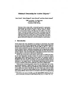
2. C O N S T R U C T I O N The sorting switch uses two laser diodes (Axcel Photonics M9-A64-0200-S50) operating at the wavelength 1064 nm, TEMoo transversal profile and maximal output power 200 mW. Each emanating beam was collimated by aspherical lens (Asphericon 15-12 HPX-S). Both elliptical laser beams with perpendicular polarization are coupled together by polarizing beam splitter (Thorlabs PBS203) and form two elliptical spots with perpendicular longer axes. Part of the sorting beam was blocked for maximal deviation of sorted cell. The dichroic mirror reflects long wavelengths into infinity-corrected microscope objective (Fig. 1).
TOP VIEW
—NE
Figure 1. Optical scheme for active sorting switch.
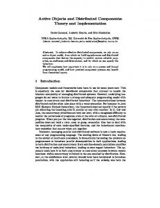
The whole optical system was investigated using Zemax software and optimized with respect to the aberra tions. Figure 2 clearly demonstrates that the beam spot can be considered as diffraction limited, e.g. focus does not significantly overfill Airy disc of the optical system. Shape of the beam spot near their focus corresponds to strongly focused astigmatic and elliptical profile of used laser diode. OBJ : 0,0000, 0.0000 OEG
! 4>
SUPFRCE: IMfl
IMR: 0.000. 0.000 MM SPOT DIRGRRM
SURFRCE: IHfl
DE F Q C U S IN / T H R O U G H F Q C U S ~ ~ S P O f DIRGRFtM
Figure 2. Spot diagram (left), through focus spot diagram (right) from Zemax optical program, scales are in micrometers.
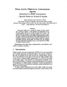
The mechanical concept of the system was designed as a module which could be easily mounted on the standard upright commercial microscope (Fig. 3). Connecting flanges could be easily changed for different microscope manufacturers. Mechanical materials were appropriately selected with respect to high mechanical and thermal stability of the system. Invariance of power and position of the laser spot in the focal volume was achieved by an active temperature stabilization of each laser diode by Peltier module (Melcor SH1.0-23-06L).
Proc. of SPIE Vol. 7762 776210-2
->90
\
( Figure 3. Isometric view of optical components from Autodesk Inventor.
3. E X P E R I M E N T S Sorting switch (Fig. 4) was tested in combination with upright microscope Olympus BX-41 and focusing water immersion microscope objective (Olympus UplanSAPO 60x). Solution of cells was pumped into home-made microfluidic chip with one input channels and two output channels. One output channel collected the sorted cells and the other the waste. The flowing medium dragged the cells into the sorting area where some of them were trapped.
Figure 4. Sorting head mounted on upright optical microscope Olympus BX-41.
Proc. of SPIE Vol. 7762 776210-3
At the same time the activity of the photosystem was investigated by fluorescence kinetics excitation and detection system (Photon System Instruments FluorCam). Cells were sorted according to the results of the Fv/Fm parameter which is widely used to indicate the maximum quantum efficiency of Photosystem II. This parameter is considered to be a sensitive indication of plant photosynthetic performance with healthy samples typically achieving a maximum Fv/Fm value close to 0.8. Cells with Fv/Fm value higher than 0.3 were selected as the vital ones and they were pushed to higher position in medium flow and aimed into the sorting channel of the microfluidic chip by the sorting beam. The cells having lower Fv/Fm ratio are unusable for later cultivation and stayed in the same flowline leading into the waste microfluidic channel. The applicability of this sorting technique was tested on three types of algae cells, Chlorella (Fig. 5), Botryococcus sudeticus (Fig. 6) and Trachydiscus minutus (Fig. 7). First set of figures shows sorting of Chlorella. This organism could be easily optically trapped and bonding of cell to microfluidic chip is minimal. Second organism (Botryococcus sudeticus) is smaller than Chlorella cell and could be also successfully trapped. Last presented organism Trachydiscus minutus could be trapped, but bonding forces between cell and microfluidic chip walls were higher and cells tended to block microfluidic channels.
Figure 5. Chlorella trapped in focused laser beam (left), cell deviated by sorting beam (right). The original position of the cell is denoted by the white cross.
Figure 6. Botryococcus sudeticus trapped in focused laser beam (left), cell deviated by sorting beam (right).
Proc. of SPIE Vol. 7762 776210-4
Figure 7. Trachydiscus minutus trapped in focused laser beam (left), cell deviated by sorting beam (right).
4. C O N C L U S I O N In this paper, we present active sorting switch for separating various biological objects. Algae cells Chlorella, Botryococcus sudeticus and Trachydiscus minutus were effectively sorted with respect to their photosystem vitality in through-flow microfluidic chip. The sorting mechanism is based on two independently driven laser diodes, which beams are focused by microscope objective with high numerical aperture. Both laser spots could be positioned in the field of view. The first beam is used to immobilize the cell during the measurement of photosystem condition. The second beam, partially blocked by obscuration for maximization of deviation, pushes cells with good vitality ratio into higher flowline of home-made microfluidic chip. 5. A C K N O W L E D G M E N T S The authors acknowledge support from MCT FR-TI1/433, MEYS 0008034 and IS IIR P AV0Z20650511. REFERENCES [1] Ashkin, A., Dziedzic, J. M., Bjorkholm, J. E., and Chu, S., “Observation of a single-beam gradient force optical trap for dielectric particles,” Opt. Lett. 11, 288-290 (1986). [2] Ashkin, A. and Dziedzic, J. M., “Optical trapping and manipulation of viruses and bacteria,” Science 235, 1517-1520 (1987). [3] Ashkin, A. and Dziedzic, J. M., “Internal cell manipulation using infrared laser traps,” Proc. Natl. Acad. Sci. USA 86, 7914-7918 (1989). [4] Neuman, K., Chadd, E., Liou, G., Bergman, K., and Block, S., “Characterization of photodamage to Escherichia coli in optical traps,” BIOPHYSICAL JOURNAL 77(5), 2856-2863 (1999). [5] Berns, M. W., Wright, W. H., and Tromberg, B., “Internal cell manipulation using infrared laser traps,” Proc. Natl. Acad. Sci. USA 86, 4539-4543 (1989). [6] Liang, H., Wright, H. W., Cheng, S., He, W., and Berns, M. W., “Micromanipulation of chromosomes in PTK2 cells using laser microsurgery (optical scalpel) in combination with laser-induced optical force (optical tweezers).,” Exp. Cell Res 204, 110-120 (1993). [7] Fu, A. Y., Spence, C., Scherer, A., Arnold, F. H., and Quake, S. R., “A microfabricated fluorescenceactivated cell sorter,” Nature Biotechnol. 17(11), 1109-1111 (1999). [8] Grover, S. C., Skirtach, A. G., Gauthier, R. C., and Grover, C. P., “Automater single-cell sorting system based on optical trapping,” J. Biomed. Opt. 6, 14-22 (2001).
[9] Arai, Y., Yasuda, R., Akashi, K., Harada, Y., Miyata, H., Kinosita, K., and Itoh, H., “Tying a molecular knot with optical tweezers,” NATU RE 399, 446-448 (JUN 3 1999). [10] Dholakia, K., MacDonald, M. P., Zemánek, P., and Cižmár, T., “Cellular and colloidal separation using optical forces,” Methods in Cell Biology 82, 467-495 (2007). [11] Buican, T. N., Smyth, M. J., Crissman, H. A., Salzman, G. C., Stewart, C. C., and Martin, J., “Automated single-cell manipulation and sorting by light trapping,” Appl. O pt 26, 5311-5316 (1987). [12] Wang, M. M., Tu, E., Raymond, D. E., Yang, J. M., Zhang, H., Hagen, N., Dees, B., Mercer, E. M., Forster, A. H., Kariv, I., Marchand, P. J., and Butler, W. F., “Microfluidic sorting of mammalian cells by optical force switching,” Nature Biotechnol. 23, 83-87 (2005). [13] Applegate, R. W., Jr., Squier, J., Vestad, T., Oakey, J., and Marr, D. W. M., “Optical trapping, manip ulation, and sorting of cells and colloids in microfluidic systems with diode laser bars,” Opt. Express 12, 4390-4398 (2004). [14] Applegate, R. W., Jr., Squier, J., Vestad, T., Oakey, J., Marr, D. W. M., Bado, P., Dugan, M. A., and Said, A. A., “Microfluidic sorting system based on optical waveguide integration and diode bar trapping,” Lab Chip 6, 422-426 (2006). [15] Applegate, R. W., Jr., Squier, J., Vestad, T., Oakey, J., and Marr, D. W. M., “Fiber-focused diode bar optical trapping for microfluidic flow manipulation,” Appl. Phys. Lett. 92, 013904 (2008). [16] Roichman, Y., Wong, V., and Grier, D. G., “Colloidal transport through optical tweezer arrays,” Phys. Rev. E 75, 011407 (2007). [17] Zemánek, P., Jonáš, A., and Liška, M., “Simplified description of optical forces acting on a nanoparticle in the gaussian standing wave,” J. Opt. Soc. Am. A 19, 1025-1034 (2002). [18] Zemánek, P., Jonáš, A., Jákl, P., Šerý, M., Ježek, J., and Liška, M., “Theoretical comparison of optical traps created by standing wave and single beam,” Opt. Commun. 220, 401-412 (2003). [19] Lekner, J., “Force on a scatterer in counter-propagating coherent beams,” J. Opt. A: Pure Appl. Opt. 7, 238-248 (2005). [20] Korda, P. T., Taylor, M. B., and Grier, D. G., “Kinetically locked-in colloidal transport in an array of optical tweezers,” Phys. Rev. Lett. 89, 128301 (2002). [21] MacDonald, M. P., Spalding, G. C., and Dholakia, K., “Microfluidic sorting in an optical lattice,” Na ture 426, 421-424 (2003). [22] Pelton, M., Ladavac, K., and Grier, D. G., “Transport and fractionation in periodic potential-energy land scapes,” Phys. Rev. E 70, 031108 (2004). [23] Ladavac, K., Kasza, K., and Grier, D. G., “Sorting mesoscopic objects with periodic potential landscapes: Optical fractionation,” Phys. Rev. E 70, 010901 (2004). [24] Reichhardt, C. and Reichhardt, C. J. O., “Directional locking effects and dynamics for particles driven through a colloidal lattice,” Phys. Rev. E 69, 041405 (2004). [25] Reichhardt, C., Reichhardt, C. J. O., and Hastings, M. B., “Nonlinear dynamics, rectification, and phase locking for particles on symmetrical two-dimensional periodic substrates with dc and circular ac drives,” Phys. Rev. E 6 9, 056115 (2004). [26] Lacasta, A. M., Sancho, J. M., Romero, A. H., and Lindenberg, K., “Sorting on periodic surfaces,” Phys. Rev. Lett. 94, 160601 (2005). [27] Libál, A., Reichhardt, C., Jankó, B., and Reichhardt, C. J. O., “Dynamics, rectification, and fractionation for colloids on flashing substrates,” Phys. Rev. Lett. 96, 188301 (2006). [28] Gleeson, J. P., Sancho, J. M., Lacasta, A. M., and Lindenberg, K., “Analytical approach to sorting in periodic and random potentials,” Phys. Rev. E 73, 041102 (2006). [29] Sun, Y. Y., Ong, L. S., and Yuan, X.-C., “Composite-microlens-array-enabled microfluidic sorting,” Appl. Phys. Lett. 89, 141108:1-3 (2006). [30] Milne, G., Rhodes, D., MacDonald, M., and Dholakia, K., “Fractionation of polydisperse colloid with acousto-optically generated potential energy landscapes,” Opt. Lett. 32, 1144-1146 (2007). [31] Čižmár, T., Šiler, M., Šerý, M., Zemánek, P., Garcés-Chávez, V., and Dholakia, K., “Optical sorting and detection of sub-micron objects in a motional standing wave,” Phys. Rev. B 74, 035105 (2006).
Proc. of SPIE Vol. 7762 776210-6
[32] Ricárdez-Vargas, I., Rodríguez-Montero, R , Ramos-García, R., and Volke-Sepúlveda, K., “Modulated op tical sieve for sorting of polydisperse microparticles,” Appl. Phys. Lett. 88, 121116 (2006). [33] Smith, R. L., Spalding, G. C., Dholakia, K., and MacDonald, M. R , “Colloidal sorting in dynamic optical lattices,” J. Opt. A: Pure Appl. Opt. 9, S134-S138 (2007). [34] Forster, A., Wang, M., Butler, W., Chachisvilis, M., Chung, T., Esener, S., Hall, J., Kibar, O., Lykstad, K., Marchand, R , Mercer, E., Pestaña, L., Sur, S., Tu, E., Yang, R., Zhang, H., and Kariv, I., “Use of moving optical gradient fields for analysis of apoptotic cellular responses in a chronic myeloid leukemia cell model,” Anal. Biochem. 327, 14-22 (APR 1 2004). [35] Gherardi, D. M., Carruthers, A. E., Čižmár, T., Wright, E. M., and Dholakia, K., “A dual beam photonic crystal fibre trap for microscopic particles,” Appl. Phys. Lett. 93 (2008). [36] Zemánek, P., Karásek, V., and Sasso, A., “Optical forces acting on Rayleigh particle placed into interference field,” Opt. Commun. 240, 401-415 (2004). [37] Jákl, P., Čižmár, T., Šerý, M., and Zemánek, P., “Static optical sorting in a laser interference field,” Appl. Phys. Lett. 92 (APR 21 2008). [38] Paterson, L., Papagiakoumou, E., Milne, G., Garcés-Chávez, V., Tatarkova, S. A., Sibbett, W., GunnMoore, F. J., Bryant, P. E., Riches, A. C., and Dholakia, K., “Light-induced cell separation in a tailored optical landscape,” Appl. Phys. Lett. 87, 123901 (2005). [39] Hayashi, Y., Ashihara, S., Shimura, T., and Kuroda, K., “Particle sorting using optically induced asym metric double-well potential,” Opt. Commun. 281, 3792-3798 (2008).
Proc. of SPIE Vol. 7762 776210-7