cially tospoviruses (Whitfield et al., 2005; Rotenberg et al.,. 2015). In screening ..... biased herbivory in Jack-in-the-pulpit (Arisaema triphyllum) by a specialist ...
DOI: 10.1111/eea.12682
S P E C I A L I S S U E : M U LT I L E V E L F E E D I N G E C O L O G Y T E C H N I C A L N OT E
An objective high-throughput screening method for thrips damage quantitation using Ilastik and ImageJ Isabella G. S. Visschers1
, Nicole M. van Dam1,2,3
& Janny L. Peters4*
1
Department of Molecular Interaction Ecology, Institute for Water and Wetland Research, Radboud University, 6525 AJ Nijmegen, The Netherlands, 2German Centre for Integrative Biodiversity Research (iDiv) Halle-Jena-Leipzig, Deutscher Platz 5e, 04103, Leipzig, Germany, 3Friedrich Schiller University Jena, Institute of Biodiversity, Dornburger-Str. 159, 07743, Jena, Germany, and 4Department of Molecular Plant Physiology, Institute for Water and Wetland Research, Radboud University, 6525 AJ Nijmegen, The Netherlands Accepted: 25 January 2018
Key words: methodology, insect resistance, leaf silvering, Frankliniella occidentalis, Capsicum, pepper, resistance breeding, vegetable crops, plant ontogeny, Thysanoptera, Thripidae, Solanaceae
INTRODUCTION Quantifying insect damage is essential for identifying resistance mechanisms in plants. For chewing insects such as caterpillars and beetles, feeding damage is commonly determined by measuring the amount of removed leaf area, using freely available software such as ImageJ Fiji (https://fiji.sc) (Schindelin et al., 2012; Neves et al., 2014; Nguyen et al., 2016) or commercial programs such as Winfolia (http://regent.qc.ca/assets/winfolia_about.html) (Joshi & Tielb€ orger, 2012). However, cell-sucking insects such as thrips, do not remove sections of the leaf lamina, but rather cause localized discoloration of the leaf surface. It is very challenging to quantify this type of damage in an objective manner using these software programs. Thrips are widespread sucking-piercing insects that are responsible for severe yield reduction in several vegetable crops such as cucumber, strawberry, melon, and pepper. Crops infested with thrips show stunted growth, deformation of the plant, and scarring of the fruits, resulting in reduced yield and marketing quality (Welter et al., 1990; Tommasini & Maini, 1995; Shipp et al., 1998). In addition, thrips are important vectors of plant viruses, especially tospoviruses (Whitfield et al., 2005; Rotenberg et al., 2015). In screening programs for host-plant resistance to thrips, the total damaged leaf area is used as a criterion to
*Correspondence: Janny L. Peters, Department of Molecular Plant Physiology, Institute for Water and Wetland Research, Radboud University, 6525 AJ Nijmegen, The Netherlands. E-mail: jl.peters@ science.ru.nl
determine resistance levels. Damage is often characterized by silvery spots that show a high contrast with the intact leaf area, though thrips feeding can also include darker areas ranging from dark green to brown. These more gradual discolorations of the leaf are too subtle to precisely quantify with ImageJ or Winfolia alone. As a result, thrips damage is commonly scored by individuals that rate the samples. Samples are classified into categories signifying the amount of damage (Mirnezhad et al., 2010; Maharijaya et al., 2011, 2012), or damage is estimated to the nearest 1 mm2 (Leiss et al., 2009; Mirnezhad et al., 2010; Maharijaya et al., 2011, 2012). These subjective measurements make comparison between studies/screening programs difficult. Moreover, they are time consuming and thus costly for breeding companies. Here we present an objective, automated protocol for the screening of thrips damage on leaves. We developed a high-throughput standardized screening method to measure leaf surface damage caused by thrips using two types of freeware, ImageJ (Schindelin et al., 2012) and Ilastik (Sommer et al., 2011). Ilastik has a wide range of applications ranging from cell biology (Fabrowski et al., 2013), where it is used to compute the amount of surface flattening of epithelial cells, to biomechanics (Bongiorno et al., 2014), where it is used to identify boundaries of human mesenchymal stem cells. It is an easy-to-use, self-learning image processing program that allows segmentation and classification of two-dimensional surfaces based on labels provided by the user (Sommer et al., 2011). ImageJ is often used to quantify the amount of removed leaf area by chewing herbivores and the total leaf surface of intact leaves (Meyer &
© 2018 The Authors. Entomologia Experimentalis et Applicata published by John Wiley & Sons Ltd on behalf of Netherlands Entomological Society Entomologia Experimentalis et Applicata 1–9, 2018 This is an open access article under the terms of the Creative Commons Attribution-NonCommercial-NoDerivs License, which permits use and distribution in any medium, provided the original work is properly cited, the use is non-commercial and no modifications or adaptations are made.
1
2 Visschers et al.
Hull-Sanders, 2008; Morrison & Lindell, 2012). However, it is rarely used to quantify feeding damage caused by thrips, as the program is limited in quantifying more gradual color differences, for which Ilastik provides a more suitable alternative. Analyzing thrips damage in a high-throughput manner requires a fast and reliable screening protocol. We chose to screen damage on leaf discs, as this is a widely applied experimental approach in pest resistance screening, for example, with mites (Adesanya et al., 2018), thrips (van Rijn et al., 1995), aphids (Sattar et al., 2016), whiteflies (Thakur et al., 2014), and even fungi (Fukino et al., 2013). The downside of working with leaf discs is the relatively high amount of damage that is inflicted to the leaf prior to the assay. This might induce resistance to the herbivore that is tested. However, several studies demonstrated that there was no difference in resistance scores between detached/attached leaves and leaf disc assays (Chaerle et al., 2007; Maharijaya et al., 2011; Eshraghi et al., 2014). Moreover, leaf discs are easily kept fresh for several days on a drop of water-agar, which also allows for standardization of the leaf side that is exposed to thrips feeding. A setup with leaf discs is space-efficient because the screening assays can be conducted in refrigerator-size climate cabinets. In addition, the screening can be physically separated from plant production, and therefore the risk of thrips contamination in the greenhouse can be minimized. We developed the leaf disc screening method using Capsicum spp. as the host plant. Capsicum, commonly known as hot or sweet pepper, suffers greatly from damage caused by various thrips species, especially in the seedling stage (Fery & Schalk, 1991). In Capsicum, thrips feeding causes deformation of the leaves, short internodes, chlorosis (Fery & Schalk, 1991), reduced photosynthesis, and yield losses (Shipp et al., 1998). The genus Capsicum contains a broad range of accessions with a wide range of resistance levels, providing a good model to develop an optimal thrips resistance screening system. We illustrate the successful application of our screening method by addressing two research questions. Based on observations in the field (Daniel et al., 1983; Culliney, 1990; Feller et al., 2002; Tree & Walter, 2009), it is widely accepted that thrips feeding occurs mostly on the abaxial leaf side. However, we are not aware of a direct quantitative comparison of feeding damage between the two leaf sides. In this study, we therefore test our method by comparing thrips feeding damage on the abaxial and adaxial leaf sides. Furthermore, we investigate whether thrips resistance changes over the course of plant ontogeny. Thrips resistance is mostly studied in young, vegetative plants. It is unclear whether resistance levels in early, vegetative stages can be extrapolated to the mature,
reproductive stage, which also suffers severely from thrips damage. Resistance to thrips may change due to ontogenetic changes that result in alterations in the plant’s metabolism and the allocation of defense metabolites toward younger leaves and flowers (van Dam et al., 1996, 2001). We applied our novel high-throughput screening method to assess whether and how the plant’s ontogenetic stage affects the level of thrips resistance.
Materials and methods Plant material
We used five Capsicum species, Capsicum annuum L., Capsicum chinense Jacq., Capsicum baccatum L., Capsicum pubescens Ruiz & Pavon, and Capsicum eximium Hunz (Solanaceae) (Table 1). Seeds of four accessions were used in experiment 1 to compare thrips damage on the abaxial and adiaxial leaf sides. In addition, nine commercially available accessions plus accession Shanshu-2001 were used in experiment 2, assessing the ontogenetic effects on thrips damage (Table 1). Experiment 1: Comparing thrips damage on the abaxial and adaxial leaf sides
Seeds of accessions A04750316, A14750547, 944750228, and DS were germinated on potting soil (Potting soil 4; Horticoop, Bleiswijk, The Netherlands) in trays (20 9 10 9 5.5 cm) in a climate cabinet (Snijders Labs, Tilburg, The Netherlands) at L16(30 °C):D8(20 °C) photo-thermoperiod. When the first two true leaves had developed, the seedlings were transplanted to pots (11 9 11 9 12 cm) containing the same potting soil. The pots were placed on tables in a greenhouse, inside an insect-free net cage (Rovero 0.30-mm gauze, 7.5 9 3 9 2.75 m) at L16:D8 photoperiod and minimum temperatures set to 20 (day) and 17 °C (night). Natural light was supplemented when below 200 Watt m 2 with Greenpower lights (400 V/1000 W; Phillips, Amsterdam, The Netherlands). Leaves for thrips assays were sampled 4 weeks after transplanting. Experiment 2: Ontogenetic effects on thrips damage
Seeds were germinated in plastic cups (7 cm diameter) on sterile glass beads (1 mm diameter) and placed in the same cabinet used in experiment 1. When the cotyledons had fully developed, seedlings were transferred to single pots as in experiment 1 and placed on tables in the greenhouse, inside the insect-free net cage. Leaves for thrips assays were harvested 3 weeks after transplantation to test resistance in the vegetative plant stage. For the flowering stage, leaves were collected after fully opened flowers had emerged on all plants. For the fruit ripening stage, leaves were collected
Screening method for thrips damage quantitation 3
Table 1 Overview of Capsicum accessions used in experiments 1 (comparing thrips damage on abaxial and adaxial leaf sides) and 2 (ontogenetic effects on thrips damage) Experiment
Species
Accession
Code
Source1
1
C. C. C. C. C.
A14750547 A04750316 944750228 DS Golden California Wonder Serrano Thai Hot Culinary Yolo Wonder Aij Crystal Fatalii Red Habanero Red Rocoto Red ShanShu-2001
547 316 228 DS GCW SR THC YW AC FR HR RR SS
SC SC SC SC PZ PZ PZ PZ PZ PZ PZ PZ VRI
2
baccatum chinense eximium pubescens 9 C. spec. annuum
C. baccatum C. chinense C. pubescens C. annuum 1
SC, Solanaceae Collection Radboud University, Nijmegen, The Netherlands (http://www.ru.nl/bgard/solanaceae-collection/databases/ solanaceae); PZ, peperzaden.nl; VRI, Vegetable Research Institute of Shanxi, Academy of Agricultural Sciences, China.
when fruit ripening reached the breaker stage. Thrips colony rearing conditions and testing conditions were kept constant over time. Insect culture
To establish a stock colony, Frankliniella occidentalis (Pergande) (Thysanoptera: Thripidae) was obtained from Wageningen University, The Netherlands. Cultures were kept in glass jars (11 cm high, 7.5 cm diameter) with lids (8.3 cm diameter) with fine polyester gauze (45 lm mesh, 6 cm diameter) for aeration. Each glass jar contained five fresh green beans (Phaseolus vulgaris L.) and a 1.5-ml Eppendorf tube with a small amount of organic pollen grains (De Traay Imkerij, Lelystad, The Netherlands) to increase oviposition rates (Kirk, 1985; Anjum et al., 2012). Three layers of filtration paper were placed on the bottom of the jars to absorb excess moist and to prevent the beans from fouling. Thrips were transferred to clean jars weekly; beans that were still looking fresh were also transferred. The jars with thrips were kept in a climate cabinet (Economic Delux 432 L with TL lightning; Snijders Labs, Tilburg, The Netherlands) at 25 °C and L16:D8 photoperiod. All experiments were performed with synchronized L1/L2 larvae that were starved for 24 h.
50 lm polyethylene ‘foliezak met druksluiting’; Vink Lisse, Lisse, The Netherlands) containing 2 ml of water, and transported to the laboratory. Using a cork borer, leaf discs (1.5 cm diameter) were punched from the leaves, avoiding the mid-vein. One leaf disc per Petri dish was placed on a drop of 1.5% slightly liquid agar (Sigma Aldrich, St. Louis, MO, USA) in the center of the Petri dish. For experiment 1, leaf discs were placed either with the abaxial or adaxial side up (n = 5 Petri dishes per leaf side, per accession), whereas for experiment 2, leaf discs were placed only with the abaxial side up (n = 3 Petri dishes per accession, per ontogenetic stage). Five thrips larvae were placed on each leaf disc using a small painting brush. The Petri dish was sealed with Parafilm M and placed in the same climate cabinet as used for insect rearing. After 48 h, the thrips were removed and digital images of the leaf discs were acquired at 1 200 dpi using an Epson Expression 11000XL scanner and Epson Scan Utility v.3.4.9.9 software. Leaf discs were placed on the scanner using a grid to ensure equal distribution of the leaf discs, which is important for processing of the acquired image. Before scanning, the leaf discs were covered with black paper to obtain a dark background providing sufficient contrast and to prevent overexposure. Scan files were stored as TIFF files until further image processing.
No-choice leaf disc assay
Leave samples, between the fourth leaf node from the bottom of the plant and below the fourth leaf node from the top of the plant (avoiding the oldest and youngest leaves), were collected in the greenhouse of the Radboud University by cutting them off at the petiole with a sharp razor. Leaves were placed in a Ziploc-like bag (18 9 25 cm,
Image analysis
Image processing and quantitation of feeding damage was performed with ImageJ Fiji (v.2.0.0 with Java 1.6.0_24) and Ilastik (v.1.1.3) (Sommer et al., 2011). Scanned images were automatically ‘cut’ in scans of individual leaf discs using ImageJ Fiji (Figure 1A). Before further
4 Visschers et al.
A
B
C
D
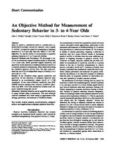
Figure 1 Example of image analyses using Illastik and ImageJ Fiji, illustrating the progression from (A) the scanned image and (B) the learning phase in Ilastik, (C) to a simple segmentation image produced in Ilastik and eventually to (D) an image that is used for calculating the thrips damage in ImageJ Fiji.
were analyzed by one-way ANOVA and, if significant differences were detected, means were subjected to post-hoc Tukey’s test.
processing in Ilastik, the program was trained to recognize damage based on color/intensity, edge (based on brightness and color gradient), and texture at the level of 1 pixel. Three labels were assigned to the various color spectra that were identified: one for thrips damage, one for undamaged leaf area, and one for background. The program was trained to recognize the three segments (thrips damage, leaf disc, and background) using four leaf discs per accession with sufficient damage until the program could precisely identify the segments in the scanned images. Whether training had been sufficient was checked by making use of the ‘life update’ function that allows the user to switch between the scan image and an overlay image that shows the three segments based on the learning process up until that point (Figure 1B). After proper training, images were converted to JPEG files that are simple segmentations of the original image in black (thrips damage), grey (leaf disc), and white (background) (Figure 1C). In ImageJ Fiji, thrips damage was extracted resulting in a TIFF image which shows thrips damage in black (Figure 1D). Total thrips damage area was easily determined using the ‘analyze particles’ command that counts and measures objects in threshold images after setting the correct scale (number of pixels : length in mm) in ImageJ Fiji. A step-by-step protocol can be found at Bio-protocol (http://www.bio-pro tocol.org/; Visschers et al., 2018).
Our protocol based on the freewares Ilastik and ImageJ resulted in effective screening for identification of thrips damage. On average, it takes ca. 4 h to process 50 images (containing a single leaf disc) of 3.08 Mb each (from scan image to a data graph in Microsoft Excel on a PC with an Intel core i7-4910MQ CPU at 2.9 GHz, and 16 GB RAM). Training is important, as the program depends on sufficient training to accurately recognize the various components in the image. Training can take up to 30 min, with a training image processing speed of ca. 0.9 MB s 1 (Intel core i7-4910MQ CPU at 2.9 GHz, 16 GB RAM). The number of leaf discs necessary for training depends on the amount of thrips damage that has been inflicted. As a reference, approximate 10 cm (with the pencil tool set to 3 pixels) of thrips damage area marking is necessary for accurate learning. Once Ilastik is properly trained for one accession of a model plant, image analysis can proceed in batch mode, allowing fast analysis of leaf disc images with the same settings.
Statistical analysis
Comparing thrips damage on the abaxial and adaxial leaf sides
All data were analyzed using SPSS v.21.0. Prior to analysis, the distribution of the feeding damage values was normalized by performing log transformation. Transformed data
To validate our standardized digital damage processing method we first analyzed whether there was a difference in thrips feeding between the abaxial and adaxial leaf sides. In
Results and discussion An effective screening method
Screening method for thrips damage quantitation 5
general, we observed more thrips damage on the abaxial than on the adaxial side of the leaf discs in all four accessions (F1,31 = 62.091, P