The evolving technology of computer auto-fabrication. ("3-D printing") now makes it possible to produce physical models for complex biological molecules and.
Augmented Reality with Tangible Auto-Fabricated Models For Molecular Biology Applications
Alexandre Gillet, Michel Sanner, Daniel Stoffler, David Goodsell, and Arthur Olson. The Scripps Research Institute. {gillet,sanner,stoffler,goodsell,olson}@scripps.edu
ABSTRACT: The evolving technology of computer auto-fabrication ("3-D printing") now makes it possible to produce physical models for complex biological molecules and assemblies. We report on an application that demonstrates the use of auto-fabricated tangible models and augmented reality for research and teaching in molecular biology, and for enhancing the scientific environment for collaboration and exploration. We have adapted an augmented reality system to allows virtual 3D representations (generated by the Python Molecular Viewer) to be overlaid onto a tangible molecular model. Users can easily change the overlaid information, switching between different representations of the molecule, displays of molecular properties such as electrostatics, or dynamic information. The physical model provides a powerful, intuitive interface for manipulating the computer models, streamlining the interface between human intent, the physical model, and the computational activity. CR Categories: J.3 [Life and Medical Sciences]: Biology and Genetics. I.3.8 [Computer Graphics]: Applications. Keywords: Molecular Modeling, Molecular Visualization, Augmented Reality. 1
Introduction
With the prevalence of structural and genomic data, molecular biology has become a human-guided, computer-assisted endeavor. The computer assists the essential human function in two ways: in exploration of scientific data, searching for and testing scientific hypotheses; and in collaboration between two or more scientists, to share knowledge and expertise. As databases grow, as our structure and process models become more complex, and as software methods become more diverse, access and manipulation of digital information is increasingly a critical issue for research in molecular biology.
Currently, exploratory research in structural molecular biology is dominated by 3-D representations via computer graphics. Collaboration, both remote and local, is aided by shared viewing of these interactive visual representations of molecular data. Yet, recent advances in the field of human-computer interfaces have not been applied to the technology used by molecular biologists -- most work in biomolecular structure and genomics is performed in front of a workstation using a mouse and keyboard as input devices. The tactile and kinesthetic senses provide key perceptual cues to our ability to understand 3-D form and to perform physical manipulations, but are currently under-utilized in molecular biology. Early structure research relied heavily on physical models: Pauling used his newly-invented spacefilling models to predict the basic folding units of protein structures and Watson and Crick used brass-wire molecular models to determine the structure of DNA, which reconciled decades of genetic data. These researchers "thought with their hands" to produce important scientific results. Current research in molecular biology now focuses on larger assemblies and more complex interactions, for which existing atomic models are inadequate. Merging physical and virtual objects into an "augmented reality" (AR) environment [1] enables new modes of interaction through the manipulation of tangible models and the complex information they represent [2]. The evolving technology of computer autofabrication ("3D printing") now makes it possible to produce physical models for complex molecular assemblies. In this paper we report on an application that demonstrates the use of auto-fabricated tangible models and AR for research in molecular biology to enhance the scientific environment for collaboration and exploration. The physical models are integrated into an augmented reality environment to streamline the interface between human intent, the physical model, and the computational activity. We have developed an AR system that allows virtual 3-D representations generated by our Python Molecular Viewer (PMV) [3] to be overlaid on an auto-fabricated model of the molecule.
1
The precise registration of the virtual objects with the real world is done using the ARToolKit library developed at the University of Washington [4]. While using the system, users can easily change the representation shown, and, for example, access information about molecular properties of the molecules. We will first describe how we create 3D tangible models of a molecular structure from a known atomic structure, then explain the integration of ARToolKit in our Python framework, and finally present some examples.
2
Design of Physical Models
We use PMV [3] both to create our virtual objects and to design our tangible models, simplifying the integration of the models with the virtual environment. PMV is a modular software framework for designing and specifying a wide range of molecular models, including molecular surfaces, extruded volumes, backbone ribbons, and atomic ball-and-stick representations. It allows the design of models at different levels of abstraction for different needs: using representations that focus on molecular shape when large systems and interactions are presented, and incorporating atomic details when needed to look at function at the atomic level. PMV is built within the interpreted language Python, which serves as a glue layer to interconnect different software components at a high level [5]. We have used Python for a number of years in this capacity because of its many desirable characteristics: it is open source and object oriented, platform independent, extensible and efficient, and has excellent introspection capabilities. PMV includes a generic 3D visualization component (DejaVu), which provides a high level interface to the OpenGL library and its geometry viewing application. To this, we have added components that provide all manner of molecular modeling and visualization functionality, including MSMS [6] for the calculation of solvent-excluded surfaces, GLE (www.linas.org/gle) for the extrusion of arbitrary 2D shapes along an arbitrary 3D path (as needed for ribbon diagrams), Babel (eyesopen.com/babel.html) for handling molecular files and coordinates, and RAPID (Gottschalk 1996) for the fast detection of intersections between polygonal models. New components have also been developed to export the resulting representations from the PMV environment in STL or VRML format for use in the rapid prototyping machinery.
for the atoms in the molecule, merges the atoms into a molecular solid model using Boolean operations, detects hydrogen bonds and prepares pockets for the magnets that simulate the bonds, computes internal smooth tubes representing the backbone, and creates an internal structure, including necessary connecting rods, as needed for hollow models. In addition, for moldmaking, the software can fit the atoms to a plane, split the model into halves, and create outer and inner models for injection molding. The ultimate goal of this work is the mass production of selected models.
3 Production of Models Newly emerging computer-automated layered fabrication machines are revolutionizing the process of fabrication. In layered fabrication (also called “rapid prototyping”), objects are built up as fused stack of thin layers of material, producing a solid or hollow object of the desire shape. The great advantage of these methods is that nearly any shape can be built—limited only by the imagination and the structural integrity of the building material. We have utilized two rapid prototyping technologies. In our testing of the methods, we have found that each has definite advantages and disadvantages for the construction of molecular models. The Z-corp process (Zcorp 406 color 3D printer) applies a pigment-binder mixture to powdered gypsum using ink-jet print heads. The parts are finished by infiltrating a strengthening agent into the model after construction. This may be wax or cyanoacrylate glue if rigid models are desired, or an elastomer to produce rubber-like flexible models. The process is relatively fast and the materials are relatively inexpensive. The major advantage is that fullcolor models may be constructed automatically. The models, however, can be fragile, and a major challenge for our future work is to overcome this fragility. Stratasys (Stratasys Prodigy Plus) uses a fused deposition method that extrudes a molten ABS plastic filament to form each layer. The process is slower and approximately twice the cost for materials and the models are monochrome. However, they are far more durable, so finer representations, such as ball-and-stick models and pre-assemble mechanical parts, can be routinely created.
In collaboration with the University of Utah, we have also developed a parser to transform atomic coordinates into a surface/feature-based representation, for use as an STL representation or as the basis for further analysis for mold making. By placing the biological model in a mechanical design context, a much richer set of modeling additions have been made possible. This conversion software creates appropriate-sized spheres
2
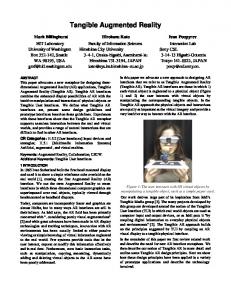
Figure 1 A number of molecular models built with different materials, showing wide range of molecular representations, scales and sizes.
4
Augmented Reality Interface
Physical molecular models, while vastly more informative and intuitive than 2D drawing or textual descriptions, are fixed in form and cannot show everything about a structure’s properties. We use
computer-based spatial tracking and rendering methods to enhance the semantic content of our models and to show dynamic properties. Augmented reality combines real world presence with virtual object presence, giving the illusion of a real interaction by leveraging the natural semantics of physical object manipulation [4, 710]. Our AR interface combines real-world user and physical model presence with computational models and data. The user manipulates a model, and the model is tracked by a video camera and displayed on the computer screen. A virtual representation (e.g., another 3D rendering of the same molecule, textual labels, or a 3D animation) is superimposed over the video display, and spatially registered with the model as the user explores the structure. The result is a quite compelling sense of virtual object realism. Our approach is based on the widely-used ARToolkit [4], an open-source software library for developing vision-based AR applications. ARToolkit is a software library that can be used to calculate the real camera position and orientation relative to physical markers in real time, allowing overlay of virtual objects onto the physical markers (see Fig 2). Some of the features of the library include: use of a single camera for position/orientation tracking, marker tracking code that uses simple black
Figure 2 The image processing pipeline used in ARToolKit. (Adapted from ARToolkit 2.33 Manual)
3
squares, pattern matching software that allows arbitrary marker patterns to be used, and fast performance for real time AR applications. We have wrapped the ARToolkit in Python to allow integration with PMV, creating PyARTK, a standalone Python module that provides a framework to manage markers, display composite images from video input, and access to the functionality of the ARToolkit library (Fig 3). It has been integrated with PMV to streamline both the design and the display of models within the same environment. A geometry manager in PyARTK assigns the geometries, animations and masks to specific AR markers or sets of markers. Changes can be made interactively as the modeling proceeds. Computer graphic objects, camera operations, clipping and lighting controls are provided in the interface, along with the video tracking and composite display. PyARTK tracks the embedded markers and then combines the video display of the model with the molecular graphics created by PMV. We have also added a basic animation facility to PyARTK, which allows run-time paging through different molecular structure representations while examining the model.
It was apparent in early tests that masking was needed to occlude the virtual object and give a better realism for systems where the virtual object interpenetrates with the physical object (Fig 4). Currently, the mask is created directly from the geometry used to build the tangible model. The mask is used to erase portions of the virtual object that should be occluded by the physical model. The tangible models are recognized and tracked using square fiducial markers placed on the surface of the model. These markers are used to register the superimposed virtual object with the manipulated real-world object. When designing the model in PMV, we add one or more small square plates to the surface of the model. Once the model is built, the markers are glued onto these plates. The transformation between the marker and the models is saved during the design phase, and later used to specify the correct registration to overlap the 3D virtual object with the tangible model. By using several markers, the AR overlay can be maintained and appropriately occluded while being arbitrarily manipulated from all angles.
Figure 3 PyARTK GUI is shown integrated with PMV. The ARViewer is the interface for managing the pattern, GeomMgr provides an interface for setting the geometry to be assigned to a pattern, and finally the PyARTK windows provide the composite graphic and video display
4
To facilitate this, we added the concept of group of markers, so that one model can carry several markers that all display the same virtual object but with different orientation depending on the location of the marker on the model.
Figure 4 3D virtual object occluded by physical object. The picture shows the use of masking to give a compelling sense of virtual object realism. The right picture shows the scene with the mask, using the geometry of the tangible model as the mask. The picture on the left show the resulting composite image when masking is not in use. Notice how the three red oxygen atoms appear to be under the protein chain in the left image.
5
that exhibits a strong electrostatic funneling effect. The user holds a tangible model of the SOD molecule built with the Stratasys printer (Fig 5), and AR enhances the monochrome tangible model with color and shows dynamic properties. . The SOD is represented as spherical harmonic surface, which shows the overall shape of the protein but smoothes out the atomic details. When a pattern is detected, the animated electrostatic field vectors show the forces on a negatively-charged oxygen free radical. A transfer function widget in the molecular viewer is used to interactively manipulate the volume rendered electrostatic potential. With this interface we can also manipulate interactions of two SOD proteins which form a dimeric complex, and thus provide an intuitive way to guide computational exploration.
Examples
With the easy auto-fabrication of tangible molecular models, we can now test our hypothesis that the perception of the complex shapes and interactions of biological molecules can be enhanced by the manipulation of augmented physical models. Through these following examples we demonstrate how this application can be useful in a collaborative environment and also be a powerful tool for teaching key molecular biology concept. HIV Protease In this first example, we integrate the physical protein backbone of HIV protease with computer graphic display of various inhibitor molecules that are effective in the treatment of AIDS. We built a backbone representation of HIV protease (Fig 4) using the Zcorp printer. The geometry is represented by an extruded tube colored by the amino acid type. To track the model in any position we placed three markers on the model, so that in any orientation, at least one marker is in the field of view of the video camera. We used AR overlay to show the bound conformation of five inhibitors within the active site of the protease (Fig 4). Each inhibitor is displayed as spacefilling representation and colored by atom types. The user can page through each inhibitor by using the animation player built within PyARTK. Notice in the figure that a mask is superimposed on the physical backbone model, providing correct occlusion of the computer graphic with the video texture. Superoxide Dismutase (SOD)
Figure 5 SOD model with and without AR. The electrostatic field is shown with small arrows that point along the local field vectors (they appear as small dots in this picture), and the potential is shown with volume-rendered clouds, with positive in blue and negative in red.
Ribosome The ribosome is a complex biomolecular machine composed of two subunits that together build proteins, by aligning tRNA molecules along an mRNA strand. We have created a tangible model of the small subunit (Fig 6) using the Zcorp printer, using a smooth spherical harmonics representation. We augment the model using virtual representation of the large subunit, to show how the two subunits assemble into the functional complex. We also show an animation of the three positions of tRNA at the translation interface of the ribosome.
Figure 6 Ribosome with and without AR. The large subunit is shown with a wire cage, and one tRNA position is shown in red in a spacefilling representation.
This example illustrates the function of superoxide dismutase (SOD), a detoxification enzyme
5
6
Conclusions
The current approach to augmented reality has been prototyped in an inexpensive, portable form, using offthe-shelf components. We routinely demonstrate the technology during presentations, at the podium, projecting the overlapped video and virtual images for the audience to view. This system may be set up in a classroom at reasonable cost. PyARTK provides facile combination of molecular modeling capabilities with input and output in the AR environment. Using PMV along with our system has proven to be fast and efficient approach to develop and test new ideas. Other input devices can be easily added to our existing system by creating appropriate interface modules.
7.Brave, S., Ishii, H. and Dahley, A., Tangible interfaces for remote collaboration and communication. In Proceedings of CSCW 98, Seattle, 1998: p. 169-178. 8.Fitzmaurice, G., Ishii, H., and Buxton, W., Bricks: laying the foundations for graspable user interfaces. In CHI'95 Proceedings, 1995: p. 442-449. 9.Gorbert, M.G., Orth, M. and Ishii, H., Triangles: tangible interface for manipulation and exploration fo digital information topography. In CHI'98 Proceedings, 1998: p. 49-56. 10.Ishii, H.a.U., B., Tangible bits: towards seamless interfaces between people, bits, and atoms. In CHI'97 Proceedings, 1997: p. 234-241.
In our future work, we plan to develop a spatially tracked “data probe’ designed to enable interaction with both physical and virtual models. Users will be able to point to different places on the tangible model and get information from the virtual model. We also plan to develop new methods for markerless spatial tracking, removing the need for fiducial tracking markers. We plan to extend the use and assessment of our augmented tangible model technologies to a wide range of grade level and settings, including K-12, undergraduate, graduate and public science exhibits.
7
Acknowledgements
We would like to thanks our collaborators Elaine Cohen, David Johnson, Rich Reisenfeld at University of Utah and Suzanne Weghorst, Mark Billingshurst, Tom Furness at University of Washington. We also acknowledge the National Institute of Health (NIH BISTI EB00798) and National Science Foundation (NSF ITR 0121282) for supporting this work. This is publication 15624-MB from the Scripps Research Institute.
REFERENCES 1.Milgram, P., Kishino, F., Taxonomy of Mixed Reality Visual Displays. IEEE Trans. On Information and Systems (Special Issue on Networked Reality), 1994. E77-D.(12): p. 1321-1329. 2.Behringer, R., Klinker, G., Mizell, D.W.,, Augmented Reality:Placing Artificial Object in Real Scenes. 1999, Naticks,Mass.: A.K.Peters, Ltd,. 3.Coon, S.I., M.F. Sanner, and A.J. Olson. Re-usable components for structural bioinformatics. in Ninth Annual International Python Conference. 2001. Long Beach, CA. 4.Billinghurst, M.a.K., H., Collaborative mixed reality. In Proceedings of International Symposium on Mixed Reality (ISMR '99). Mixed Reality--Merging Real and Virtual Worlds, 1999: p. 261-284. 5.Sanner, M.F., Python: a programming language for software integration and development. J Mol Graph Model, 1999. 17(1): p. 57-61. 6.Sanner, M.F., A.J. Olson, and J.-C. Spehner, Reduced surface: An efficient way to compute molecular surfaces. Biopolymers, 1996. 38: p. 305-320.
6