863
Journal of Alzheimer’s Disease 40 (2014) 863–868 DOI 10.3233/JAD-131847 IOS Press
Short Communication
Yueqing Hu, Jingyao Liang and Shengyuan Yu∗
CO
PY
High Prevalence of Diabetes Mellitus in a Five-Generation Chinese Family with Huntington’s Disease Department of Neurology, Chinese PLA General Hospital, Beijing, P.R. China
Accepted 27 December 2013
OR
Handling Associate Editor: Yong Shen
TH
Abstract. Huntington’s disease (HD) is associated with diabetes mellitus (DM) in population studies, but no case has been reported in a large HD family. We report a case of a five-generation Chinese family who is afflicted by both HD and DM. The prevalence of DM in HD of this family was high (72.7%). The diagnosis of HD in 11 family members was confirmed by the genetic test of the proband who had 42 CAG repeats. Furthermore, the proband’s daughter had abnormal locus at G3460T in MT-ND1 among mtDNA genome. Our case report suggests a genetic link between HD and DM.
INTRODUCTION
AU
Keywords: Diabetes mellitus, genes, Huntington disease, pathogenesis
Huntington’s disease (HD) is an autosomal dominant neurodegenerative disorder caused by an abnormally expanded CAG repeat in the first exon of the huntingtin gene [1]. Individuals with more than 36 CAG repeats will manifest the symptoms of HD. Its severity is dictated by the length of the trinucleotide repeats [2, 3]. HD is one of several genetic syndromes found to be associated with diabetes [4]. In clinical studies, an increased incidence of diabetes mellitus (DM) has been reported in HD [5, 6]. However, pathological examination of the pancreatic tissue at different disease states of HD patients and mice showed that insulin levels were similar to that of the controls [7]. Moreover, there is no large family case report to sug∗ Correspondence to: Shengyuan Yu, PhD, MD, Department of Neurology, Chinese PLA General Hospital, Fuxing Road 28, Haidian District, Beijing 100853, P.R. China. Tel.: +86 10 55499118; Fax: +86 10 88626299; E-mail:
[email protected].
gest a genetic link between HD and DM. We report here a five-generation Chinese family manifested with symptoms of HD with autosomal dominant inheritance and with high prevalence of DM. CLINICAL DETAILS The proband, a 59-year-old woman, first noticed mild involuntary movement of her limbs at age 49. Her daily activities were not affected because these symptoms were mild at onset, so she had not sought medical care until she developed dysarthria, mood swings, and rapid, abrupt, and unintentional movements of her limbs, trunk, and neck. These abnormal movements were absent at sleep and were exacerbated by stress. She did not have a history of drug abuse or medication allergies. Examination revealed impaired cognition (Mini-Mental State Examination, 19/30), mild anxiety (Self-rating Anxiety Scale, 54/100), and depression (Self-rating Depression Scale, 58/100), hypomyotonia,
ISSN 1387-2877/14/$27.50 © 2014 – IOS Press and the authors. All rights reserved
Y. Hu et al. / Huntington’s Disease and Diabetes Mellitus
AU
TH
OR
CO
PY
864
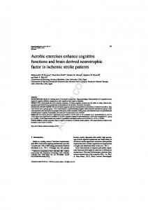
Fig. 1. A) Axial T1 and T2-weighted MRI of the proband revealed atrophy of the cerebral cortex, basal ganglia structures, and white matter. The ventricular system expanded as well. An arrow indicated the caudate nucleus. B) Capillary electrophoretogram of the proband showed forty-two CAG repeats, higher than the negative. C) Chromatogram of the variant G3460T in MT-ND1 among mtDNA for proband’s daughter was presented in the top right corner. The genetic defects were demonstrated by a black border. D) The pedigree of the five-generation Chinese family was showed at the bottom of the figure. The meaning of the symbols was attached in the left of the pedigree.
Y. Hu et al. / Huntington’s Disease and Diabetes Mellitus
865
Table 1 Demographics of the diabetic patients in the family Gender
Age at onset of DM
Duration of DM
Age at death
F F M M F F F M M F M F F
45 43 40 48 50 48 52 40 50 52 47 45 48
NA 7 9 7 8 7 2 5 2 4 6 3 6
NA 50 49 55 58 – – 45 52 – – 48 –
DNT, diabetic nutritional therapy; NA, not available.
Treatment for DM DNT and metformin DNT and metformin DNT, metformin, and insulin DNT, metformin, and insulin DNT, metformin, and insulin DNT, metformin, and insulin DNT and metformin DNT, metformin, and insulin DNT and metformin DNT and metformin DNT, metformin, and insulin DNT and metformin DNT and metformin
years [9]. Moreover, six HD patients died from injuries secondary to repeated falls and two from pulmonary infection. Deglutition barrier and psychosis might be other causes of death. Her parents did not have a history of HD (Fig. 1D). Her father died from cerebrovascular disease at the age of 50 years old, probably before the manifestation of HD symptoms. Her mother was 85 years old with normal neurological examination. This study was approved by the Ethics Committee of the Chinese PLA General Hospital, Beijing and consent was received by the family.
AU
TH
OR
and grade IV myodynamia in all extremities. Slitlamp examination was negative for Kayser-Fleischer ring in both eyes. Brain magnetic resonance imaging (MRI) revealed bilateral atrophy of the caudate nucleus and putamen with enlarged ventricles, consistent with HD (Fig. 1A). The laboratory tests did not show any evidence of Wilson’s disease, systemic lupus erythematosus, hyperthyroidism, or acanthocytosis. DNA analysis showed the presence of 42 CAG trinucleotide repeats on chromosome 4 in the proband and 17 CAG repeats in her daughter (Fig. 1B). The complete mtDNA sequence testing of the proband was normal. But the locus of G3460T on MT-ND1 of her daughter’s mtDNA was abnormal (Fig. 1C). It was identified as a candidate gene for Leber hereditary optic neuropathy (LHON) [8]. She had a strong family history of HD (Fig. 1D). We collected her family history by interviewing the living family members. For the deceased, the history was obtained by chart reviewing or interviewing of their first or second degree relatives. There were 30 members in this five-generation Chinese family. Among them 11 members suffered from HD (11/30), 13 type 2 (noninsulin-dependent) diabetes mellitus (T2DM, 13/30), and 8 both diseases (8/30). The prevalence of T2DM in HD patients was 72.7% (8/11). The demographics were shown in the Table 1. Insulin sensitizers, such as metformin, were used as first line treatment. Some of them failed to respond and were subsequently treated with insulin. However, no data was available to evidence whether anti-diabetic agents help or accelerate the symptoms of HD, which might be attributed to lack of neurology follow-up after the diabetes treatment. Additionally, nine HD patients died from age 43 to 58 years old, consistent with the average death age of Chinese HD patients which was 45.6 ± 13.5
PY
I-2 II-2 II-3 III-3 IV-1 IV-4 IV-5 IV-7 IV-8 IV-12 IV-13 IV-14 IV-17
CO
Patient number
DISCUSSION We reported a HD pedigree of a five-generation family also affected with DM. This is the first Chinese family reported with this comorbidity. This pedigree showed that HD was a neurodegenerative disorder of autosomal dominant inheritance. Although abnormal carbohydrate metabolism has been documented in patients with HD [5], there was no study in the Chinese population. Several reasons may contribute to the dearth of study, including the low prevalence of HD and lack of recognition of the association between HD and DM. The prevalence of DM in this five-generation HD family was much higher than the estimated 3.9% (urban 5.2%, rural 2.9%) prevalence of diabetes in China in 2009 [10]. In our report, the brain MRI of the proband showed mild atrophy of the caudate nuclei with marked dilatation of the ventricles in the hypothalamus regions. In fact, impaired glucose metabolism is observed radiographically in the caudate nucleus of the asymptomatic subjects [11], prior to atrophy of this nucleus. The hypothalamus integrates signals of the energy level
Y. Hu et al. / Huntington’s Disease and Diabetes Mellitus
reduction in insulin mRNA [27]. Insulin exocytosis is reduced in R6/2 pancreatic -cells owing to a dramatic reduction in the number of intracellular secretory vesicles [31]. Mhtt was reported to disrupt intracellular transport and insulin secretion by direct interference with microtubular beta-tubulin [34]. Taken together, insulin dysfunction in HD is likely associated with mhtt. We did not find variation in the proband’s mtDNA genome, however, a mutant locus was detected at G3460T in MT-ND1 (subunit one of complex INADH dehydrogenase) gene among her daughter’s mtDNA, which was identified as one of candidate genes for LHON [35, 36]. Of note, maternally inherited LHON with infantile bilateral striatal neurosis patients manifest basal ganglia degeneration and associated movement disorders similar to those seen in HD patients [37]. Moreover, biochemical analysis of HD patients’ blood platelet and muscle mitochondria has revealed a Complex I defect [38, 39]. Subsequently, impaired mitochondrial function and increased oxidative stress are commonly observed in both diseases [40–42]. These similarities in the two diseases suggest that there may be a link between HD and LHON.
AU
TH
OR
CO
from peripheral organs and is one of the main regulators of energy metabolism. Previous studies have shown atrophy and cell death of the hypothalamus in HD patients and animal models [12, 13]. This change also has been reported to associate with the alteration in endocrine function of HD, like high incidence of impaired glucose tolerance [5, 14], abnormalities of growth hormone (GH) [15–17] and disorder of cortisol levels [18–20]. GH of HD patients is raised to abnormally higher levels than the control subjects during the glucose tolerance test [14, 15]. Dysregulation of GH can contribute to some signs and symptoms in HD patients, which is possibly due to an imbalance among the biogenic amines involved in hypothalamic secretion of growth hormone-inhibiting factor and growth hormone-releasing factor [15, 21]. The increase in dopamine or serotonin activity could disrupt neurotransmission in the hypothalamus and lead to a rise in GH level. Furthermore, high GH could suppress the utilization of plasma glucose, subsequently leading to hyperglycemia. Thus hypothalamus dysfunction may play an important role in abnormal glucose metabolism of HD patients. Though we did not have data available to test whether the anti-diabetics agents helped or accelerated the symptoms of HD patients, the benefits from several hypoglycemic agents have been found in HD mice. Exendin-4 (an FDA-approved antidiabetic glucagon-like peptide 1 receptor agonist), metformin, glibenclamide, and insulin-like growth factor-1 might show efficacy in HD models, including: (i) improved glycemic control and normalized the insulin levels; (ii) suppressed brain and pancreatic pathologies; (iii) ameliorated motor function and extended survival; and (iv) decreased htt aggregate accumulation [22–25]. Prospective, placebo controlled studies are warranted to address the treatment for abnormal carbohydrate metabolism in patients with HD. Mutant htt (mhtt) was believed to impair neuron cell in cerebral cortex and striatum [26] and proved to be widely expressed in most tissues [27, 28], including the pancreas [28, 29]. The impairment of pancreatic islet cells may be involved in the development of glucose intolerance and insulin-deficient diabetes observed in HD patients and animal models. The pancreatic levels of insulin and insulin mRNA were reduced in R6/2 mice [27, 30]. Moreover, htt aggregates were latter identified to be expressed in -cells [31–33], which explained why -cells become dysfunctional in HD patients. The appearance of htt aggregates coincided with impaired mRNA expression of the key regulators of insulin gene expression and with a progressive
PY
866
CONCLUSIONS Our case report of a five-generation family of HD has shown high prevalence of DM, suggesting a genetic link between the two diseases. Further genome wide association studies are warranted to explore the underlying mechanism of this comorbidity. ACKNOWLEDGMENTS We would like to thank Dr. Yiming Mu (Department of Endocrinology, Chinese PLA General Hospital, Beijing, China) for reviewing the paper. Authors’ disclosures available online (http://www.jalz.com/disclosures/view.php?id=2085). REFERENCES [1]
[2]
MacDonald ME, Ambrose CM, Duyao MP, Myers RH, Lin C, Srinidhi L, Barnes G, Taylor SA, James M, Groot N (1993) A novel gene containing a trinucleotide repeat that is expanded and unstable on Huntington’s disease chromosomes. Cell 72, 971-983. Rubinsztein DC, Leggo J, Coles R, Almqvist E, Biancalana V, Cassiman J-J, Chotai K, Connarty M, Craufurd D, Curtis A (1996) Phenotypic characterization of individuals with 30–40 CAG repeats in the Huntington disease (HD) gene reveals HD cases with 36 repeats and apparently normal elderly individuals with 36–39 repeats. Am J Hum Gene 59, 16.
Y. Hu et al. / Huntington’s Disease and Diabetes Mellitus
[8]
[9]
[10]
[11]
[12]
[13]
[14]
[15]
[16]
[17]
[18]
[19]
[20]
[21]
[24]
[25]
PY
[7]
[23]
Martin B, Golden E, Carlson OD, Pistell P, Zhou J, Kim W, Frank BP, Thomas S, Chadwick WA, Greig NH, Bates GP, Sathasivam K, Bernier M, Maudsley S, Mattson MP, Egan JM (2009) Exendin-4 improves glycemic control, ameliorates brain and pancreatic pathologies, and extends survival in a mouse model of Huntington’s disease. Diabetes 58, 318-328. Ma TC, Buescher JL, Oatis B, Funk JA, Nash AJ, Carrier RL, Hoyt KR (2007) Metformin therapy in a transgenic mouse model of Huntington’s disease. Neurosci Lett 411, 98-103. Hunt MJ, Morton AJ (2005) Atypical diabetes associated with inclusion formation in the R6/2 mouse model of Huntington’s disease is not improved by treatment with hypoglycaemic agents. Exp Brain Res 166, 220-229. Duarte AI, Petit GH, Ranganathan S, Li JY, Oliveira CR, Brundin P, Bjorkqvist M, Rego AC (2011) IGF-1 protects against diabetic features in an in vivo model of Huntington’s disease. Exp Neurol 231, 314-319. Sharp AH, Loev SJ, Schilling G, Li S-H, Li X-J, Bao J, Wagster MV, Kotzuk JA, Steiner JP, Lo A (1995) Widespread expression of Huntington’s disease gene (IT15) protein product. Neuron 14, 1065-1074. Andreassen OA, Dedeoglu A, Stanojevic V, Hughes DB, Browne SE, Leech CA, Ferrante RJ, Habener JF, Beal MF, Thomas MK (2002) Huntington’s disease of the endocrine pancreas: Insulin deficiency and diabetes mellitus due to impaired insulin gene expression. Neurobiol Dis 11, 410-424. Sathasivam K, Hobbs C, Turmaine M, Mangiarini L, Mahal A, Bertaux F, Wanker EE, Doherty P, Davies SW, Bates GP (1999) Formation of polyglutamine inclusions in non-CNS tissue. Hum Mol Genet 8, 813-822. Schilling G, Sharp AH, Loev SJ, Wagster MV, Li S-H, Stine OC, Ross CA (1995) Expression of the Huntington’s disease (IT15) protein product in HD patients. Hum Mol Genet 4, 1365-1371. Hurlbert MS, Zhou W, Wasmeier C, Kaddis FG, Hutton JC, Freed CR (1999) Mice transgenic for an expanded CAG repeat in the Huntington’s disease gene develop diabetes. Diabetes 48, 649-651. ˚ Bj¨orkqvist M, Fex M, Renstr¨om E, Wierup N, Peters´en A, Gil J, Bacos K, Popovic N, Li J-Y, Sundler F (2005) The R6/2 transgenic mouse model of Huntington’s disease develops diabetes due to deficient -cell mass and exocytosis. Hum Mol Genet 14, 565-574. Bates G (2003) Huntingtin aggregation and toxicity in Huntington’s disease. Lancet 361, 1642-1644. Ye C-F, Li H (2009) HSP40 ameliorates impairment of insulin secretion by inhibiting huntingtin aggregation in a HD pancreatic  cell model. Biosci Biotechnol Biochem 73, 17871792. Smith R, Bacos K, Fedele V, Soulet D, Walz HA, Oberm¨uller ¨ S, Lindqvist A, Bj¨orkqvist M, Klein P, Onnerfjord P (2009) Mutant huntingtin interacts with -tubulin and disrupts vesicular transport and insulin secretion. Hum Mol Genet 18, 3942-3954. Howell N, Bindoff L, McCullough D, Kubacka I, Poulton J, Mackey D, Taylor L, Turnbull D (1991) Leber hereditary optic neuropathy: Identification of the same mitochondrial ND1 mutation in six pedigrees. Am J Hum Gene 49, 939. Huoponen K, Vilkki J, Aula P, Nikoskelainen EK, Savontaus M (1991) A new mtDNA mutation associated with Leber hereditary optic neuroretinopathy. Am J Hum Gene 48, 1147. Novotny EJ, Singh G, Wallace DC, Dorfman LJ, Louis A, Sogg RL, Steinman L (1986) Leber’s disease and dystonia a mitochondrial disease. Neurology 36, 1053-1060.
CO
[6]
[22]
[26]
[27]
OR
[5]
TH
[4]
Brinkman R, Mezei M, Theilmann J, Almqvist E, Hayden M (1997) The likelihood of being affected with Huntington disease by a particular age for a specific CAG size. Am J Hum Gene 60, 1202. American Diabetes, Association (2013) Diagnosis and classification of diabetes mellitus. Diabetes Care 36, S67-S74. Podolsky S, Leopold N, Sax D (1972) Increased frequency of diabetes mellitus in patients with Huntington’s chorea. Lancet 299, 1356-1359. Farrer LA (1985) Diabetes mellitus in Huntington disease. Clin Genet 27, 62-67. ˚ Luts L, Maat-Schieman Bacos K, Bj¨orkqvist M, Peters´en A, ML, Roos RA, Sundler F, Brundin P, Mulder H, Wierup N (2008) Islet -cell area and hormone expression are unaltered in Huntington’s disease. Histochem Cell Biol 129, 623-629. Puomila A, Viitanen T, Savontaus M-L, Nikoskelainen E, Huoponen K (2002) Segregation of the ND4/11778 and the ND1/3460 mutations in four heteroplasmic LHON families. J Neurol Sci 205, 41-45. Guo X, Zhang S, Burgunder J-M, Shang H (2010) Clinical features of Huntington disease in 243 Chinese patients. Neural Regen Res 5, 102-107. Pan C, Shang S, Kirch W, Thoenes M (2010) Burden of diabetes in the adult Chinese population: A systematic literature review and future projections. Int J Gen Med 3, 173-179. Mazziotta JC, Phelps ME, Pahl JJ, Huang S-C, Baxter LR, Riege WH, Hoffman JM, Kuhl DE, Lanto AB, Wapenski JA (1987) Reduced cerebral glucose metabolism in asymptomatic subjects at risk for Huntington’s disease. N Engl J Med 316, 357-362. Kremer H, Roos R, Dingjan G, Bots GTA, Maran E (1990) Atrophy of the hypothalamic lateral tuberal nucleus in Huntington’s disease. J Neuropathol Exp Neurol 49, 371-382. Kremer H, Roos R, Dingjan G, Bots G, Bruyn G, Hofman M (1991) The hypothalamic lateral tuberal nucleus and the characteristics of neuronal loss in Huntington’s disease. Neurosci Lett 132, 101-104. Podolsky S, Leopold N (1977) Abnormal glucose tolerance and arginine tolerance tests in Huntington’s disease. Gerontology 23, 55-63. Keogh H, Johnson R, Nanda R, Sulaiman W (1976) Altered growth hormone release in Huntington’s chorea. J Neurol Neurosurg Psychiatry 39, 244-248. Phillipson O, Bird E (1976) Plasma growth hormone concentrations in Huntington’s chorea. Clin Sci Mol Med 50, 551-554. Caraceni T, Panerai AE, Parati EA, Cocchi D, M¨uller EE (1977) Altered growth hormone and prolactin responses to dopaminergic stimulation in Huntington’s chorea. J Clin Endocrinol Metab 44, 870-875. ˚ Bacos K, Isaacs J, Norl´en P, Gil J, Bj¨orkqvist M, Peters´en A, Popovic N, Sundler F, Bates GP, Tabrizi SJ (2006) Progressive alterations in the hypothalamic-pituitary-adrenal axis in the R6/2 transgenic mouse model of Huntington’s disease. Hum Mol Genet 15, 1713-1721. Leblhuber F, Peichl M, Neubauer C, Reisecker F, Steinparz F, Windhager E, Maschek W (1995) Serum dehydroepiandrosterone and cortisol measurements in Huntington’s chorea. J Neurol Sci 132, 76-79. Aziz NA, Pijl H, Fr¨olich M, van der Graaf AM, Roelfsema F, Roos RA (2009) Increased hypothalamic-pituitary-adrenal axis activity in Huntington’s disease. J Clin Endocrinol Metab 94, 1223-1228. Podolsky S (1979) Hormone studies in patients with Huntington’s disease. Age 2, 17-22.
AU
[3]
[28]
[29]
[30]
[31]
[32] [33]
[34]
[35]
[36]
[37]
867
[41]
transgenic mouse model of Huntington’s disease. J Neurochem 79, 1246-1249. Schapira A (2002) Primary and secondary defects of the mitochondrial respiratory chain. J Inherit Metab Dis 25, 207-214. Carelli V, Rugolo M, Sgarbi G, Ghelli A, Zanna C, Baracca A, Lenaz G, Napoli E, Martinuzzi A, Solaini G (2004) Bioenergetics shapes cellular death pathways in Leber’s hereditary optic neuropathy: A model of mitochondrial neurodegeneration. Biochim Biophys Acta 1658, 172-179.
PY
[42]
CO
[40]
OR
[39]
Parker WD, Boyson SJ, Parks JK (1989) Abnormalities of the electron transport chain in idiopathic Parkinson’s disease. Ann Neurol 26, 719-723. Arenas J, Campos Y, Ribacoba R, Mart´ın MA, Rubio JC, Ablanedo P, Cabello A (1998) Complex I defect in muscle from patients with Huntington’s disease. Ann Neurol 43, 397400. Bogdanov MB, Andreassen OA, Dedeoglu A, Ferrante RJ, Beal MF (2001) Increased oxidative damage to DNA in a
TH
[38]
Y. Hu et al. / Huntington’s Disease and Diabetes Mellitus
AU
868