Benchmarks cad locus may influence quantitative traits in chicken. J. Exp. Zool. 263:303-308. 4.Khatib, H., D. Bercovich, T. Ratz, Y. Plotzki, A. Fainsod and Y. Gruenbaum. 1995. Mapping the CdxA gene into a new linkage group in chicken. Anim. Genet. 26:211. 5.Khatib, H. and Y. Gruenbaum. 1996. Chicken red blood cells as a substrate for direct polymerase chain reaction. Anim. Genet. 27:53-54. 6.Khatib, H., N. Sagiv and Y. Gruenbaum. 1998. Fresh and Frozen pools of chicken red blood cells as a substrates for direct polymerase chain reaction. Poult. Sci. 77:902-904.
Address correspondence to Prof. Yosef Gruenbaum, Department of Genetics, The Institute of Life Sciences, The Hebrew University of Jerusalem 91904, Israel. Internet:
[email protected] Received 17 November 1998; accepted 10 March 1999.
Figure 1. PCR amplification of CdxA alleles from chicken blood cells. (A) The upper map shows the position of the homeobox (striped boxes) and the BamHI sites (B1-B3) in the CdxA gene. The lower map shows an enlargement of the region around the polymorphic site B1 (arrow). The position of the different primers that were used for the amplification is also marked with arrows. Primers 1 (5′-CGCTCCGGTTCGTCACTGCT-3′), 4C (5′-GCTGTCGGCCACCTGTTGTG-3′) and 1F (5′-GTGGCACGGACAGGGGATCC-3′) were used for the detection of the F allele and primers 1, 4C and 1S (5′-GTGGCACGGACAGGGGATCG-3′) were used for the detection of the S allele. Removal of the internal primer (1S or 1F) resulted in a slight increase of amplification (data not shown). The bar below the lower map represents 450 bp for the upper map and 100 bp for the lower map. (B) The PCR products were separated on 2% agarose gel and stained with ethidium bromide. Each pair of lanes represents PCR amplification of one blood sample (1 and 2, 3 and 4, 5 and 6, 7 and 8) or one DNA sample (9 and 10). Samples were amplified either with primers 1, 4C and 1F (lanes 1, 3, 5, 7, 9) or with primers 1, 4C and 1S (lanes 2, 4, 6, 8, 10). Lanes 1, 2, 5 and 6 show amplification from fresh blood cells; lanes 3, 4, 7 and 8 show amplification from frozen blood cells. Lanes 1–4 show results of amplification according to the previously published technique (1,2); lanes 5–8, amplification according to the method presented here; whereas lanes 9 and 10 are from control amplifications using 50 ng of isolated chicken DNA. Lane M shows size markers of 100-bp ladder.
Dani Bercovich1,2, Yoram Plotsky1 and Yosef Gruenbaum2 1MIGAL—Galilee Technological Center Kiryat Shmona 2The Hebrew University of Jerusalem Jerusalem, Israel
Bridge-Overlap-Extension PCR Method for Constructing Chimeric Genes BioTechniques 26:1082-1086 (June 1999)
methods work only with a very limited range of blood-cell concentrations (between 0.125–1.0 µL of blood; see References 5 and 6). One difference between the original vs. the modified method is degradation of hemoglobin, or heme compound, by boiling in NaOH that results in a stronger inhibitory effect on the Taq DNA polymerase (2,4). In addition, it is possible that repeated boiling of the sample before DNA amplification increases the accessibility of the DNA to the primers.
REFERENCES 1.Akane, A. 1996. Hydrogen peroxide decomposes the heme compound in forensic specimens and improves the efficiency of PCR. BioTechniques 21:392-394. 2.Akane, A., K. Matsubara, H. Nakamura, S. Takahashi and K. Kimura. 1994. Identification of the heme compound co-purified with deoxyribonucleic acid (DNA) from bloodstains, a major inhibitor of polymerase chain reaction (PCR) amplification. J. Forensic Sci. 39:362-372. 3.Iraqi, F., H. Khatib, G. Shani, A. Davarsi, A. Frumkin, G. Zeitlin, M. Soller, A. Fainsod and Y. Gruenbaum. 1992. CHox-
Since the discovery of polymerase chain reaction (PCR) (4), innumerable innovations have heralded several important advances in modern molecular biology. One such application is the introduction of predetermined alterations in the sequences of the target genes, for example, site-specific mutations and hybrid gene constructions. In the latter development, chimeric gene constructs are synthesized by an overlap-extension technique, termed splicing by overlap extension (SOE) (1). In this technique, PCR products corresponding to the genes to be spliced are synthesized
Benchmarks using complementary primers that contain sequences representing the junction of two genes to be spliced, which allows the annealing of the two PCR products synthesized from the half reactions. Following annealing, a chimeric PCR product is obtained by using the two external primers. The megaprimer approach utilizes three oligonucleotides. In the first step, the mutant primer is used along with a reverse primer to synthesize a megaprimer product, which then serves as a primer, along with the forward primer, for synthesis of the complete gene containing the mutation in the second PCR step (5). However, in our laboratory, especially while using the classical SOE technique for site-directed mutagenesis, we have frequently found the efficiency of correct recombinants to be less than 10%. The reason for this was not quite clear, but it might be a result of the reannealing of oligonucleotides from the templates produced by the mutant oligonucleotide to the wild-type template (which is apparently never com-
pletely removed, despite isolation of products of half reactions from the agarose gel), thus reducing the amplification of the mutant double-stranded DNA molecules. Similar problems are encountered while constructing chimeric genes. Here, we have sought to obviate this problem by a modification that allows the construction of a precise chimera between two genes or portions of two genes, although it might restrict its application to cases in which one of the gene segments is of smaller size. This is achieved by using two bridging-
overlap PCRs, as outlined in Figure 1. The experiment described here involves the construction of a chimera between the secretory signal of Plus (P)-factor (2) of Schizosaccharomyces pombe and streptokinase from Streptococcus sp. (3). In the experimental design, the first PCR is performed using the forward oligonucleotide B (Figure 1) corresponding to the junction between the 3′ end of the P-factor (2) and the 5′ sequence of the processed form of streptokinase (SK) gene (3), together with reverse oligonucleotide C representing
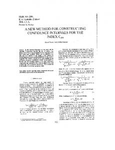
Figure 1. A schematic representation of the experimental design and the steps involved in the bridge-overlap-extension PCR approach, as applied to the synthesis of a chimeric gene between the S. pombe P-factor secretory signal and SK. (A) Shown are the two segments of the proposed chimera, namely the P-pheromone segment and the processed form of SK. Also shown are the different oligonucleotides, namely A, B, C and compAB. These oligos are shown below the regions of P-factor and/or SK gene to which they correspond, as described in detail in the text. The sequences of the oligonucleotides were: oligonucleotide A, 5′-GATGGATCCTCGAGGATGAAGATCACCGCTGTCATTGCCCTTTTATTCTCACTTG-3′; oligonucleotide compAB, 5′-ACACCAGGATCGGCAACTGGAATAGGTGAGGCAGCAAGTGAGAATAAAAGG-3′; oligonucleotide B, 5′-GTTGCCGATCCTGGTGTGGTTTCAGTTAGCAAGATTGCTGGACCTGAGTGGCTG-3′; and oligonucleotide C, 5′-GGAGCTCGAGATGAAAAATTACTTATCTTTTGG-3′. (B) Shown are the 3 PCR steps proposed to carry out the synthesis of the P-factor/SK chimera. In step 1, DNA from boiled cells of Streptococcus along with oligonucleotides B and C are used for PCR to yield the product D. In step 2, PCR is carried out using the oligonucleotides A and compAB in 10:1 molar ratio in absence of any DNA to yield the product E. In step 2, PCR product E is shown on top of the step 1 product D to depict the partial complementarity of the product D with the product E. Step 3 is carried out with the gel-purified product D from step 1 and the unpurified product E of step 2 and the oligonucleotide C and yields the product F representing the chimera between the P-factor and SK. The conditions of PCR, including the concentrations of DNA, oligonucleotides, different PCR products and cycling parameters, are described in detail in the text.
the C-terminal end of SK. The second PCR step is carried out using the bridging oligonucleotide compAB (Figure 1, A and B) and the forward oligonucleotide A (Figure 1, A and B), which corresponds to the N-terminal region of the P-secretory signal (Figure 1). The bridging oligonucleotide compAB has partial complementarity both at its 3′ end to the 3′ end of the proximal forward primer, oligonucleotide A, and also near its 5′ end to the 5′ end of the distal sense primer, oligonucleotide B. The middle region of the oligonucleotide spans the gap between the proximal and distal segments not covered by oligonucleotides A and B. The second PCR is carried out using a 10:1 molar excess of oligonucleotide A to produce significant amounts of the hybrid product E (Figure 1B). This product now incorporates the region of P-factor sequence not present in oligonucleotide A, because of the length of the complete sequence of P-factor (Figure 1A) and also contains a region of complementarity with the 5′ end of the PCR product generated by oligonucleotide pair B and C. Then, the asymmetrically amplified PCR product E is used in combination with the oligonucleotide C and the oligonucleotide-free PCR product D from the first step in the
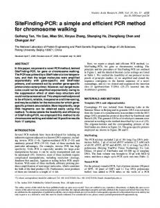
final PCR step to yield the chimeric product F (Figure 1B, step 3). Figure 1 outlines all these steps. The resulting PCR products from the three steps are visualized by agarose gel electrophoresis. Step 1 yielded a 1.32-kb band representing the junction of the distal part of the P-factor and the complete sequence of the SK gene (Figure 2A, lane 2 and Figure 1B, product D). The product of asymmetric PCR from step 2 (Figure 1B, product E) is shown separately (Figure 2B, lane 3) along with the control mixture of oligonucleotides used for the reaction (Figure 2B, lane 2), as these had to be run on a 2.5% agarose gel for proper resolution. We observe that this step yields a smeary band of approximately 90 bases (Figure 2B, lane 3) with slower mobility than the starting oligonucleotides of 65 bases (Figure 2B, lane 2). Step 3 yields a product of the predicted length of 1.39 kb (Figure 2A, lane 3). The product was cloned into a suitable expression vector. Sequencing of five independent clones indicated that all had the correct sequence (data not shown) indicating 100% efficiency of construction of the chimeric gene. Moreover, all the recombinant clones carrying the chimeric construct in the expression vector expressed SK efficiently, as monitored by the halo
Figure 2. Agarose gel electrophoresis of the PCR products representing the P-factor/SK chimeras. Products of the 3 PCR steps depicted in Figure 1B are shown. (A) Lane 1, size markers; lane 2, step 1 product D; lane 3, step 3 product F. A 1.5% agarose gel was run to resolve the products. (B) Lane 1, size markers; lane 2, mixture of oligonucleotides A and compAB; lane 3, step 2 product E. These samples were run on a 2.5% agarose gel to obtain better resolution of the oligonucleotides and the short asymmetric PCR product D.
Benchmarks assay on skim milk plates (3). The detailed conditions for the PCRs are described below. For step 1, DNA (50 ng) obtained after boiling the Streptococcus cells in TE buffer (10 mM Tris-HCl, pH 8.0, 1 mM EDTA) were used in combination with oligonucleotides B and C and 2.5 U Taq DNA Polymerase (PE Biosystems, Foster City, CA, USA) in a final volume of 50 µL. Buffer conditions were as recommended by the manufacturer (PE Biosystems). Cycling conditions were: denaturation at 94°C for 3 min, followed by 30 cycles of denaturation at 94°C for 1 min, annealing at 58°C for 30 s and extension at 72°C for 2 min and a final extension step of 72°C for 5 min. Product D was resolved by agarose gel electrophoresis and purified away from oligonucleotides by GENECLEAN procedure (BIO 101, Vista, CA, USA). In step 2, 100 ng of oligonucleotide A and 10 ng of oligonucleotide B were used with similar cycling conditions, except that 20 cycles involving 30-s steps of denaturation, annealing and extension were carried out, and the annealing temperature was 55°C to yield product E. In the third step, 50 ng of the oligonucleotide-free step 1 product D and 100 ng each of product E and reverse oligonucleotide C were used. It was not necessary to purify product E. Cycling conditions were the same as step 1, except that the extension at 72°C was for 1 min, and 0.01 U Vent polymerase (New England Biolabs, Beverly, MA, USA) was included to generate blunt-ended chimeric product. The temperatures for the annealing steps for different oligonucleotides were determined by using the Gene Runner Program Version 3.02 (Hastings Software, Hastings-On-Hudson, NY, USA). We understand that this bridge-overlap-extension PCR approach can have several applications, such as insertion of epitope tags, nuclear localization signals, secretory or proteolytic signals, insertion of DNA-binding regions or other functional elements into proteins of interest. The sequence element to be inserted can be of small size (30 bp) or as long as 150 bp, both being within the limits of efficient oligonucleotide synthesis. A distinct feature of this method is that by using overlapping oligonu-
cleotides and bringing about a bridging step (Figure 1, step 2), we carry out the synthesis of a longer primer that contains an overlap with the hybrid PCR product of step 1, in the absence of any wild-type template. This helps in avoiding the use of template DNA and, therefore, the problem of re-annealing by oligonucleotides to wild-type template during the PCR steps altogether. The logic is similar to the megaprimer-mediated, site-directed mutagenesis method described previously (5). However, in contrast to the megaprimer approach, which utilizes three oligonucleotides, our method uses four oligonucleotides. Another apparent shortcoming of the present method is the limits to the lengths of the proximal gene segment, which is determined by the limits of the lengths of the oligonucleotide synthesizer. However, some companies nowadays provide facilities to synthesize oligonucleotides that are approximately 140 bases or longer, which can extend the limit of the smaller gene segment to approximately 250 bp and beyond. Nevertheless, this peculiar feature could make our method especially suitable where shorter sequences such as secretory signals, nuclear localization signals, phosphorylation sites, epitope tags, DNA-binding regions or interprotein interaction domains have to be incorporated. The success of the technique is indicated by the 100% efficiency of obtaining the correct clones. Moreover, a high level of expression of precursor (fusion of P-factor and SK) and mature SK was observed when the hybrid gene construct was cloned into an expression vector and transformed in suitable strains of S. pombe (data not shown). Therefore, this new approach could serve as an alternative method for construction of chimeric genes. REFERENCES 1.Horton, R.M., H.D. Hunt, J.K. Pullen and L.R. Pease. 1989. Engineering hybrid genes without the use of restriction enzymes: gene splicing by overlap extension. Gene 77:61-68. 2.Imai, Y. and M. Yamamoto. 1994. The fission yeast mating pheromone P-factor: its molecular structure, gene structure, and ability to induce gene expression and G 1 arrest in the mating partner. Genes Dev. 8:328-338. 3.Malke, H., B. Roe and J.J. Ferretti. 1985. Nucleotide sequence of the streptokinase gene
from Streptococcus equisimilis H46A. Gene 34:357-362. 4.Mullis, K.B. and F. Faloona. 1987. Specific synthesis of DNA in vitro via a polymerasecatalyzed chain reaction. Methods Enzymol. 155:335-350. 5.Sarkar, G. and S.S. Sommer. 1990. The “megaprimer” method of site-directed mutagenesis. BioTechniques 8:404-407.
We are grateful to Vintoo Goel for help in typing, editing and artwork. This work was supported by the Council of Scientific and Industrial Research and Department of Science and Technology, New Delhi, India. Address correspondence to Dr. Jagmohan Singh, Yeast Developmental Genetics Laboratory, Institute of Microbial Technology, Sector 39A, Chandigarh-160036, India. Internet:
[email protected] Received 13 October 1998; accepted 9 March 1999.
Raj Kumar Mehta and Jagmohan Singh Institute of Microbial Technology Chandigarh, Punjab, India
Microsatellite Locus Amplification Using Deer Antler DNA BioTechniques 26:1086-1088 (June 1999)
The study of population genetics has been greatly enhanced by the use of microsatellite loci. Polymerase chain reaction (PCR)-based (5) genotyping of these ubiquitous, extremely polymorphic loci allows rapid estimates of genetic variation, population substructure and differentiation and effective population size. These techniques have been used to describe the population genetics of a wide variety of mammals [e.g., bears (7), mice (1) and wombats (11)]. Unfortunately, almost all population studies are limited to present-day populations. The study of population change over time must be done by inference from data derived from existing populations. Museum specimens hold some