Free-nuclease Distilled water. 3.1.3 Tools & Equipment: 1. Tube centrifuge; (Hettich ..... E MgCl2. 1.5 μM. F Stabilizer and tracking Dye. DNA template sample.
Chapter Three
3.1
Materials & Methods
Materials and Equipment:-
3.1.1 Kits:The following diagnostic kits were used during the study: 1. Stool DNA extraction kit; (BioNeer, Korea). 2. PCR Pre-mix-Accu Power (master mix tube); (BioNeer, Korea). Accu power PCR Premix delivered by BioNeer Company each kit contain 96-eppindorf tube with total volume reaction 20 µl. 3. Forward (EH1) and reverse primer (EH2) for E. histolytica trophpzoit and cyst 16S-like ribosomal RNA gene; (BioNeer Oligo, Korea). 4. Forward (S-f) and reverse primer (S-r) specific for Shigella dysenteriae (ial) gene sequence; (BioNeer Oligo, Korea). 5. DNA ladder 100-2000-base pairs; (BioNeer, Korea). 6. DNA Loading Dye. 7. SYPER Green DNA Dye, Ready to use, (iNtRON Biotechnology, Inc).
3.1.2 Reagents & Other Diagnostic Facilities: 1. TBE buffer (10X); (Promega, U.S.A.). 2. Agarose powder; (Promega, U.S.A.). 3. Mineral oil; (Sacace, Italy). 4. Ethidium bromide; (BDH, England). 5. Absolute ethanol (100%); (VWR, U.S.A.). 6. Absolute isopropanol (C3H8O); (Applichem, Germany). 7. Gram stain; (Vaccines and Sera institute, Iraq). 8. Methylene blue and iodine solution. 9. Free-nuclease Distilled water.
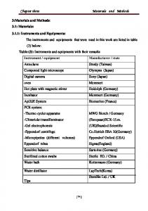
3.1.3 Tools & Equipment: 1. Tube centrifuge; (Hettich EDA20, Japan). 2. Water bath; (Clifton, England).
40
Chapter Three
Materials & Methods
3. Spin centrifuge, mini-spin (plus); (eppindorf AG, Germany). 4. Desktop micro centrifuge, Microfuge; (eppindorf AG, Germany). 5. Vortex shaker; (Genie U.S.A. bohemia NY 11716). 6. Fume hood; (Lab tech, Korea). 7. UV spectrophotometer; (Unico, U.S.A.). 8. Thermal cycler; (Esco, Canada). 9. Autoclave; (Tꞌuttanuer EZ 10k). 10. Oven; (GallenKamp B.S). 11. Refrigerator; (Admiral / NRC food master). 12. Deep freeze; (Frolab, France). 13. UV transilluminator; (Viper Lourmat, made in EEC). 14. Burner, (Germany). 15. Electronic balance; (Sartorius, Germany). 16. Distill water; (Daihan, Turkey). 17. Electrophoresis power supply; (Ms Major Science). 18. Electrophoresis tank; (Ms Major Science). 19. Automatic micropipette; (100-1000 µl); (Islamed, Germany). 20. Automatic micropipette (10-100 µl); (Islamed, Germany). 21. Automatic micropipette (2-20 µl); (Eppindorf, Germany). 22. Automatic micropipette (1.5-2.5 µl); (Eppindorf, Germany). 23. Automatic micropipette (10-100 µl); (Eppindorf, Germany). 24. Sterile disposable syringes, 5 and 10 ml; (QG, Qater). 25. Printer; (Epson, Japan). 26. Disposable collection cup; (China). 27. Eppindorf tubes 1.5 ml; (Germany). 28. Pipette tips blue; (Germany). 29. Para film; (Mco, Germany). 30. Disposable surgical gloves, (powder less); (Enana, Malaysia).
41
Chapter Three
Materials & Methods
31.Wooden applicator sticks; (China). 32. Pipette tips yellow; (Germany). 33. Gel champers; (Ms Major Science). 34. Magnetic stirrer; (Taiwan). 35. Digital camera; (HX1); (Japan). 36. Sterile cotton; (Iraq). 37. Foil papers; (local market). 38. Ice block bags; (India). 39. Flask (500 ml and 1000 ml); (Germany). 40. Incubator; (Carbolite, England). 41. Jar; (Oxoid, U.S.A.). 42. Microscopy; (Olympus, Japan). 43. Plastic petri dish; (China). 44. Slide and cover slid. 45. Sterile plain tube; (Malaysia).
3.1.4 Culture Media: The following culture media were engaged in the extant work: 1. Xylose-lysine-deoxychlate (XLD); (Himedia, India). 2. Deoxycholate citrate agar (DCA); (Himedia, India). 3. MacConkey agar; (Oxoid, England). 4. Salmonella-Shigella agar (SS-agar); (Oxoid, England). 5. Hekton Enteric agar.
3.1.5 Study Samples. 3.1.5.1 Patients: One Hundred forty-five stool samples were obtained from children admitted to Maternity and Children Teaching Hospital in Ramadi provinceIraq with acute diarrheal diseases (80 male and 65 female) within the mean
42
Chapter Three
Materials & Methods
age (2.168±12.571 months). They were enrolled in the study from January 2014 to March 2015. These admitted patients with dysentery symptoms were raised by their physician for diagnostic stool sample in Maternity and Child Teaching Hospital in Ramadi city. After the informed consent was obtained from the parents of the subjects, a pediatrician filled out the information relevant to clinical symptoms and illness onset on a standardized questionnaire, the rest of the questionnaire was filled by the parents of the children see Appendix-1while Appendix -2- for colonies appearance of Shigella dysenteriae for complete listing of the items on this questionnaire. Diarrhea was defined as the passage of three or more loose or watery stools in the preceding 24 hours. A questionnaire was completed for each patient containing the following information: age, gender, resident, water source, feed source.
3.2
Methods.
3.2.1 Collection of Stool Samples: Stool samples were collected from patients using sterile disposable wide mouth cups. Neonatal stool samples were excluded in this study.
3.2.2 Examination of Stool Samples: Macroscopic examination:- Stool specimens were inspected for consistency, color, and for the presence of blood, mucus…etc. Microscopic examination (Parasitic detection):- Stool samples were investigated by native-Lugol examination. Lugol’s iodine was added to the stool smear and covered with a cover slip. Stool smears with saline or iodine examined microscopically at low (10X) and high (40X)
43
Chapter Three
Materials & Methods
magnifications within 15 minutes. The identification of E. histolytica/ dispar trophozoites was made by the characteristic movement of the protozoan and the presence of phagocytized red blood cells. The identification of amebic cysts (E. histolytica/ dispar) was based on morphologic characteristics, (10-15μm, spherical form, mature tetranucleated cysts having a central endosome). Examination of the cellular exudates of diarrheal stools may give an indication of the organism involved (World Health Organization, 2001):-
o Clumps of PMNs> 50 cells hpf, macrophages and erythrocytes are typical of shigellosis.
o Small number of PMNs< 20 cells hpf, are found in salmonellosis, and invasive E. coli.
o In amoebic dysentery, the cells are mostly degenerated (ghost cells).
o The presence of CLC (Chrcot-Leyden Crystals) in the stool give an indication of the amoebic dysentery.
o Leukocytes and erythrocytes are also found in about half cases of diarrhea due to Campylobacter spp.
o Few leukocytes (2-5 cells per high-power field) are present in cases of cases of cholera, enterotoxigenic and enteropathogenic E.coli (ETEC, EPEC), and viral diarrhea. The percentages viability of erythrophagocytic trophozoites was higher (65% and 95%) during the first and third hours in stool sample. This was probably due to the antioxidant molecules (superoxide dismutase, catalase, glutathione, peroxirredoxin and vitamin E) present in the engulfed erythrocytes (Kuypers, 2007).
44
Chapter Three
Materials & Methods
3.2.3 Culture of Stool Samples: The following is a procedure for the culture and isolation of Shigella dysenteriae from feces. Selective enrichment suppresses fecal-flora while allowing the target pathogen to grow, these media also utilize various phenotypic characteristics to preliminarily differentiate potential pathogens from fecal-flora. Samples consist of fecal preserved by one of two preservatives materials (e.g. formalin or PVA) or unpreserved feces. The unpreserved fecal sample must be submitted in a clean, container with no soap or disinfectant residue. The sample must be kept cold and transported to the lab within 8 hours of collection. If the sample cannot reach the laboratory within specified time (48 hours in transport media/ 8 hours without media), the sample should be frozen at -70 °C or stored on dry ice. Procedures: 1. All culture media were labeled with the laboratory accession number before inoculating media. 2. All samples manipulation were performed in a biological safety cabinet. 3. Small amount of feces was collected from an area with visible blood or mucous, if present. 4. Rolled swab over the first quadrant of the MacConkey & Hektoen plate or MacConkey & XLD plate. 5. Sterile inoculating loop or inoculating needle was used for streaking the isolates.
45
Chapter Three
Materials & Methods
3.2.3.1 Examination of Shigella spp.: Following overnight (18-24 hours) incubation, plates are examined for Shigella-like colonies, as in (Table 3.1.): MacConkey Agar (MAC): Shigella produce colourless (lactose negative) colonies (2-4 mm) on MAC. Most coliforms produce red colonies. If no Shigella-like colonies are present, no additional, testing is performed on this plate. The results on "stool culture worksheet", were recorded. Hektoen Enteric Agar (HE): Shigella spp. typically produce colonies on HE which range in color from clear to white/ pale green. If all colonies are not clear/ pale green, no additional testing is performed on this plate. Record results on "stool culture worksheet". Xylose Lysine Desoxycholate Agar (XLD): Shigella spp. typically produce colonies on XLD which range in color from clear to white/ pale-red. If all colonies are not clear/ pale red, no additional testing is
performed
on
this
plate.
While
E.
coli will
ferment
the lactose and sucrose present in the medium to an extent that will prevent pH reversion by decarboxylation and acidify the medium turning it yellow. Table 3.1. Morphology of Shigella spp. on various selective and differential media after 18-24 incubation at 36 ºC. Media
Shigella spp.
MacConkey Agar (MAC)
Smooth, colourless colonies, 2-3mm.
Xylose Lysine Desoxycholate Agar (XLD)
Red colonies, 1-2mm.
Hektoen Enteric Agar (HEA)
Clear/ green colonies, 2-3mm
46
Chapter Three
Materials & Methods
3.2.3.2 Other Identification Tests: which achieved during our study for identification of Shigella spp., after incubation period used gram stain procedure (Shigella spp. gram negative rods arranged singly in pairs and in chains) after that which used: A. Oxidase test: It is used for identification of gram negative bacteria. For instance to identify members of the family Enterobacteriaceae, which are oxidase negative, except members of the genus Plesiomons (oxidase positive). Members of the family Pseudomonadaceae, and the genera Aeromonas and Campylobacter are oxidase positive. B. Urease test: By growing the bacteria in urease medium containing a pH indicator, it can be determined if the bacteria express urease. If the bacteria have an urease, urea will be converted to ammonium carbonate and the medium will turn alkaline. Thus, the color will change to red. Positive result mean Non-Shigella spp. while negative result mean Shigella spp. C. Carbohydrate fermentation test (Mannitol): negative result confirm present Shigella dysenteriae while positive result mean other types of Shigella, all this tests shown in the (Table 3.2.) according to: (Public Health England, 2015).
47
Chapter Three
Materials & Methods
Table 3.2. The Standard Procedures for Identification of Shigella dysenteriae (Public Health England, 2015). Clinical Specimen Primary Isolation Plate XLD and / or DCA agar incubated at 35-37°C for 18-24hr in air. Colony appearance and morphology varies. Purity Plate On A Non-Selective Plate Gram stain on pure culture Gram negative rods arranged singly in pairs and in chains
Oxidase test
Urease test
+
ـــ
Non-Shigella spp.
+
ـــ
Shigella spp.
Shigella spp.
Non-Shigella spp.
Carbohydrate fermentation test
+ S. flexneri/ boydii/ sonnei
S. dysenteriae
Agglutination with polyvalent antiserum
Agglutination with polyvalent antiserum S. dysenteriae
++
Shigella sonnei (1) Shigella flexneri (1-6, x,y) Shigella boydii (1-6, 7-11, 12-15)
ـــ
ـــ
+
Pure culture specific agglutination with Further identification, if clinically
48
ـــ
Not
S. dysenteriae
Chapter Three
Materials & Methods
3.2.4 Molecular Investigation. 3.2.4.1 Extraction Of DNA from Stool. 3.2.4.1.1
DNA Extraction Kit (BioNeer, Korea):
A. Components and volumes: The components and volumes of DNA extraction kit were included the following:1. Stool Lysis Buffer (50 ml). 2. Proteinase K (25 mg). 3. Elution Buffer (25 ml). 4. Binding Buffer (50 ml). 5. Washing Buffer 1 (40 ml). 6. Washing Buffer 2 (20 ml). B. Procedure: The AccuPrep Stool DNA extraction kit can quickly and conveniently extract up to 5µg of DNA from about 100 mg of stool. Proteins and other contaminants are removed through washing steps, and the DNA isolated and eluted in the final elution step. The process does not require the use of dangerous organic solvents or Ethanol precipitation steps. In addition, DNA is efficiently extracted regardless of the condition of the stool. 1. Twenty l proteinase k was added to 1.5 ml micro-centrifuge tube. 2. About 100-200 mg of the stool sample was added to the tube. 3. Four-hundred l SL buffer was added to the tube and mixed by light overtaxing for about 30 seconds. Then, this tube was incubated for 10 min at 60 ºC. 4. After 10 min, the tube was centrifuged at 12,000 rpm for 5 min, then the supernatant was transferred to a new tube.
49
Chapter Three
Materials & Methods
5. The supernant was transferred to a new tube and 400 l of binding buffer was added. Then, the tube was incubated again for 10 min at 60 ºC. 6. One-hundred l of isopropanol was added and lightly overtaxing for about 5 seconds, then spinning down for 10 seconds to down the liquid clinging to the walls and lid of the tube. 7. The binding column was appropriated into the 2 ml collection tube and the liquid was transferred into the binding column, must take care to not let the lid get wet. 8. Carefully, the lid was closed and centrifuged for 1 min at 8,000 rpm. 9. After centrifugation, the binding column was transferred to a new 2ml collection tube. 10. Five-hundred l of Washing buffer 1 was added to the column, taking care that the sides do not get wet; lid was closed, and centrifuged for 1 min at 8,000 rpm. 11. After centrifugation, the binding column was transferred to a new 2 ml collection tube. 12. Five-hundred l of Washing buffer 2 was added, taking care that the sides do not wet; lid was closed, and centrifuged for 1 min at 8,000 rpm. 13. Spinning down once more at 13,000 rpm for 1 min was achieved for removing of ethanol. Checking that there were not any droplets hanged from the bottom of the binding column. Outstand Washing buffer 2 left in the binding column can hinder the following steps. 14. The binding column was transferred to a 1.5 ml collection tube and 200 l of Elution buffer was added, and let stand for 1 min to allow the buffer to permeate the column.
50
Chapter Three
Materials & Methods
15. Spinning down at 8,000 rpm for 1 min was achieved. About 180l of elute can be recovered after used 200 l of Elution buffer. The elute DNA solution can directly be used, or stored at 4 ºC, or -20 ºC longer storage periods. 5 g of total DNA (2.5 ng/l) was obtained from about 100 mg of stool when used 200 l Elution buffer.
3.2.4.2 Measurement of Extracted DNA amount. A. UV Spectrophotometric measurement of DNA Concentration and Purity: DNA samples equipped from stool samples was quantified by ultra violet spectrophotometer (260UV/Vis spectrophotometer Unico, USA). The optical densities were performed at 260 and 280 nm. All samples were kept at -20oC until used. Amount of DNA was measured as follow: 1. The DNA for valuation was removed from cold storage placed for 20 minutes at room temperature. 2. The samples were then centrifuged at 8000 rpm for 1 minutes. 3. The blank was prepared by taking 1000 l of sterile distilled water and used to zero the spectrophotometer by first Quartz cuvette. 4. In the second Quartz cuvette, 10 l of DNA sample was mixed with 990l
distilled
water
(sample
cuvette).
After
zeroing
the
spectrophotometer on the first cuvette was carried into place to obtain an absorbance reading. 5. After the spectrophotometer was zeroed on the first cuvette, readings of DNA samples in second cuvette were attained at wave length of 260 nm (OD260) for alcohol contamination of DNA sample and 280 nm (OD280) for protein concentration of DNA sample. The spectrophotometer was re zeroed between each wave length reading.
51
Chapter Three
Materials & Methods
6. The ratio between the readings (OD260/ OD280) delivered an estimation of sample purity. The concentration of DNA (g/l) was estimated by using the following calculation: DNA Concentration (g /l)= OD in 260nm×(dilution factor) × constant 50g 1000l
B. DNA Quantification by Agarose Gel Electrophoresis: Electrophoresis of DNA extracted (Pre-PCR) on agarose gel was surveyed by visualization after staining with SYBER Green or ethidium bromide (a fluorescent dye that intercalates into the DNA), it is one of the simplest methods of amplicons detection. After staining, ultraviolet transillumination was used for visualization of the DNA bands in the gel. 1. Equipment that used in Electrophoresis in the Present Study Consist of: Ethidium bromide. 1X TBE buffer. Agarose gel with 0.7% concentration. SYBER Green Dye. Blue Loading DNA Dye. 2. Preparation of Equipment: o 0.7% of agarose gel was prepared by boiling 0.7 gm from agarose powder in 100 ml of 1X TBE buffer up to the solution becomes clear. The solution was permitted to cool to below (60 ºC). Then agarose solution was poured into taped plate to make gel. o 1X TBE buffer was prepared by mixing 10 ml from 10X concentrated TBE solution with 90 ml of distilled water.
52
Chapter Three
Materials & Methods
SYPER Green solution was used as dye and loaded solution for DNA directly which mixes with amplified DNA without adding any material into the gel. 3. Procedure of Electrophoresis: After solid the agarose gel (about 2530 min at room temperature), the comb and tape were removed. The gel was placed into the gel chamber that was filled with 1X TBE buffer. The gel bucket should be completely covered with buffer. Ten l from the total DNA was mixed with 10l from Loading DNA
i.
dye on paraffin film then loaded on to the gel bucket. ii.
The electrophoresis was then carried out for about 1 hour with the following conditions (5 volt/cm, 100 watt, and 60 mA).
iii.
When the electrophoresis was completed, the gel was placed in a container of staining containing ethidium bromide with concentration of 0.5g/ml for one hour.
iv.
After that the gel was placed on UV transilluminator (suitable face protection against UV radiation was worn).
v.
A digital picture was made for evaluation documentation of the results (Sambrook et al., 2010).
o Also in the present study, usage SYPER Green (3l) that mix with (10 l) of DNA and loaded directly on gel bucket without using ethidium
bromide.
3.2.4.3 Polymerase Chain Reaction (PCR). A. Optimization of Polymerase Chain Reaction: PCR is very sensitive, suitable measures were taken to avoid contamination from other DNA which may be found in the lab environment (bacteria, viruses, own DNA, etc.). The DNA sample preparation reaction mixture assemblage and the PCR process, in addition to the following product analysis were performed in separated area. For the preparation of reaction
53
Chapter Three
Materials & Methods
mixture, a laminar flow cabinet with UV lamp was used; fresh gloves used for each PCR step as well as micropipettes with sterile tips. The reagent for PCR was prepared separately in ice and used only for this purpose and also used optimum concentration. Aliquots were stored separately from other DNA samples (Mülhardt, 2007). B. Preparation of Primers: The primers were supplied by BioNeer company, Korea, as a product of different picomols concentrations and the re-suspension using deionized water to reach a final concentration of suspension by using the tools: ((C 1 × V1 = C2 × V2)) reflected in (Table 3.3.) for E. histolytica, and (Table 3.4.) reflected to S. dysenteriae according to BioNeer Company. Table 3.3. The preparation of final concentration of the study PCR primers of E. histolytica 16S-Like rRNA gene. Primer Name
Sequences
Con. Pmole
Required S.D.W (l)
Final conc. Pmolel
Eh-1 (forward)
5'-AAG CAT TGT TTC TAG ATC TGA G-3'
138.2l
138.2l
100l
Eh-2 (reveres)
5'-AAG AAG TCT AAC CGA AAT TAG-3'
134.0l
134.0l
100l
Table 3.4. The preparation of final concentration of the study PCR primers of Shigella dysenteriae (ial) gene. Con.
Primer Name
Sequences
S-1 (forward)
5'-AGA CTG CTA CGG GAG GCA GCA GT-3'
S-2 (reveres)
5'-GTT GCG CTC GTT GCG GGA CTT AA-3'
Required S.D.W (l)
Final conc. pmole/ l
131.2l
131.2l
100l
140.8l
140.8l
100l
Pmole
54
Chapter Three
Materials & Methods
In the present study, to get the required volume for each PCR primers, 200l working reagent with final concentration of five pmoll was prepared as: C1 × V1 = C2 × V2 V1= 1000 pmole/ 100 pmol/l
100 pmol/l × V1 = 5 pmol/l × 200 l V1= 10l .
Then (10 l) from stock solution was added to (190l of sterile distilled water and (1 l) from this solution was drawn for each reaction. One pair of primers was used in this study. These primers and their characters with all details are listed in (Table 3.5.) and (Table 3.6.) for E. histolytica, (Table 3.7.) and (Table 3.8) for S. dysenteriae according to BioNeer Company, South Korea. Table 3.5. EH1 primer: sequence:5'-AAGCATTGTTTCTAGATCTGAG-3' O.D
Total (nmol)
scale (umoles)
length
Tm
GC content
3.0
13.8
0.025
22mer
47.3 36.4%
Molecular weight 6764.3 g/mole
Table 3.6. EH2 primer: sequence: 5'-AAGAAGTCTAACCGAAATTAG-3' O.D
Total (nmol)
scale (umoles)
length
Tm
GC content
3.0
13.4
0.025
21mer
44.4 33.3%
Molecular weight 6471.1 g/mole
Table 3.7. S1 primer:sequence:5'-AGACTGCTACGGGAGGCAGCAGT-3' O.D 3.0
Total (nmol)
scale (umoles)
length
Tm
GC content
13.1
0.025
23mer
62.2 60.9%
Molecular weight 7138.5 g/mole
Table 3.8. S2 primer: sequence: 5'-GTTGCGCTCGTTGCGGGACTTAA-3' O.D 3.0
Total (nmol)
scale (umoles)
Lengt h
Tm
14.1
0.025
23mer
65.8 56.5%
55
GC content
Molecular weight 7086.5 g/mole
Chapter Three
Materials & Methods
C. Target Genes of Entamoeba histolytica and Shigella dysenteriae: Entamoeba histolytica: conventional PCR targeting 16S-like ribosomal RNA gene was used to genetically characterize E. histolytica. Amplification was achieved using specific primer sets Eh-1 and Eh-2 to detect E. histolytica (439-bp) (Ngui et al., 2012). Shigella dysenteriea: polymerase chain reaction was used for direct isolation and identification of S. dysenteriae from feces. Target DNA was amplified with a primer pair specific for a gene coded on the invasion-associated locus (ial) of the large virulence plasmid of S. dysenteriae. A 320-bp DNA fragment was generated by specific primer (Mokhtari et al., 2012). D. Procedure: The working mixture was prepared in pre-mix tube as shown in the following (table) according to BioNeer company. Table 3.9. The novel reagents and final concentrations used in procedure. Component
Concentration
Volume
PCR-Premix master mix tube contain A
Taq DNA polymerase
1U
B
Each dNTPs(dATP, dCTP, dGTP, and dTTP)
250 M
C
Tris-HCL (pH9)
10M
D
KCL
30M
E
MgCl2
1.5 M
F
Stabilizer and tracking Dye
DNA template sample
2l
Forward primer
5 pmol/l
1l
Revers primer
5 pmol/l
1l
Sterile distilled water
15.5l
Final volume
20 l
56
Chapter Three
Materials & Methods
Then all the components in tube mix well by vortex. All tubes were centrifuged for 30 second at 10000 rpm according to manufacture company. The following procedure was performed according to (Ngui et al., 2012) for E. histolytica and (Mokhtari et al., 2012) for S. dysenteriae:1. All tubes were transferred into thermal cycler. 2. The PCR was started as in following program: A. Program of PCR for E. histolytica: a. Initial denaturation for 2 min at 96 ºC. b. Thirty cycles of: Denaturation at 92 ºC for 1 min. Annealing at 48 ºC for 1 min. Extension at 72 ºC for 1 min. c. Final extension at 72 ºC for 7 min. d. Hold temperature at 4 ºC. B. Program of PCR for S. dysenteriae. Twenty six cycles of: Denaturation at 94 ºC for 1 min. Annealing at 43 ºC for 2 min. Extension at 72 ºC for 3 min.
3.2.4.4 Agarose Gel Electrophoresis (Post-PCR). The PCR products (Amplicons) were detected and separated by agarose gel electrophoresis, followed by detection of the specific bands (439-bp for E. histolytica) and (320-bp for S. dysenteriae) under Ultra Violet Light. 1) Preparation of Materials:
a. 2% of agarose gel was prepared by boiling 2gm from agarose powder in 100 ml of 1X TBE buffer for small tank, 5gm dissolved in 250 ml of 1X TBE buffer for large tank until the solution become clear.
57
Chapter Three
Materials & Methods
The two most frequently used buffers are Tris-acetate-EDTA buffer (TAE) and Tris-borate-EDTA buffer (TBE). TBE has a much better load carrying ability >10 V/cm and permits a much faster electrophoresis (Mülhardt, 2007).
b. DNA ladder 100-bp kit provided by BioNeer Company is ideal for the determination of the size of double-stranded DNA from 100-2000-base pairs. The ladder contains:
Thirteen double-strand DNA fragments ranging in size from 100-1000bp in 100-bp increments, and additional fragments of 1200, 1600, 2000-bp. The 500, 1000, and 2000-bp are two to three times brighter for easy identification.
Blue Loading DNA Dye, this dye is used to loading DNA sample into gel electrophoresis well and tracking migration during electrophoresis, with present ethidium bromide in gel.
SYPER Green Dye use to loading DNA sample and dyeing the sample in same time without use ethidium bromide. Correct size: 5l of the 100-bp DNA ladder were mixed with one l of SYPER Green and subjected to electrophoresis on a 2% agarose gel with 1X TBE. 2) Loading DNA Samples & Agarose Gel Electrophoresis: After 20 min from the agarose polymerized, the comb and tape were removed and the gel was placed into gel tank that was filled with 1X TBE buffer. The gel should be completely covered with buffer. 1. Tenl from each amplicons were added to designed well.
58
Chapter Three
Materials & Methods
2. Sixl from 100-bp DNA prepared ladder was added to first well on the gel. 3. The electrophoresis was then carried out for one hour, with following condition 5volt/ cm, 100 watt, and 60 mA. 4. When the electrophoresis was completed, the gel was located on UV transilluminator. 5. A digital picture was made for evaluation and documentation of the result (Partha et al., 2013).
3.3
Statistical Analysis: All data were analyzed by using percentage with only standard
deviation and mean of age of children under five years (Simon, 2006).
59