anomalies, such as large vestibular aqueduct syndrome and other inner ear ... frequencies, and because the hair cells reside on top of the basilar membrane, ...
Chapter two-----------------------------------------------------literature review
CHAPTER TWO LITERATURE REVIEW 2.1 The Anatomy and Physiology of the Auditory System The goal of this is to describe the basic anatomic and physiologic principles of the auditory apparatus and thus provide a basis for understanding the complex disorders that can affect this sensory organ (Rudolf et. al., 2006) .
2.1.1 Anatomy Hearing in humans plays a central role in social communication , while also serving as a warning and orientation system that functions in all spatial directions (Rudolf
et al., 2006) . The following systems are responsible for
carrying out these functions: 2.1.1.1The peripheral auditory system ( the “ear”) : The ear consists of the external ear; the middle ear, or tympanic cavity; and the internal ear, or labyrinth, which contains the organs of hearing and balance. A) The external Ear: has an auricle and an external auditory meatus. The auricle has a characteristic shape (Fig.2-1) and collects air vibrations. It consists of a thin plate of elastic cartilage covered by skin. It possesses both extrinsic and intrinsic muscles, which are supplied by the facial nerve . The external auditory meatus is a curved tube that leads from the auricle to the tympanic membrane (Fig.2-2). It conducts sound waves from the auricle to the tympanic membrane (TM) (paul et al., 2010) .The framework of the outer third of the meatus is elastic cartilage, and the inner two thirds is bone, formed by the tympanic plate. The meatus is lined by skin, and its outer third is provided with
5
Chapter two-----------------------------------------------------literature review hairs and sebaceous and ceruminous glands. The latter are modified sweat glands that secrete a yellowish brown wax. The hairs and the wax provide a sticky barrier that prevents the entrance of foreign bodies. The sensory nerve supply of the lining skin is derived from the auriculotemporal nerve and the auricular branch of the vagus nerve. The lymph drainage is to the superficial parotid, mastoid, and superficial cervical lymph nodes ( Snell , 2007 ) .
Fig.( 2-1) Surface anatomy of the auricle (Cummings, 2010).
B) The middle ear: is an air-filled, mucous membrane-lined space in the temporal bone between the tympanic membrane laterally and the lateral wall of the internal ear medially (Gray’s , 2007) . These communicate with the nasopharynx via the eustachian tube. The principal middle ear space is the tympanic cavity. It is separated from the external auditory canal by the TM., which in turn is mechanically linked to the inner ear by a chain of three small bones, called auditory ossicles. These bones are named for their shapes: the hammer, anvil, and stirrup ( slowik ,2011) . Also connected to the two intra-aural muscles,the stapedius and tensor tympani (Rudolf , 2006).
6
Chapter two-----------------------------------------------------literature review B) The inner ear :is a fluid-filled chamber divided into two parts: The vestibular labyrinth, which is the portion of the inner ear that functions as part of the body's balance mechanism. The cochlea, which contains the hearingsensing nerve. The cochlea is a hollow tube inside the inner ear that is coiled to resemble a snail's shell. It contains thin fluid and a highly specialized structure called the organ of Corti, which contains thousands of minute, sensory, hair-like cells ( Slowik , 2011) .
FIG (2-2 ) The peripheral auditory system can be divided into three parts: the external ear (blue), middle ear (green), and inner ear (red). The vestibulocochlear nerve is shown in yellow. ( Basic Otorhinolaryngology 2006).
The hair cells are arranged in four rows: three rows of outer hair cells lateral to the tunnel formed by the rods of Corti, and one row of inner hair cells medial to the tunnel, The organ of Corti functions as the switchboard of the hearing system. It is to the cochlea that sound vibrations picked up by the middle ear are carried
7
Chapter two-----------------------------------------------------literature review ( Ganong , 2010) . The acoustic nerve (also called auditory nerve or cochlear nerve ) a trunk of the cochleovestibular or eighth cranial nerve, contains afferent fibers transmitting auditory information from inner and outer hair cells to the brainstem.i.e leads from the inner ear to the brain, serving as the pathway for the nerve impulses that the brain will interpret as sound ( Slowik . 2011). The cell bodies of these afferent neurons are located within the spiral ganglion of the cochlea. Spiral ganglion neurons are bipolar, with one process extending toward inner and outer hair cells and the other projecting to the brainstem. Approximately 90% to 95% of the traversing axons are large myelinated fibers, and the remaining 5% to 10% are thinner, unmyelinated axons ( Paul et al , 2010) . 2.1.1.2 The central auditory system: Begins in the brainstem at the cochlear nucleus, where the cochlear nerve terminates Central auditory pathways involve all ascending and descending neuronal projections interconnecting the auditory nerve, brainstem, midbrain, thalamus, and cerebral cortex (Fig. 2-3). All peripheral auditory information enters the central auditory system through the cochlear nucleus. Within the cochlear nucleus, initial auditory processing occurs, and fibers are distributed to higher brainstem centers ( Paul et al , 2010) .The cochlear nucleus, unlike many other nuclei, receives its afferents entirely from one side, fast the cochlear nucleus, the auditory pathways runs mainly but not exclusively via the two inferior olivary complexes (Rudolf, 2006) . The inferior colliculus is a midbrain structure that receives extensive innervation from higher and lower brain regions. Almost all ascending and descending auditory pathways between the brainstem and forebrain synapse within the inferior colliculus. Principal functions of the inferior colliculus involve sound localization, frequency determination, and integration of auditory with nonauditory systems ( Paul et al , 2010) .
8
Chapter two-----------------------------------------------------literature review The inferior colliculus of the midbrain, and the thalamus to the contralateral areas of the auditory cortex, which are located mainly in the temporal lobe (Rudolf , 2006) .
Fig ( 2-3 ) The central auditory system illustration of the major central ascending auditory pathways for sound entering via the right cochlea. Commissural pathways and descending feedback projections from higher centers are not depicted. DAS, dorsal acoustic stria; IAS, intermediate acoustic stria; VAS, ventral acoustic stria. ( Cummings,2010)
9
Chapter two-----------------------------------------------------literature review
2.1.2 Physiology: Physiology of hearing involves a complex chain reaction within the ear. Sound creates vibrations in the air somewhat similar to the rippling waves created when a stone is thrown into a pond. (peter , 2006). The amplitude of a sound wave can be expressed in terms of the maximum pressure change at the eardrum, but a relative scale is more convenient. The decibel scale is such a scale. The intensity of a sound in bels is the logarithm of the ratio of the intensity of that sound and a standard sound. A decibel (dB) is 0.1 bel. Therefore,
Number of dB=10 log* Intensity of sound / intensity of standard sound Sound intensity is proportionate to the square of sound pressure. Therefore,
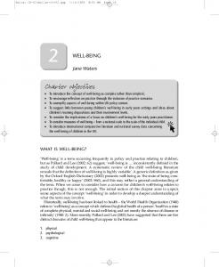
Number of dB=20 log*pressure of sound / pressure of standard sound In Figure (2-4) the decibel levels of various common sounds are compared. It is important to remember that the decibel scale is a log scale ( Ganong , 2010) .
Fig (2-4) Decibel scale for common sounds ( Ganong , 2010)
The outer-ear "trumpet" collects these sound waves, and they are funneled down the external ear canal to the tympanic membrane (Peter , 2006). As the sound
10
Chapter two-----------------------------------------------------literature review waves strike the eardrum (TM) , they cause it to vibrate. The vibrations of sound are transmitted through the middle ear over the bony bridge formed by the hammer, anvil, and stirrup in air gathered at the tympanic membrane to the fluid of the inner ear at the oval window( Irwin, 2006 ). The motion of the fluid in the inner ear excites the nerve cells in the organ of Corti , producing electrochemical impulses that are gathered together and transmitted to the brain along the acoustic nerve. Although the cochlea is usually thought of as having two mobile windows (the oval and round windows), the possibility of a third mobile window has been proposed ( Guyton, 2012). More recently, a select group of patients with an air-bone gap on audiologic testing (which is typically associated with middle ear pathology) who do not have any middle ear pathology on intraoperative exploration has been described ( Al Muhaimeed et al., 2002). It has been shown that the air-bone gap in this group of patients can be explained by a pathologic “third window” in the inner ear ( Merchant and Rosowski, 2008). Perhaps the most well-studied example of this phenomenon is seen in superior semicircular canal dehiscence Patients with superior semicircular canal dehiscence often complain of autophony, aural fullness, sound-induced or pressure-induced vertigo, and hearing impairment (Merchant et al., 2007). It is thought that the dehiscence in the superior semicircular canal acts as a third mobile window of the inner ear (in addition to the oval and round windows), shunting acoustic energy away from the cochlea, resulting in a decreased sensitivity to airconducted sound and an air-bone gap seen on audiologic testing ( Minor et al., 2003). This third window is also theorized to decrease cochlear input impedance at the oval window, increasing the pressure gradient across the cochlear partition, and resulting in hypersensitivity to bone-conducted sound (Rosowski et al., 2004). Repair, or plugging of superior semicircular canal dehiscence in some cases results in improvement of the preoperative air-bone gap . The third window hypothesis has also been used to explain the air-bone gap associated with other temporal bone
11
Chapter two-----------------------------------------------------literature review anomalies, such as large vestibular aqueduct syndrome and other inner ear malformations ( Merchant et al., 2007) . As the cochlear partition is deflected in response to the compressional wave initiated by the stapes, it causes a shearing force between the stereocilia of the hair cells and the tectorial membrane. This shearing force causes a deflection of the hair cell stereocilia. The hair cell stereocilia are arranged in rows, and the rows are arranged in an orderly fashion by height . The tip of each stereocilia is connected from one row to another by an elastic filament called the tip link (Wade and Daniel, 2010). It is thought that as the stereocilia is deflected toward the direction of the tallest row, it causes the tip links to stretch. The stretch of the tip links causes the opening of stretch-sensitive cationic channels located on the stereocilia . Because there is a large electrochemical gradient across the apical surface of the hair cells (with a large positive endocochlear potential on one side, and a large negative intracellular potential on the other side), the opening of these stretch-sensitive cationic channels on the stereocilia causes a large influx of cationic current, which leads to hair cell depolarization ( Paul et al , 2010) . As the stereocilia is deflected away from the tallest row, the tip links relax, decreasing the probability of ion channel opening; this leads to hyperpolarization of the hair cell. The relationship between the degree of stereocilia deflection and hair cell depolarization or hyperpolarization is not symmetric or linear. Stereocilia deflection in the depolarization direction produces a greater response than deflection in the hyperpolarization direction .The deflection of the hair cell stereocilia and the resulting hair cell depolarization or hyperpolarization represents an important step in the signal transduction process of the hair cell i.e., by converting a mechanical signal (inner ear fluid wave) into an electrochemical signal (Paul et al , 2010) . Because potassium is the major cation in the endolymph, it is believed that potassium current plays an important role in triggering the signal transduction
12
Chapter two-----------------------------------------------------literature review process in hair cells. When inner hair cells are depolarized, voltage-gated calcium channels open. These voltage-gated calcium channels are clustered in several “hot spots” along the basolateral surface of the inner hair cells, where synaptic contacts with primary afferent auditory nerve fibers are located. The calcium current mediated by these voltage-gated ion channels are important for triggering neurotransmitter release across the synapse, which leads to activation of the auditory nerve fibers. The neurotransmitter involved in this process has not been definitively identified, but is believed to be a molecule closely related to glutamate.( Wade and Daniel , 2010) . In contrast to the inner hair cell, an outer hair cell can also change its length in response to voltage changes; it contracts with depolarization and elongates with hyperpolarization. The molecular motor that is associated with rapid changes in outer hair cell length is a voltage-dependent , integral membrane protein called prestin (Zheng et al., 2000). The change in outer hair cell length in response to voltage changes is believed to add energy into the basilar membrane motion through a mechanical feedback scheme. In other words, the outer hair cell acts as a cochlear amplifier, augmenting the signals transmitted into the inner ear by the stapes vibration (Wade and Daniel , 2010) . The importance of prestin in hearing is supported further by the finding that in animal studies in which prestin has been knocked out or altered, the hearing sensitivity and frequency selectivity are impaired ( Dallos et al., 2008) . Because different regions of the basilar membrane are tonotopically tuned to specific frequencies, and because the hair cells reside on top of the basilar membrane, it is logical to assume that the hair cells from different regions are also tonotopically tuned to specific frequencies. The frequency tuning curves for outer and inner hair cells have been recorded in guinea pigs in response to various frequencies, and the hair cells in different regions along the basilar membrane are tonotopically tuned to specific frequencies that correspond to the tonotopic arrangement of the basilar membrane (Paul et al , 2010). The frequency to which a hair cell is most sensitive
13
Chapter two-----------------------------------------------------literature review is called the characteristic frequency. As discussed subsequently, this tonotopic arrangement is essential for the processing of auditory information, and is preserved throughout the entire auditory pathway (Wade and Daniel, 2010) .
2.2 Hearing Impairment Sensorineural hearing impairment is reported to be a prevalent childhood condition that may result in significant language and academic deficits (Melissa et al., 2006) It is obvious that hearing disorders may cause problem in our life ( Mohammad and Afsaneh, 2010).. Although it has been posited that slight or mild HI (i.e., _15 and _35 dB) may also affect development, and there is conflicting evidence about this (Melissa et al., 2006 ; Tharpe , 2008). HI is caused by a disturbance of the conduction of sound to the inner ear, the perception of sound by the sensory cells of the cochlea, or the processing of sound in the cochlear nerves, the auditory pathway, or the cortical auditory centers ( Thomas , 2011).
2.2.1 Pathophysiology Hearing impairment can be classified as conductive, sensorineural, or both (mixed impairment). Conductive hearing impairment occurs secondary to lesions in the external auditory canal, TM, or middle ear. These lesions prevent sound from being effectively conducted to the inner ear (Merck, 2010) . Sensorineural hearing impairment is caused by lesions of either the inner ear (sensory) or the auditory (8th) nerve (neural), SNHI accounts for about 90% of all HI (Timothy , 2010) . The distinction is important because sensory HI is sometimes reversible and is seldom life threatening. A neural HI is rarely recoverable and may be due to a potentially life-threatening brain tumor—commonly a cerebellopontine angle tumor. Mixed impairment may be caused by severe head injury with or without fracture of the skull or temporal bone, by chronic infection, or by one of many genetic
14
Chapter two-----------------------------------------------------literature review disorders. It may also occur when a transient CHI, commonly due to otitis media (OM) , is superimposed on a SNHI (Merck , 2010) . Central auditory disorder results from problems in the processing of sound in higher auditory areas of the brain. This type of auditory problem affects more complex auditory processes such as understanding speech when there is background noise. Hearing sensitivity and physiologic tests such as OAE and ABR, are normal in children with a central auditory disorder ( Clinical Practice Guideline , 2007).
2.2.2 Etiology 2.2.2.1 Non-genetic Causes The predominant etiology of HI in children has evolved with advances in medical knowledge and therapeutics. Historically, infectious disorders such as OM. that lead to significant reduction in the hearing sensitivity (Sandeep and Jayaram , 2008) . maternal rubella infections, bacterial meningitis (it is the most common cause of both acquired profound SNHI in childhood and labyrinthitis ossificans)
( Shervin , et al., 2005). Mumps are the most common cause of one-
sided total deafness in the United States. Other childhood infections, such as scarlet fever, may also affect hearing, particularly by destroying the eardrum and damaging the middle ear bones ( Slowik , 2011) . Besides, environmental factors such as intrauterine teratogenic exposure or ototoxic insult are the dominant causes of congenital and acquired hearing impairment. The introduction of antibiotics and vaccines, along with improved knowledge and enhanced awareness about teratogens, has led to a decline in hearing impairment resulting from infections and environmental agents (Anil , 2007) .
15
Chapter two-----------------------------------------------------literature review
2.2.2.2 Genetic Causes (Eisen and Ryugo ,2007) provide an excellent review of the molecular pathophysiology of genetic HI , they have found that more than 4000 infants born deaf each year, more than half have a hereditary disorder. Hereditary disorders must be differentiated from acquired HI . Not all hereditary HI is present at birth; some children inherit the tendency to develop hearing impairment later in life. Genetic SNHI includes a broad range of disorders that affect infants, children, and adults. Affected individuals may have unilateral or bilateral HI ranging from mild to profound disorders .Genetic SNHI
includes the following: congenital,
progressive, and adult onset; conductive, sensory, and neural; syndromic and nonsyndromic; high-frequency, low-frequency, or mixed frequency; and mild or profound. Genetic HI may show patterns of recessive, dominant, or sex-linked inheritance and may be a result in mutation of both cellular or mitochondrial DNA (and RNA, in the case of mitochondrial genes) (Van and Smith , 1999). Dysfunctional proteins have been identified in the impaired molecular-physiologic processes of potassium and calcium homeostasis (Brini et al., 2007 ; Xing et al., 2007). Apoptotic signaling stereocilia linkage (El-Amraoui and Petit ,
2005).
Mechanicoelectric transduction, electromotility, and other processes ( Eisen and Ryugo , 2007).
2.2.2.3 Etiological causes of SNHI in Children : Family history Represented important causes for SNHI in addition to all type of HI ( Rachael et al., 2012 ) . prenatal causes (Gallo et al., 2011): 1.Mother medication during pregnancy ( methyl mercury, retinoic acid, thalidomide, trimethadione) . 2.Mother infection during pregnancy ( cytomegaloviral (CMV) infections, herpes, rubella, syphilis, toxoplasmosis, varicella) .
16
Chapter two-----------------------------------------------------literature review Perinatal causes A history of prematurity, low birth weight, anoxia and/or low Apgar scores, hyperbilirubinemia, or sepsis should prompt an evaluation of hearing because these conditions may also result in SNHL ( Scott-Brown's, 2008) . Postnatal causes Childhood infections, such as meningitis or mumps, may result in SNHI. Treatment with ototoxic medications, such as aminoglycosides or furosemide can lead to SNHI .Otitis media or major head injury may cause SNHI or CHL (Aarno et al., 2009) . Unknown causes Many children are presented with SNHI without obvious etiological causes or risk factors . It important to emphasize that genetic causes cannot be easily excluded ,because there are children with recessive SNHI without family history of hearing impairment (Janka et al., 2009) .
2.2.3 Prevalence of SNHI in Children Different studies agree that one or two of every 1000 newborns have a HI that on current evidence warrants treatment or observation, i.e., permanent HI with a lowering of the absolute threshold of hearing for speech perception by at least 35 dB ( Ptok , 2011). Half of the neonates with HI show no risk factor(RF) ( Zamani et al., 2004) .The incidence of significant HI is 10 times greater for infants with one or more RF than for those with no RF ( Paul et al , 2010) . These infants are at-risk for HI at birth . Studies have shown increased risk in this population for progressive and/or late onset HI. In a comprehensive study of extremely low birth-weight children, the incidence and severity of HI increase significantly from 5% to 13% over a 24 year period. As these infants are identified as being at-risk for progressive or late-onset HI, it is essential that they are not only screened at birth, but are followed regularly for audiological assessment and are not lost to follow-up ( Synnes et al ., 2012).
17
Chapter two-----------------------------------------------------literature review The universal newborn hearing screening ( UNHS ) study groups have concentrated on detecting such HI as early as possible ( Ptok , 2004); SNHI is reported to be a prevalent childhood condition that may result in significant language and academic deficits ( Melissa et al., 2004). In united states (US) National Health and Nutrition Examination Survey (NHANES) ( Melissa et al., 2005) , 1.5% - 3% of school aged children had bilateral HI of slight/mild degree . National ascertainment of deaf children has demonstrated that the prevalence of bilateral deafness by 9 years of age has risen to 1.65/1000 and could be as high as 2/1000 (Watkin and Baldwin , 2010). A population-based study of the prevalence and effect of slight-mild bilateral SNHI in school-aged children was undertaken (Melissa et al., 2006).
2.2.4 Diagnosis of Hearing Impairment 2.2.4.1 Patient history In infants who fail hearing screening tests, a directed history to identify potential sources of HI is appropriate (Kim et al., 2002 ; Paul et al , 2010). A careful family history looking for HI , pregnancy, birth, and early medical history were obtained prospectively through parental questionnaires and medical records (Amanda et al., 2011) . The questioning was looking for RF. Then, the child was sent for diagnosis towards an ENT specialist or a specialized structure (Roman et al ., 2011).
2.2.4.2 Physical Examination A majority of newborns with HI do not have abnormal physical features ( Hone and Smith , 2002 ; Paul et al , 2010). Nevertheless, a complete physical examination is essential. Presence of abnormal iris color, are potential signs of a syndromic disorder. For example , Wardenburg's syndrome, may explain HI identified through infant hearing screening. Alternatively, these physical signs may prompt careful follow-up investigation and imaging to identify progressive SNHI or CHI, which may occur in neurofibromatosis or craniosynostosis. as well as the otoscopy which are essential to detect and diagnose a hearing disorder. Screening
18
Chapter two-----------------------------------------------------literature review tests help to identify the children with risk of HI . Then, the child is sent for diagnosis towards an ENT specialist or a specialized structure. In spite of the frequency of the CHI, in case of the slightest doubt, a SNHI must be always looked for by a PTA examination via the headphones (Roman et al., 2011).The electrophysiological measurement is the most employed tool to identify and characterize the HI in the population of infants and children (Ana et al., 2010)
2.2.4.3 Laboratory Studies : Depending on the patient's history and physical findings, biochemical evidence or genetic testing may help to determine the etiology of deafness if a genetic syndrome is suspected. Some have recommended that any child with a diagnosis of SNHI should have blood studies to search for evidence of thyroid and renal disease( Rahul et al., 2011).
2.2.4.4 Imaging Studies : In the past, the benefit of imaging studies has been questioned. Although a positive finding on magnetic resonance imaging (MRI) or computed topography (CT) scanning may occasionally help to explain the defect, it does not lead to treatment options. However, some abnormalities uncovered during imaging (eg, enlarged vestibula aqueduct) may indicate a child with a sensitive ear in whom minor head trauma could worsen his or her hearing. However, MRI may help in identifying a malformation of the cochlea or the cochlear nerve .Such information may be critical when cochlear implants are being placed in profoundly deaf individuals. Recent work suggests the superiority of MRI in preoperative planning for candidates for cochlear implants (Parry et al ., 2005).
2.2.4.5 Audiolgical Examination : 2.2.4.5.1 Free field hearing tests During this test, examiner own voice is used as a sound stimulus, while the patient's non-test ear is masked by rubbing the finger over the tragus. This
19
Chapter two-----------------------------------------------------literature review produces some sound, which helps to ‘mask’ that ear. Practice is essential as the positioning for this procedure can be awkward (corbridge et al., 2006). Procedure •
Shield the patient's eyes with your hand.
•
Tragal rub with your other hand to the non-test ear.
•
Whisper a number at approximately 60cm from the test ear.
•
If the patient cannot hear, use a normal volume spoken voice, followed by a
shout if necessary. •
Repeat with opposite ear.
Patients should be 50% accurate at repeating your words to pass the test. If they hear your whisper at a distance of 60cm, then their hearing is better than 30dB ( Corbridge, et al 2006) . The test is then repeated with a whispered voice. A whisper at 1 meter is around 15 dB . If this is accurately repeated, then the hearing is probably within the normal range at least in the lower frequencies. If the conversational voice is not heard at arms' length, then the opposite ear should be masked with a Barany box (90 dB masking if held opposite the ear and 100 dB if placed in the canal) and the examiner uses a loud voice to test the ear. If no voice is heard, the patient has a significant hearing impairment and a probable dead ear on the tested side can be found. This should be confirmed with formal audiometry and speech audiometry (Peter, 2007).
2.2.4.5.2 Auditory brainstem response Auditory brainstem response is a neurologic test of auditory brainstem function in response to auditory (click) stimuli. First described by Jewett and Williston in 1971, ABR audiometry is the most common application of auditory evoked responses. Test administration and interpretation is typically performed by an audiologist (Neil , 2011) . ABRs provide an objective and noninvasive means
20
Chapter two-----------------------------------------------------literature review for examining (Erika and Nina , 2010) This form of testing is not painful, although you may need to be sedated in certain situations . Auditory brainstem response is an auditory evoked potential extracted from ongoing electrical activity in the brain and recorded via electrodes placed on the scalp. ABR audiometry typically uses a click stimulus transmitted from an acoustic transducer in the form of an insert earphone or headphone. The elicited waveform response is measured by surface electrodes typically placed at the vertex of the scalp and ear lobes. The amplitude (microvoltage) of the signal is averaged and charted against the time (Ms), much like an EEG. that generates a response from the basilar region of the cochlea. The signal travels along the auditory pathway from the cochlear nuclear complex proximally to the inferior colliculus. ABR waves I and II correspond to true action potentials. Later waves may reflect postsynaptic activity in major brainstem auditory centers that concomitantly contribute to waveform peaks and troughs. The positive peaks of the waveforms reflect combined afferent (and likely efferent) activity from axonal pathways in the auditory brain stem ( Neil , 2011). The resulting recording is a series of vertex positive waves of which I through V are evaluated. These waves, labeled with roman numerals in Jewett and Williston convention, occur in the first 10 Ms after onset of an auditory stimulus. The ABR is considered an exogenous response because it is dependent upon external factors ( Burkard et al., 2007; Hall, J. III , 2007) .The auditory structures that generate the ABR are believed to be as follows: ( Burkard et al., 2007; DeBonis et al ., 2008) .
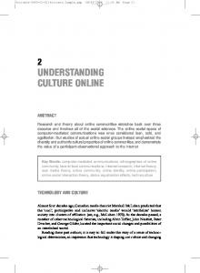
2.2.4.5.2.1 Waveform components: (figure 2.5) Wave I: The ABR wave I response is the far-field representation of the compound auditory nerve action potential in the distal portion of cranial nerve (CN) VIII. The response is believed to originate from afferent activity of the CN VIII fibers (first-order neurons) as they leave the cochlea and enter the internal auditory canal.
21
Chapter two-----------------------------------------------------literature review Wave II: The ABR wave II is generated by the proximal VIII nerve as it enters the brain stem. Wave III: The ABR wave III arises from second-order neuron activity (beyond CN VIII) in or near the cochlear nucleus. Literature suggests wave III is generated in the caudal portion of the auditory pons. The cochlear nucleus contains approximately 100,000 neurons, most of which are innervated by 8th nerve fibers. Wave IV: The ABR wave IV, which often shares the same peak with wave V, is thought to arise from pontine thirdorder neurons mostly located in the superior olivary complex, but additional contributions may come from the cochlear nucleus and nucleus of lateral lemniscus. Wave V: Generation of wave V likely reflects activity of multiple anatomic auditory structures. The ABR wave V is the component analyzed most often in clinical applications of the ABR. Although some debate exists regarding the precise generation of wave V, it is believed to originate from the vicinity of the inferior colliculus. The second-order neuron activity may additionally contribute in some way to wave V. The inferior colliculus is a complex structure, with more than 99% of the axons from lower auditory brainstem regions going through the lateral lemniscus to the inferior colliculus. Wave VI and VII: Thalamic (medial geniculate body) origin is suggested for generation of waves VI and VII, but the actual site of generation is uncertain (Rudolf , 2006) .
22
Chapter two-----------------------------------------------------literature review
Fig . ( 2-5 ) The curve shows the typical ABR waveform, which consists of 5or 6 waves (numbered I through VI). The potentials reflect the acoustically induced activity of the auditory nerve and auditory neurons. The waves are produced by dipole generators in the various anatomical structures .( Basic Otorhinolaryngology 2006 )
2.2.4.5.2.2 Applications Auditory brainstem response’s clinical applications can be divided into neurologic and audiologic, with the main goal of identifying abnormalities in the auditory nerve and the brain stem, and estimate the electrophysiological auditory threshold, based on the presence of responses to different levels of stimulus intensity ( Boone et al., 2005 ). ABR’s can be used as an auditory screening test for high risk newborns ( Lieu and Champion , 2006). It is used for intraoperative monitoring during ponscerebellar angle surgery ( James and Husain, 2005). One of the most studied applications of this type of test is to diagnose vestibular nerve schwannomas (Edmir et al., 2008). However, there are controversies regarding the test’s sensitivity and specificity (Cueva , 2004).
23
Chapter two-----------------------------------------------------literature review
2.2.4.5.2.2.1 Identification of retrocochlear pathology Auditory brainstem response audiometry is considered an effective screening tool in the evaluation of suspected retrocochlear pathology such as an acoustic neuroma or vestibular schwannoma. Still, an abnormal ABR finding suggestive of retrocochlear pathology indicates the need for MRI of the cerebellopontine angle (Neil ,2011).
2.2.4.5.2.2.2 Newborn Hearing Screening Auditory brainstem response technology has been used in testing newborns for the past 15 years. The newborn hearing screening program (NHSP) (Watkin and Baldwin , 2010 ) has been nationally implemented since 2006 and has been justifiably considered a ‘quite astonishing achievement (Kennedy and Cann , 2004 ) . Approximately 1 of every 1000 children is born deaf. Many more are born with less severe degrees of HI, while others may acquire HI during early childhood Historically, only infants who met 1 or more criteria on the high-risk register were tested. Universal hearing screening has been recommended because about 50% of the infants later identified with HI are not tested when newborn hearing screening (NHS) is restricted to high-risk groups . Recently, hospitals across the United States have been implementing universal newborn hearing screening program (UNHSP) . These programs are possible because of the combination of technological advances in ABR and OAE testing methods and equipment availability, which enables accurate and cost-effective evaluation of hearing in newborns. As far as the benefits of the UNHS are concerned, it is now commonly accepted that early identification of HI gives children the opportunity to develop significantly improved language skills compared with those children that are diagnosed later (Cao-Nguyen et al., 2007 ; Korres et al., 2008 ) . It is now quite commonly accepted that the internationally recommended age for the diagnosis of HI is before 3 months of age, with intervention starting as soon as possible (Kabra et al., 2006 ; Rohlfs et al., 2010 ). Click-evoked ABR is highly correlated with hearing sensitivity in the frequency range from 1000-4000 hertize (Hz). ABR test
24
Chapter two-----------------------------------------------------literature review for the presence or absence of wave V at soft stimulus levels no operator interpretation is required . ABR can be used on the ward and during oxygen therapy without disturbance from ambient noise. The 2007 Joint Committee on Infant Hearing ( JCIH ) has recommended that infants with at least 1 of the following risk factors indicators for progressive or delayed-onset hearing impairment who may have passed the hearing screening should, nonetheless, receive audiologic monitoring every 6 months until the age of 3 years (American Speech-Language-Hearing Association (ASHA) 2007 ) : 1. Admission to a neonatal intensive care unit (NICU) for 48 hours or longer(Charlene et al., 2009). 2. Family history of permanent childhood sensorineural hearing impairment 3. In utero infection such as cytomegalovirus, herpes, toxoplasmosis, or rubella 4. Postnatal infections associated with hearing impairment (such as bacterial meningitis). 5. Exposure to ototoxic medications such as aminoglycoside ( Robertson et al., 2006) ,antibiotics, cisplatin ,chemotherapy agents, and certain loop diuretics . 6. Craniofacial anomalies, especially those with abnormalities of the ear or ear canal . 7. Neonatal indicators: • Birth weight less than 1,500 grams • Hyperbilirubinemia requiring exchange transfusion • Persistent pulmonary newborn hypertension requiring mechanical ventilation • Conditions requiring the use of extracorporeal membrane oxygenation (ECMO). 8. Findings associated with a syndrome known to include or be high risk for hearing impairment: • Syndromes associated with sensorineural and/or conductive hearing impairment (such as Waardenburg syndrome)
25
Chapter two-----------------------------------------------------literature review • Syndromes associated with progressive hearing impairment (such as neurofibromatosis and osteopetrosis) . • Genetic conditions that are likely to have associated with hearing impairment (such as Down syndrome and Usher syndrome) • Neurodegenerative disorders (such as Hunter syndrome) or sensory motor neuropathies 9. Head trauma (especially with fracture of the temporal bone) 10. Reanil or persistent otitis media with effusion (OME) for at least 3 months 11. Parental or caregiver concern regarding hearing, speech, language, and/or developmental delay . Other independent, neonatal, risk factors have been reported. These risk factors include a need for ventilation, use of oxygen supplementation, respiratory failure, low Apgar scores, acidosis, use of ototoxic drugs including furosemide (especially with high serum creatinine levels), treatment for hypotension, patent ductus arteriosus ligation, hyponatremia ,and noise ( Fanaroff et al., 2006 ; Hille et al., 2007; Kobaly
et. al., 2008). ABRs may be used to detect auditory
neuropathy or neural conduction disorders in newborns. Because ABRs are reflective of auditory nerve and brainstem function, these infants can have an abnormal ABR screening result even when peripheral hearing is normal. Infants that do not pass the newborn hearing screenings do not necessarily have hearing problems. When hearing impairment is suspected because of an abnormal ABR screening result, a follow-up diagnostic threshold ABR test is scheduled to determine frequency-specific hearing status. Estimation of hearing at specific frequencies may be obtained through use of brief tone stimulation, such as a tone burst (niel,2011).
2.2.4.5.2.3 Auditory brainstem response evaluation In addition to retrocochlear pathologies, many factors may influence ABR results, including the degree of SNHI, asymmetry of hearing impairment, test
26
Chapter two-----------------------------------------------------literature review parameters, and other patient factors. These influences must be factored in when performing and analyzing an ABR result. Findings suggestive of retrocochlear pathology may include any 1 or more of the following: •
Absolute latency interaural difference wave V - Prolonged
•
I-V interpeak interval interaural difference - Prolonged
•
Absolute latency of wave V - Prolonged as compared with normative data
•
Absolute latencies and interpeak intervals latencies I-III, I-V, III-V -
Prolonged as compared with normative data •
Absent auditory brainstem response in the involved ear
In general, ABR exhibits a sensitivity of over 90% and a specificity of approximately 70-90% ( Neil , 2011). Sensitivity for small tumors is not as high. For this reason, a symptomatic patient with a normal ABR result should receive a follow-up audiogram in 6 months to monitor for any changes in hearing sensitivity or tinnitus. The ABR may be repeated if indicated. Alternatively, MRI with gadolinium enhancement, which has become the new criterion standard, can be used to identify very small (3-mm) vestibular schwannomas (Cueva , 2004 ).
2.2.4.5.3 Audiology Pure-Tone Testing The auditory system can be stimulated via sound energy that is sent through air to the ear drum (air conduction) or by placing a bone vibrator against the skull (bone conduction) ( Ross et al., 2007) . Pure-tone-audiometry (PTA) is a behavioral test used to measure hearing sensitivity. This measure involves the peripheral and central auditory systems. Pure-tone thresholds (PTTs) indicate the softest sound audible to an individual at least 50% of the time. Hearing sensitivity is plotted on an audiogram, which is a graph displaying intensity as a function of frequency (Joe et al., 2010) .
2.2.5 Degrees of hearing impairment Classification of severity is usually based on the average hearing impairment in the frequency range of normal speech (fig.2-6).Thus the hearing
27
Chapter two-----------------------------------------------------literature review impairment is described solely in terms of the absolute threshold of hearing. However, the principal function of hearing is to detect rapid changes of frequency and intensity in acoustic signals above the threshold, and thus to understand speech ( Ptok , 2011). The WHO classification of the severity of hearing impairment as follow
dBHL
NH (0-25 dB): At this level, hearing is within normal limits.
Fig . (2-6) Graphic representation of categories of hearing impairment (Cummings, 2010)
Mild HI (26-40 dB): may cause inattention, difficulty suppressing background noise, and increased listening efforts. Patients with this degree of impairment may not hear soft speech. Children may be fatigued after listening for long periods. Moderate HI (41-55 dB): may affect language development, syntax and articulation, interaction with peers, and self-esteem. Patients with this degree of impairment have trouble with hearing some conversational speech. Moderate-severe HI (56-70 dB): may cause difficulty with speech and decreased speech intelligibility. Patients with this degree of impairment do not hear most conversational-level speech.
28
Chapter two-----------------------------------------------------literature review Severe HI (71-90 dB): may affect voice quality. Profound HI (>90 dB): With profound HI (defiance) , speech and language deteriorate (Thomas , 2011) .
2.2.6 Types of hearing impairment by PTA: •
Conductive
Conductive hearing impairment has normal bone-conduction thresholds, but
air-conduction thresholds are poorer than normal by at least 10 dB.
Conductive hearing impairment is secondary to an outer ear or middle ear
abnormality, which can include abnormalities of the TM. The abnormality reduces the effective intensity of the air-conducted signal reaching the cochlea, but it does not affect the bone-conducted signal that does not pass through the outer or middle ear.
Examples of abnormalities include occlusion of the external auditory canal
by cerumen or a mass, middle ear infection and/or fluid, perforation of theTM, or ossicular abnormalities. Pure-tone air-conduction thresholds are poorer than boneconduction thresholds by more than 10 dB as show in fig . (2-7) (paul et al.,2010).
. Fig . (2-7) Conductive hearing impairment( current, 2006)
29
Chapter two-----------------------------------------------------literature review •
Sensorineural
Sensorineural hearing impairment has bone- and air-conduction thresholds
within 10 dB of each other, and thresholds are higher than 25 dB HI figure (2-8). SNHI is secondary to cochlear abnormalities and/or an abnormality of the auditory nerve or central auditory pathways. Because, in this type of HI, the outer ear and middle ear do not reduce the signal intensity of the air-conducted signal, both airand bone-conducted signals are effective in stimulating the cochlea. Pure-tone airand bone-conduction thresholds are within 10 dB.
Examples included presbycusis, noise-induced HI, Ménière disease, and
retrocochlear lesions such as vestibular schwannoma ( Scott-Brown's, 2008).
Fig. (2-8) Audiogram depicting a high-frequency sloping SNHI in the Bilateral ears. (current 2006)
30
Chapter two-----------------------------------------------------literature review •
Mixed
Mixed HI has SN and conductive components. Pure-tone air-conduction thresholds are poorer than bone-conduction thresholds by more than 10 dB, and boneconduction thresholds are less than 25 dB as in figure (2-9) (Anil ,2007) .
Fig. (2-9) Audiogram depicting a mixed sloping HI. (current ,2006)
2.2.7 Treatment of hearing impairment Over 30 states in the United States require newborn hearing screenings treating hearing impairment early can allow many infants to develop normal language skills without delay. In infants born with hearing impairment, treatments should start as early as the age of 6 months. There is a general consensus that children with a bilateral, moderate, permanent hearing impairment should be treated in order to prevent significant impairment of speech and language development.
31
Chapter two-----------------------------------------------------literature review Patients with mild, moderate, and severe sensorineural hearing impairment are rehabilitated regularly with hearing aids; In the event that a hearing aid provides inadequate rehabilitation, cochlear implants are appropriate . Children with congenital and acquired profound hearing impairment are also appropriate candidates for cochlear implantation (Anil , 2007). o
Hearing aids : should be prescribed for moderate to severe hearing
impairment, while cochlear implants are indicated for deafness or profound hearing impairment that does not benefit from hearing aids. After the hearing aids have been fitted, their function must be regularly tested by parents and educators because initially the child is unable to detect and report malfunctions. But if the hearing aids are of definite benefit the child will soon ask for them himself and later will make it known if the devices are not functioning properly ( Verhaert et al., 2008 ) .
o
Cochlear implants : are used in patients who have cochlear damage and
an auditory nerve that is responsive to stimulation. They are mainly an option for two categories of hearing-impaired children (Daniel and Moshe 2010) . Congenital deafness or profound HI: The earlier cochlear implantation is performed in these children, the better the device can exploit the natural adaptability and receptivity of the central auditory structures. Most children treated early with a cochlear implant learn to understand speech. Many can even understand speech without lip reading (open speech Comprehension . It is equally important to train the child's speaking ability. Cochlear implantation after puberty very rarely provides open speech comprehension or a significant improvement of speech ( Spencer et al., 2004) . Bilateral acquired deafness (eg .after meningitis): Cochlear implantation should be performed as soon as possible so that existing auditory development is not lost and can be exploited with the cochlear implant during rehabilitation. In practice, this means that cochlear implantation should be scheduled as soon as the child is diagnosed with bilateral deafness or profound hearing impairment that is
32
Chapter two-----------------------------------------------------literature review not treatable with hearing aids. As a rule, hearing aids should be tried for several months before this determination is made (corbridge et al., 2006). Cochlear implantation surgery is a low-risk procedure even in children. It is very rarely contraindicated for medical reasons (Thomas ,2011) .
2.2.7.1 Treatment of sensorineural hearing impairment Previously, SNHI has been treated with hearing aids, which amplify sounds at preset frequencies to overcome a SNHI in that range; or cochlear implants, which stimulate the cochlear nerve directly. Some research suggests that idebenone alone or combined with vitamin E may delay the onset of HI or perhaps reverse it (Sergi et al., 2006). The use of these agents for this purpose is considered experimental now. Some audiologists and ENTs have reported if severe noise-induced HI (exposures exceeding 140dB) is treated immediately (within 24 hours) with a course of steroids, it can often be almost completely reversed. This,however,is a new field without proven success (Haynes et al .,(2009).
2.2.7.2 Speech processor fitting: The speech processor is fitted to the child on an individual basis in sessions that are consistent with pedagogic and audiologic principles. Like hearingaid fitting, it is a process that requires a great deal of patience, empathy, and experience. Education services whenever a child is diagnosed as having a significant, permanent HI, the child should receive special education services from a trained, licensed teacher . Education and counseling should also be provided to parents and other family members on dealing with issues of deafness within the family. For small children, these services are generally provided in the house. Auditory perception is trained and practiced in a play setting, while parents are taught behavioral rules and assistive measures. The goal of early education services is to integrate the hearing-impaired child into public schooling and thus into the society of normal-hearing persons. Specialized kindergartens and schools are available for children who cannot be mainstreamed. Some institutions
33
Chapter two-----------------------------------------------------literature review specialize in a total communication approach that combines oral communication with other modalities such as sign language (corbridge et al., 2006).
2.2.8 Prognosis : Sensorineural hearing impairment is a condition that is generally irreversible. Given the poor prognosis for most causes of sensorineural hearing impairment, the primary goals in management are the prevention of further impairment and functional improvement with amplification and auditory rehabilitation (Anil ,2007) .
34