2009 International Nuclear Atlantic Conference - INAC 2009 Rio de Janeiro,RJ, Brazil, September27 to October 2, 2009 ASSOCIAÇÃO BRASILEIRA DE ENERGIA NUCLEAR - ABEN ISBN: 978-85-99141-03-8
CHARACTERIZATION OF SOME DOSIMETRIC PARAMETERS OF 125 I SEEDS USED FOR PROSTATE IMPLANTS USING MONTE CARLO SIMULATIONS Lucas P. Reis1, Alessandro Facure2, Simone C. Cardoso1 and Ademir X. da Silva3 1
Laboratório de Física da Radiação Gama - Instituto de Física Universidade Federal do Rio de Janeiro Av. Marechal Trompowsky s/n 21941-972 Rio de Janeiro, RJ
[email protected] 2
3
Comissão Nacional de Energia Nuclear, CNEN Rua General Severiano, 90 – 4° andar 22294-900 Rio de Janeiro, RJ
[email protected]
Instituto Alberto Luiz Coimbra de Pós Graduação e Pesquisa de Engenharia – PEN/COPPE Universidade Federal do Rio de Janeiro Av. Horácio Macedo, 2030 – Bloco G – Sala 206 21945-970 Rio de Janeiro, RJ
[email protected]
ABSTRACT In the prostate cancer treatment, there is an increasing interest in the permanent radioactive seeds implant technique, where 125I seeds are inserted into the patient’s prostate, allowing the delivery of high doses and preserving nearby organs at risk. For the calculation of dose distributions, treatment planning systems (TPS) makes use of the calculation proposed by the protocol TG-43. According to the document, data of dose distribution should be made more precise, either experimentally or by computational simulations, to be used in the TPS. Several authors used Monte Carlo simulations to generate the parameters of brachytherapy sources (seeds) that are recommended by TG-43 protocol, which are used for dose calculations in the TPS. For a single seed, there are variations in the geometry chosen to calculate the dosimetric parameters recommended and in the medium where the dose distributions are calculated (liquid water or solid water) and also in the Monte Carlo code used. The TPS consider the sources as point entities and do not consider the attenuation effects among the seeds. In this work, computational simulations of the geometry of one of the most used seeds in permanent prostate implants, the Amersham model 6711, were performed through the Monte Carlo method using the MCNP5 code. The dosimetric parameters radial dose function g(r) and anisotropy function F(r,θ) were simulated and the results show good agreement with other works. This model can be used in the future to study the impact of the approaches and other problems in the implant procedure.
1. INTRODUCTION The prostate cancer, among the types of cancer, is the second most common cause of death from tumors in males. According to the Brazilian National Cancer Institute (INCA), the number of new cases of prostate cancer estimated to Brazil in the year 2008 is 49.530 [1]. In the prostate cancer treatment, there is an increasing interest in the permanent radioactive seeds implant technique, where 125I seeds are inserted into the patient’s prostate, allowing the delivery of high doses and preserving nearby organs at risk.
The dosimetric treatment planning has an important role in modern prostate brachytherapy. It allows the team to formulate a plan that will ensure the coverage of the whole target volume with the prescribed dose, while keeping the doses in the rectum and urethra and bladder (main organs at risk) within acceptable limits, and control the dose inhomogeneity and maintain the implant technically as simple as possible. In brachytherapy, generally, the dose calculation made by the treatment planning system (TPS) is based on an approach that considers the radioactive source as being punctual. Furthermore, the patient is considered a homogeneous medium and the effects among the radioisotope seeds, in case of implants, is neglected. However, the seeds that are used in brachytherapy implants are far from meeting the requirements of point sources. This approximation is valid in determining the dose distributions only in certain conditions of use of sources. Another assumption in the dose calculations - the radioactive seeds, individually, contribute to the dose at a given point, with the influence of neighboring seeds not being considered - in the case of multiple seed implants, is very important. Thus, with the great development of the brachytherapy today, the distribution of the seeds can reach high complexity, and it is necessary to perform the calculations using computational tools. The simulation using the Monte Carlo method can produce a more accurate determination of dose values in the tumor volume, thus contributing to increase the quality of the radiotherapy treatment. According to the document TG-43 of the American Association of Physicists in Medicine [2], data of dose distribution should be made more precise, either experimentally or by computational simulations, to be used by the TPS. Several authors [3] [4] [5] used Monte Carlo simulations to generate the parameters of brachytherapy sources (seeds) that are recommended by TG-43 protocol, which are used for dose calculations in the TPS. For a single seed, there are variations in the geometry chosen to calculate the dosimetric parameters recommended and in the medium where the dose distributions are calculated (liquid water or solid water) and also in the Monte Carlo code used. The values recommended by the review protocol [2], for each type of source, were derived from multiple published data, both as experimental measurements as Monte Carlo simulations. Within this context, this work was performed using the Monte Carlo MCNP version 5 [6], in order to validate the simulation of the geometry of the Amersham model 6711 seed, the most widely used in permanent prostate implants. Values of the anisotropy function F(r,θ) and the radial dose function g(r) were obtained, in order to validate the simulations.
2. MATERIALS AND METHODS 2.1. The Dose Calculation Formalism The dose calculation formalism recommended by the TG-43 protocol [7] requires dose rate input data of the real source in a tissue equivalent phantom. The protocol introduces a number of new quantities as the anisotropy function F(r,θ), the dose rate constant Λ, the geometric factor G(r,θ), the radial dose function g(r) and the air-Kerma strength Sk. The sources are considered to be symmetrically cylindrical. For such sources, the dose distribution is two-dimensional and can be described in terms of a polar coordinate system with origin at the center of the source, where r is the distance to the point of interest and θ is
INAC 2009, Rio de Janeiro, RJ, Brazil.
the angle with respect to the longitudinal axis of the source. The dose rate at a point of coordinates (r,θ) can be written as:
D& (r ,θ ) = Sk ⋅ Λ ⋅ (G (r ,θ ) / G (r 0,θ 0)) ⋅ g (r ) ⋅ F (r ,θ )
(1)
The reference point (r0,θ0) is chosen in the transverse axis of the source at a distance of 1 cm from its center, ie, r0 = 1 cm and θ0 = π/2. The dosimetric parameters considered in our work are described below. 2.1.1. Radial Dose Function The radial dose function takes into account effects of absorption and scattering in the medium along the transverse axis of the source. This function defines the fall of dose rate along the transverse axis due to absorption and scattering in the medium. It can also be influenced by the filtration of photons by the encapsulation and the material source. It is defined as:
g (r ) = D& (r ,θ 0)G (r 0,θ 0) / D& (r 0,θ 0)G (r ,θ 0)
(2)
2.1.2. Anisotropy Function The anisotropy function takes into account the anisotropy of the dose distribution around the source, including effects of absorption and scattering in the medium. This function provides the two-dimensional variation of the dose as a function of polar angle on the transverse plane due to the self-filtration, oblique filtration of primary photons through the encapsulating material and scattering of photons in the medium. It is defined as:
F(r,θ ) = D& (r ,θ )G (r ,θ 0) / D& (r ,θ 0)G (r ,θ )
(3)
2.1.3. Geometric Factor The geometric factor is used both in the calculation of g(r) as the F(r,θ) and represents the hypothetical relative dose distribution due only to the radioactivity distribution and ignores the effects of absorption and scattering in the source or in the medium. It is defined as:
G (r ,θ ) = β / L r senθ where L is the length of the active source and β is the angle that subtends the length of the active source with respect to point (r,θ).
INAC 2009, Rio de Janeiro, RJ, Brazil.
(4)
2.2. The 125I Seed Model 6711 The model 6711 has been the most used source for permanent implant since its introduction in 1983. It consists of a Titanium capsule of 4.6 mm in length and 0.05 mm thick. The capsule contains a Silver rod (which serves as a marker for X-rays) of 3 mm in length where the 125I is adsorbed. The geometry of the seed, recommended by the TG-43U1 protocol [2], is illustrated in Figure 1.
Figure 1. Seed Geometry, Amersham model 6711 [2]
2.3. Simulation of Dose Distribution Generated by 125I Seeds With the objective of calculating some dosimetric parameters of the prostate brachytherapy source Amersham, model 6711, the Monte Carlo code MCNP was used. The medium where the source was considered to be immersed was liquid water. The geometry of the seed, as shown in Figure 1, can initially be described as a silver rod of 0.3 cm in length and radius of 0.025 cm. The radionuclide 125I, was considered to be homogeneously distributed on the silver rod, in a thin layer. Other authors took into account similar assumptions in their work [3] [4]. The titanium encapsulation of the source was also taken into account, with total length of 0.47 cm, thickness of 0.005 cm and radius of 0.04 cm [2]. The semi-spherical end welds of the seed have thickness of 0.03 cm. The emission spectrum of the source is listed in Table 1, and the densities and compositions of all materials used in the simulations are described in Table 2.
INAC 2009, Rio de Janeiro, RJ, Brazil.
Table 1. Typical Parameters for 125I Seeds Used in Brachytherapy [2] Half-life: 59,40 ± 0,01 days Photon Energy (keV) Photons per disintegration 27,202 0,406 27,472 0,757 30,98 0,202 31,71 0,0439 35,492 0,0668 Mean energy: 28,37 (keV) Total: 1,476 125 I Γ5keV = 0,0355 µGy . m² . h-1 . Bq-1
Table 2. Description of Materials Used in Simulations Material *Ag *Ti
Atomic fraction 1 1
N14 Air O16 Ar40 Water *H 2 *O 1 * natural occurrence elements
Weight fraction 0.755 0.232 0.013
Density (g/cm³) 10,5 4,54 0,001205 1
2.3.1. Determination of F(r,θ) and g(r) Values It was considered that the seed was placed in the center of a water sphere with 30 cm radius. For the determination of the anisotropy function values (3), which is a function of radius and angle, F(r,θ), arrays of spherical water detectors were used. For the determination of its parameters, for each distance r (r ranging from 0,5 to 5 cm), nine spherical detectors were simulated, each corresponding to an angle θ (θ ranging from 10° to 90°), as illustrated in Figure 2. The spherical water detectors have volumes of 1 mm³.
INAC 2009, Rio de Janeiro, RJ, Brazil.
Figure 2. Illustration of the spherical array of detectors for determination of F(1,θ), in the xz plane
This arrangement was prepared for four quadrants: +xz, -xz, +yz, -yz. The detectors volumes were chosen as 1 mm3, a typical volume for detectors used in both simulations by Monte Carlo or experimental measurements. The spherical geometry of the detectors allows the calculation of the anisotropy function for each angle, with no recorded dose for the adjacent angles, which does not happen in the case of cylindrical rings or spherical shells detectors. The tally *F8 was used to determine the energy deposited in the volume of each sphere. Taking the mean energy deposited in the detectors of each quadrant corresponding to the same pair (r,θ), the values of F(r,θ) were determined. To verify the values of the radial dose function (2), the same arrangement for the determination of F(r,θ) was used but only were used the energies deposited in detectors corresponding to (r,90) points, since the function g(r) is defined in the transverse axis of the source. The distance r ranged from 1 to 7 cm. These values of distances were chosen, especially in the F(r,θ) case, in order to compare the results with data presented by other authors, as well as the protocol recommended values.
3. RESULTS The recommended values for the anisotropy and radial dose fuctions of the TG-43 protocol were derived from multiple sets of published data, both experimental and Monte Carlo calculated results, for each type of seed on the market. In this study, to validate the results for F(r,θ), a comparison was made both with the values recommended in TG-43U1 [2] and those published by Taylor and Rogers [5]. For the radial dose function g(r), is also compared with the values published by Rodríguez et al [4].
INAC 2009, Rio de Janeiro, RJ, Brazil.
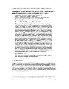
3.1. Anisotropy Function Values Calculated With the MCNP Code Figure 3 illustrates a comparison of the anisotropy function obtained by simulations with the MCNP code, implemented in this work, with the TG-43U1 [2] and the Monte Carlo code EGSnrc [5] anisotropy functions. The results are shown for radii ranging from 0,5 to 5 cm. The values for the anisotropy function had an error of about 10-7 for r values of 0,5, 1, 2 and 3 cm. For r values of 4 and 5 cm, the errors were about 10-6. Comparing the obtained results with the values of F(r,θ) calculated by the Monte Carlo code EGSnrc [5], it can be noticed that the biggest difference between both data is about of 8%. Most of the results presented a difference lower than 10% when compared with the recommended by TG-43U1 protocol [2], except for some values: F(0,5,10), F(0,5,20), F(0,5,30), F(1,10) and F(1,20), whose differences were 30, 25, 16, 24 and 12% respectively. These differences may be explained by the fact that the region near the longitudinal axis of the seed has a great variation in the doses distribution, due to higher photons attenuation at the ends of the source. 3.2. Radial Dose Function Values Calculated With the MCNP Code Figure 4 illustrates a comparison of the radial dose function obtained by simulations with the MCNP code, implemented in this work, with the TG-43U1 protocol [2], the Monte Carlo code EGSnrc [5] and the Monte Carlo code PENELOPE [4].
INAC 2009, Rio de Janeiro, RJ, Brazil.
Figure 3. Anisotropy function for the 125I source model 6711, calculated in this work (MCNP) compared with the results of the TG-43U1 [2] and EGSnrc [5]
INAC 2009, Rio de Janeiro, RJ, Brazil.
1.2 1
g(r)
0.8
MCNP PENELOPE
0.6
TG-43U1 EGSnrc
0.4 0.2 0 1
2
3
4
5
6
7
r (cm)
Figure 4. Radial dose function g(r) for the 125I source 6711 model, calculated in this work (MCNP) compared with the results of the TG43U1 [2], EGSnrc [5] and PENELOPE [4]
The values of the radial dose function had an error of about 10-7. When compared with the Monte Carlo code PENELOPE [4] the relative error was about 5%. In comparison with the results provided by the code EGSnrc [5] the biggest relative error was 7% and in comparison with the values recommended by the TG-43U1 protocol [2] the biggest relative error was 9%.
4. CONCLUSIONS The values found for the anisotropy function F(r,θ) were very satisfactory and, when compared to published data in the literature, validate the geometry of the simulated seed as well as the simulations with the MCNP5 code. Differences greater than 10% were found, when comparing the obtained data with the values recommended by the TG-43U1 protocol. These discrepancies can be explained by the large anisotropy of the dose distribution at the ends of the seed. Since the recommended protocol values are a compilation of several values, generated in different ways, the differences are more pronounced in this region. The values for the radial dose function g(r) were also quite satisfactory, no more than a 10% difference was obtained in any of the comparisons made with published data. The results show that the 125I seed Amersham, model 6711, simulated in this work with the MCNP5 code, presented dosimetric parameters values compatible with those obtained in other studies and therefore may be used as a tool for future evaluation of the dose distributions in brachytherapy. In 1998, the AAPM addressed a recommendation that at least one experimental and one Monte Carlo determination of the TG-43 dosimetric parameters be published in the peerINAC 2009, Rio de Janeiro, RJ, Brazil.
reviewed literature before using new low-energy photon-emitting sources [2]. This Monte Carlo determination, for sure, should include a rigorous uncertainty analyses. It has to be emphasized, however, that in this work the uncertainty analyses were made by simply error propagation, since the aim of this study was not validating the source for clinical use but for performing future researches about brachytherapy treatments. Some improvements on the simulated seed still need to be made to further improve the results, since some approximations in the simulations were made. Our perspective is to use the simulated model to evaluate, through Monte Carlo simulations, the technical issues relevant to permanent seed implantation in prostate, which may affect the outcome. The differences caused in the dose distribution between the use of point sources (approximation made by the TPS) and use of extensive sources (seeds) will be evaluated, as well as the attenuation effects that a seed can cause in photons emitted by another seed; the impact of the prostate swelling caused by the procedure in the doses distribution and the differences that may occur or not when the medium used in the calculations is biological tissue or water.
REFERENCES 1. INCA, Estimativas 2008: Incidência de Câncer no Brasil, Ministério da Saúde, Rio de Janeiro Brasil (2007). 2. M. J. Rivard et al, “Update of AAPM Task Group No. 43 Report: A revised AAPM protocol for brachytherapy dose calculations”, Med. Phys., 31 (3), pp. 633-674 (2004). 3. Jeffrey F. Williamsom, “Comparison of measured and calculated dose rates in water near I-125 and Ir-192 seeds”, Med. Phys., 18 (4), pp. 776-786 (1991). 4. E. A. V. Rodríguez et al, “Dosimetric Parameters estimation using PENELOPE MonteCarlo simulation code: Model 6711 a 125I brachytherapy seed”, Applied Radiation and Isotopes, 63, pp. 41-48 (2005). 5. R. E. P. Taylor and D. W. O. Rogers, “An EGSnrc Monte Carlo-calculated database of TG-43 parameters”, Med. Phys., 35 (9), pp. 4228-4241 (2008). 6. X-5 Monte Carlo Team, MCNP—A General Monte Carlo Nparticle Transport Code, Los Alamos National Laboratory, USA (2003). 7. R. Nath et al, “Dosimetry of interstitial brachytherapy sources: Recommendations of the AAPM Radiation Therapy Committee Task Group No. 43”, Med. Phys., 22 (2), pp. 209234 (1995).
INAC 2009, Rio de Janeiro, RJ, Brazil.