Original Article
Clarification of functional differences between the hallux and lesser toes during the single leg stance: immediate effects of conditioning contraction of the toe plantar flexion muscles
J. Phys. Ther. Sci. 27: 2701–2704, 2015
Junya Saeki, RPT1, 2)*, Michio Tojima, RPT, PhD3), Suguru Torii, MD3) 1) Graduate
School of Sport Sciences, Waseda University, Japan Health Sciences, Graduate School of Medicine, Kyoto University: 53 Kawahara-cho, Shogoin, Sakyo-ku, Kyoto 606-8507, Japan 3) Faculty of Sport Sciences, Waseda University, Japan 2) Human
Abstract. [Purpose] The purpose of this study was to determine the functional differences of the plantar flexion muscles of the hallux and lesser toes during the single leg stance by comparing postural sway in different conditioning contraction interventions. [Subjects] Thirty-four healthy, young males and females participated in this study. [Methods] The front-back and right-left direction components of maximal displacement and postural sway velocity during the single leg stance were measured in various conditioning contraction interventions for the plantar flexion muscles of the hallux or lessor toes. [Results] The main findings of this study were as follows: 1) the front-back direction component of maximal displacement was reduced by conditioning contraction of the plantar flexion muscles of the hallux, and 2) the front-back direction component of the postural sway velocity was reduced by conditioning contraction of the plantar flexion muscles of the lesser toes during the single leg stance. [Conclusion] The plantar flexion muscles of the lesser toes control the postural sway velocity. Furthermore, the plantar flexion muscles of the hallux appear to control the amplitude of postural sway. Key words: Toe plantar flexion muscles, Hallux, Lesser toes (This article was submitted Apr. 13, 2015, and was accepted May 25, 2015)
INTRODUCTION In humans, the structure of the hallux is independent of the lesser toes. It has been reported that the toe flexor muscles have varying morphologies (e.g., fiber length and physiological cross-sectional area [PCSA])1, 2), and that the amplitude of the muscle force exhibition is dependent on the PCSA (i.e., the total number of muscle fiber), while the velocity of the muscle contraction is dependent on the fiber length (i.e., the total number of sarcomeres, which are arranged in series3). Based on these previous studies, it can be hypothesized that differences in morphology between the hallux and lesser toes affect the differences in muscle function. In fact, it has been reported that there are different effects between hallux and lesser toe exercises4). The toe plantar flexion muscles contribute to controlling posture during the single leg stance, and previous studies have reported that the ability to control posture is
*Corresponding author. Junya Saeki (E-mail: saeki.
[email protected]) ©2015 The Society of Physical Therapy Science. Published by IPEC Inc. This is an open-access article distributed under the terms of the Creative Commons Attribution Non-Commercial No Derivatives (by-ncnd) License .
significantly improved after toe plantar flexion exercise5). The postural sway during the single leg stance is correlated with the toe plantar flexion strength6) and can be used as an index of the ability to control posture. The peripheral area and maximal displacement of the postural sway trajectory correspond to the sway amplitude, and the length of the postural sway trajectory per a unit of time indicates the sway velocity. These values increase with age7), and dancers and gymnasts, who require a high ability to control posture, have been demonstrated to show smaller sway8, 9). Taken together, these previous studies indicate that the toe plantar flexion muscles contribute to controlling posture during the single leg stance and that there are differences in morphology between the plantar flexion muscles of the hallux and lesser toes, suggesting that these muscles differ in function during the single leg stance. However, it remains unclear whether contraction of the plantar flexion muscles of the hallux and lesser toes can differentially affect the performance of the single leg stance. Of note, there is a phenomenon known as postactivation potentiation, in which the gradient of the force production increases after high-intensity, short-duration contractions (intervention of the efferent pathway)10, 11). The muscle activity during postactivation potentiation is called conditioning contraction11). Improvements in force production ability occur approximately one to four minutes after muscle
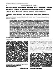
2702 J. Phys. Ther. Sci. Vol. 27, No. 9, 2015 contraction, owing to the fact that, immediately after conditioning contraction, the effect of the postactivation potentiation is countered by muscle fatigue12). After a conditioning contraction, it is seems that the muscles enable production of a required force more quickly, as the gradient of force production is increased. With this in mind, the purpose of this study was to determine the functional differences of the plantar flexion muscles of the hallux and lesser toes during the single leg stance by comparing the postural sway in different conditioning contraction interventions. SUBJECTS AND METHODS Thirty-four healthy young males and females (17 males and 17 females) participated in this study. The mean values ± standard deviations for age, height, and body weight were 22.8 ± 2.4 years, 170.0 ± 4.5 cm, and 62.3 ± 6.2 kg for males and 21.8 ± 1.8 years, 159.7 ± 3.0 cm, and 54.8 ± 5.2 kg for females, respectively. The exclusion criteria of this study included participants with lower limb or toe orthopedic disorders and neurological disorders. This study was approved by the Ethics Committee on Human Research of Waseda University (approval number: 2014-013). The purpose and methods of this study were explained to the subjects by the authors, revealing all the details of the study protocol, and informed consent was obtained from all participants. The experimental protocol is shown in Fig. 1. Before the experiments, all subjects underwent Chapman’s dominant leg test to determine their dominant leg13). During the experiments, the subjects were instructed to stand on a stabilometer (GP6000; Anima, Tokyo, Japan) using a single leg for 30 seconds, and the center of pressure was recorded on a personal computer at 20 Hz. During the single leg stance, the arms of the subjects were folded across the chest, and their eyes were open. The experiment was conducted in a limited visual and auditory disturbance room. After the single leg stance, the subjects performed conditioning contractions of the plantar flexion muscles of the hallux (Ex-1) or lesser toes (Ex-2) in the standing position or stood upright for one minute (control). In the Ex-1 and Ex-2 interventions, the subjects performed maximal voluntary isometric contractions of plantar flexion of the hallux or lesser toes for six seconds with visual feedback from a plantar pressure distribution measurement system (F-scan II; Nitta, Osaka, Japan). One minute after the intervention, the subjects performed the single leg stance for 30 seconds, and the center of pressure was recorded again. The experiments were conducted for the nondominant leg first, followed by the dominant leg. After at least a one-day interval, another intervention was conducted by using the same procedure. The types and order of interventions were randomly decided. The front-back and right-left direction components of maximal displacement (mm) and the total length (mm) of the postural sway trajectory were calculated. The maximal displacement value was normalized by the subject’s height. The postural sway velocity (m/s) was calculated as the value of the total length (mm) of the postural sway trajectory divided by the measurement time. Descriptive data are presented as means ± standard deviations. Two-way repeated measure analysis of variance with a
Fig. 1. Experimental protocol of this study All subjects performed all interventions with at least a one-day interval.
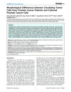
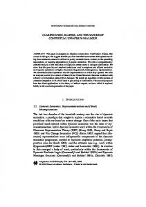
Bonferroni’s post hoc test was used to test the effects of the type of intervention on the maximal displacement and velocity of the postural sway trajectory. For all tests, statistical significance was set at p < 0.05. RESULTS For the front-back direction component of maximal displacement, there was a significant reduction after intervention Ex-1 (p < 0.01) (Table 1). However, no significant differences were observed in the Ex-2 and control interventions. For the right-left direction component of the maximal displacement, there were no significant differences in all interventions. On the other hand, the front-back direction component of the postural sway velocity was significantly reduced after intervention Ex-2 (p < 0.05) (Table 2), while no significant differences were seen in the Ex-1 and control interventions. For the right-left direction component of the postural sway velocity, no significant differences were observed in all interventions. DISCUSSION The main findings of this study were as follows: 1) the front-back direction component of maximal displacement was reduced by conditioning contraction of the plantar flexion muscles of the hallux, and 2) the front-back direction component of postural sway velocity was reduced by conditioning contraction of the plantar flexion muscles of the lesser toes during the single leg stance. From these results, it seems that the plantar flexion muscles of the lesser toes control the postural sway velocity. Furthermore, since maximal displacement corresponds to the amplitude of postural sway, the plantar flexion muscles of the hallux appear to control the amplitude of postural sway. The amplitude of muscle force exhibition is dependent on the PCSA, and the velocity of muscle contraction is dependent on the fiber length3). In two previous studies, the PCSA and fiber length of the plantar flexion muscles of the hallux (flexor hallucis longus, abductor hallucis, flexor hallucis brevis, adductor hallucis) and of the lesser toes (flexor digitorum longus, flexor digitorum brevis, quadratus plantae, lumbrical, abductor digiti minimi, flexor digiti minimi bre-
2703 Table 1. Maximal sway displacement in the interventions Direction Front-back
Right-left
Exercise
Pre (%)
Control Ex. 1 Ex. 2 Control Ex. 1 Ex. 2
2.19 ± 0.67 2.32 ± 0.67 2.22 ± 0.63 1.49 ± 0.34 1.46 ± 0.33 1.53 ± 0.36
Post (%) 2.10 ± 0.68 2.11 ± 0.47** 2.12 ± 0.61 1.46 ± 0.34 1.47 ± 0.28 1.49 ± 0.32
**p < 0.01
vis) were reported1, 2). On the basis of these previous reports on the fiber length and PCSA of the plantar flexion muscles of the toes1, 2), we calculated the theoretical fiber lengths for each group of hallux and lesser toe plantar flexion. First, we calculated the percentage of the PCSA of the respective muscle out of the total muscle group (contribution rate). Next, the contribution rate of each muscle was multiplied by the respective fiber length. Finally, we calculated the total sum of all multiplication results for fiber length and contribution rate. Therefore, the theoretical fiber length of the plantar flexion muscle of the hallux group was 15.6 mm, and that of the lesser toes was 19.0 mm. Since the normalized fiber length of the plantar flexion muscles of the lesser toes was longer than that of the hallux, it seems that the velocity of the muscle contraction was also faster for the lesser toes than for the hallux. According to previous studies, the total PCSA of the plantar flexion muscles of the hallux was larger than that of the lesser toes, and it has furthermore been reported that the plantar flexion muscles of the hallux can produce more torque than those of the lesser toes14). Overall, since the plantar flexion muscles of the hallux muscle are considered suitable for producing larger forces and the contraction velocity is slower, it appears that displacement is greatly controlled by activation of the plantar flexion muscles of the hallux during postural sway. On the other hand, since the muscle contraction velocity of the plantar flexion muscles of the lesser toes is faster, it seems that the postural sway velocity is controlled by quick activation of the plantar flexion muscles of the lesser toes before postural sway. However, in this study, there were no significant differences in any of the interventions for the right-left direction components of maximal displacement and postural sway velocity. Many toe plantar flexion muscles run on the sagittal plane, and the center of mass is shifted toward the rear by contraction of the toe plantar flexion muscles14). Therefore, we speculate that the right-left direction postural control during the single leg stance may instead be controlled at the ankle or more proximal joints. Previous studies have demonstrated that, although dancers and gymnasts require a high ability to control posture, their total trajectory length (sway velocity) was approximately equal to that of other athletes, while the peripheral area of their postural sway trajectory (sway amplitude) was conversely smaller than that of other athletes8, 9). In this study, the front-back direction sway amplitude was reduced by contraction of the plantar flexion muscles of the hallux, and it was accordingly suggested that facilitation of hallux
Table 2. Sway velocity in the interventions Direction Front-back
Right-left
Exercise
Pre (cm/s)
Post (cm/s)
Control Ex. 1 Ex. 2 Control Ex. 1 Ex. 2
3.02 ± 0.88 3.18 ± 1.02 3.16 ± 0.98 3.56 ± 0.93 3.65 ± 0.98 3.80 ± 0.98
2.98 ± 0.99 3.07 ± 0.83 3.04 ± 0.86* 3.61 ± 0.99 3.70 ± 0.98 3.66 ± 0.91
*p < 0.05
function is important for controlling sway amplitude during actions performed under high loads. On the other hand, although weakness of the plantar flexion muscles of the lesser toes was observed, no weakness of those of the hallux was seen in a previous study on elderly people prone to falling15). Moreover, it has been reported that the postural sway velocity during the single leg stance increases with aging7). In this study, it was suggested that the plantar flexion muscles of the lesser toes control the postural sway velocity, indicating that reducing the postural sway velocity by facilitating lesser toe functions may prevent falls in elderly people. The main limitation of this study was that we did not measure the exerted force and muscle contraction velocity, and future studies are needed to determine the plantar pressure distribution and the muscle reaction times during the single leg stance. Differences in the immediate effects of conditioning contraction between the plantar flexion muscles of the hallux and lesser toes were observed during the single leg stance, indicating that the functions of the hallux and lesser toes differ during this task. However, future studies are needed to confirm these findings and their implications. REFERENCES 1) Ward SR, Eng CM, Smallwood LH, et al.: Are current measurements of lower extremity muscle architecture accurate? Clin Orthop Relat Res, 2009, 467: 1074–1082. [Medline] [CrossRef] 2) Kura H, Luo ZP, Kitaoka HB, et al.: Quantitative analysis of the intrinsic muscles of the foot. Anat Rec, 1997, 249: 143–151. [Medline] [CrossRef] 3) Fukunaga T: Science Dictionary of Muscle. Tokyo: Asakura Publishing, 2002, pp 37–64 (in Japanese). 4) Shiroshita T, Fukubayashi T: Comparison of towel-gathering exercise and toe exercises for the painful accessory navicular. J Phys Ther Sci, 2011, 23: 455–458. [CrossRef] 5) Lynn SK, Padilla RA, Tsang KK: Differences in static- and dynamic-balance task performance after 4 weeks of intrinsic-foot-muscle training: the short-foot exercise versus the towel-curl exercise. J Sport Rehabil, 2012, 21: 327–333. [Medline] 6) Katayama Y, Senda M, Hamada M, et al.: Relationship between postural balance and knee and toe muscle power in young women. Acta Med Okayama, 2004, 58: 189–195. [Medline] 7) Amiridis IG, Hatzitaki V, Arabatzi F: Age-induced modifications of static postural control in humans. Neurosci Lett, 2003, 350: 137–140. [Medline] [CrossRef] 8) Asseman FB, Caron O, Crémieux J: Are there specific conditions for which expertise in gymnastics could have an effect on postural control and performance? Gait Posture, 2008, 27: 76–81. [Medline] [CrossRef] 9) Gerbino PG, Griffin ED, Zurakowski D: Comparison of standing balance between female collegiate dancers and soccer players. Gait Posture, 2007, 26: 501–507. [Medline] [CrossRef]
2704 J. Phys. Ther. Sci. Vol. 27, No. 9, 2015 10) Stewart M, Franks-Skiba K, Cooke R: Myosin regulatory light chain phosphorylation inhibits shortening velocities of skeletal muscle fibers in the presence of the myosin inhibitor blebbistatin. J Muscle Res Cell Motil, 2009, 30: 17–27. [Medline] [CrossRef] 11) Sale DG: Postactivation potentiation: role in human performance. Exerc Sport Sci Rev, 2002, 30: 138–143. [Medline] [CrossRef] 12) Miyamoto N, Kanehisa H, Kawakami Y: Potentiation of maximal voluntary concentric torque in human quadriceps femoris. Med Sci Sports Exerc, 2012, 44: 1738–1746. [Medline] [CrossRef]
13) Chapman JP, Chapman LJ, Allen JJ: The measurement of foot preference. Neuropsychologia, 1987, 25: 579–584. [Medline] [CrossRef] 14) Saeki J, Tojima M, Torii S: Relationship between navicular drop and measuring position of maximal plantar flexion torque of the first and secondfifth metatarsophalangeal joints. J Phys Ther Sci, 2015, 27: (in press). 15) Menz HB, Morris ME, Lord SR: Foot and ankle risk factors for falls in older people: a prospective study. J Gerontol A Biol Sci Med Sci, 2006, 61: 866–870. [Medline] [CrossRef]