Biochimica et Biophysica Acta 1218 (1994) 297-307. BB. Biochi~ic~a et Biophysica ~ta. Cloning and expression of a complementary DNA encoding the bovine.
BB ELSEVIER
Biochimica et Biophysica Acta 1218 (1994) 297-307
etBiochi~ic~a Biophysica~ta
Cloning and expression of a complementary DNA encoding the bovine receptor for pituitary adenylate cyclase-activating polypeptide (PACAP) Yasunori Miyamoto, Yugo Habata, Tetsuya Ohtaki, Yasushi Masuda, Kazuhiro Ogi, Haruo Onda *, Masahiko Fujino Discovery Research Laboratories I, Discovery Research Division, Takeda Chemical Industries, Ltd., Wadai 10, Tsukuba, lbaraki 300-42, Japan Received 18 November 1993
Abstract A cDNA encoding a pituitary adenylate cyclase-activating polypeptide (PACAP) receptor was cloned from a bovine brain cDNA library using a synthetic oligonucleotide probe corresponding to the partial N-terminal amino acid sequence of the PACAP receptor purified from the bovine brain. The cloned cDNA encoded a polypeptide of 513 amino acid residues with seven putative transmembrane domains. The deduced amino acid sequence exactly matched the N-terminal amino acid sequence of the purified PACAP receptor. It also shared an apparent similarity with the vasoactive intestinal peptide (VIP), secretin, growth hormone releasing hormone, calcitonin, and glucagon receptors, suggesting that the PACAP receptor is a member of the secretin receptor subfamily of the guanine nucleotide-binding regulatory protein-coupled receptor family. Northern blot analysis showed that the size of the major mRNA band which hybridized with the cDNA was about 7 kb in the bovine cerebral-cortex and hippocampus. An expression vector containing the cloned cDNA for the PACAP receptor was introduced into Chinese hamster ovary (CHO) cells. The affinity of PACAP receptors expressed on the transfected CHO cells was quite similar to that of natural PACAP receptors on the bovine brain membranes. Competitive binding experiments showed that PACAP38 displaced the binding of 125I-labeled PACAP27 to the receptors on the CHO cells more efficiently than PACAP27, while VIP was less effective. In addition, both of PACAP27 and PACAP38 elevated the levels of cAMP and inositol phosphates in the transformed CHO cells. These results indicate that the PACAP receptors encoded by the cloned cDNA are identical to the purified PACAP receptors, and that they can stimulate dual signaling cascades.
Key words: PACAP receptor; cDNA cloning; G-protein; Gene expression; Inositol phosphate; (Bovine)
I. Introduction * Corresponding author. Fax: + 81 298 645000. The nucleotide sequence data reported in this paper have been submitted to the DDBJ, EMBL, and GenBank Nuclotide Sequence
Databases under the accession number D17290. Abbreviations: PACAP, pituitary adenylate cyclase activating polypeptide; VIP, vasoactive intestinal peptide; GHRH, growth hormone releasing hormone; BSA, bovine serum albumin; G-protein, guanine nucleotide-binding regulatory protein; CHO, Chinese hamster ovary; SSC, standard saline citrate (1.5 M NaCI and 0.15 M sodium citrate); SSPE, saline-sodium phosphate-EDTA (0.15 mM NaCI, 10 mM NaH2PO4 and 4 mM EDTA); PMSF, phenylmethylsulfonyl fluoride; BSOCOES, bis(2-(succinimidooxycarbonyloxy)ethyl)sulfone; PAGE, polyacrylamide gel electrophoresis. 0167-4781/94/$07.00 © 1994 Elsevier Science B.V. All rights reserved
SSDI 0 1 6 7 - 4 7 8 1 ( 9 3 ) E 0 3 0 7 - A
Pituitary adenylate cyclase activating polypeptide ( P A C A P ) was first identified as a novel hypothalamic h o r m o n e that stimulates adenylate cyclase in pituitary cells [1]. Two forms of P A C A P have b e e n described: one has 38 amino acid residues with an a m i d a t e d C-terminus ( P A C A P 3 8 ) and the o t h e r is a truncated form with the same N-terminal 27 residues ( P A C A P 2 7 ) [2]. P A C A P shows sequence similarity with m e m b e r s of the secretin family, especially with vasoactive intestinal peptide (VIP) [1]. P A C A P has a variety of biological
298
Y. Miyamoto et al. / Biochimica et Biophysica Acta 1218 (1994) 297-307
actions, e.g., stimulation of the secretion of growth hormone (GH), prolactin, adrenocorticotropin, and luteinizing hormone from pituitary cells [1,3], interleukin-6 from folliculo-stellate cells [4], catecholamines from adrenal chromaffin cells [5], and insulin from the pancreas [6]. In addition, the PACAP gene is highly conserved between sheep [7], rats [8] and humans [9,10]. Taken together, these facts suggest that PACAP plays pivotal roles in vivo as a biologically active substance. Binding assays using radioisotope-labeled PACAP revealed that PACAP specifically binds to receptors in tissues such as the brain [11-14], lung [11] and pancreas [14]. PACAP receptors have been classified into two types, type I and type II, based on their ligand-binding specificities [15]: the type I receptor is a PACAP-specific receptor, whereas the type II receptor binds VIP as well as PACAP. The type II receptor appears to be identical to a receptor designated as the VIP receptor because the receptor expressed on animal cells transfected with VIP receptor cDNA shows the same ligand-binding specificities as the type II PACAP receptor [16]. The molecular mass of the type I receptor from bovine brain was estimated to be 57 kDa by affinity labeling with I25I-PACAP27 [13]. The affinity of the type I PACAP receptor was modulated by guanine nucleotides [13], suggesting that it is a member of the guanine nucleotide-binding regulatory protein (G-protein) coupled-receptor family. Signal transduction through the PACAP receptor was studied principally on the activation of adenylate cyclase using a variety of cell types in culture [17-19], or membrane fractions prepared from a variety of tissues [14,20]. On the other hand, Deutsch and Sun have found that PACAP38 activated the inositol lipid cascade in PC12 cells more potently than PACAP27 [21]. In addition, PACAP elevated cytosolic Ca 2+ concentration in PC12h cells [5]. However, at the moment it is unclear whether the single type of PACAP receptor was involved in both signal transduction pathways, the activation of adenylate cyclase and inositol lipid cascade. The type I PACAP receptor is abundant and widely distributed in the brain [22]. Therefore, we purified the type I PACAP receptor to homogeneity from a membrane fraction of the bovine brain using a combination of several chromatographic techniques, including affinity chromatography, and then determined its N-terminal amino acid sequence [23]. These results motivated us to clone a cDNA encoding the PACAP receptor with the chemically synthesized oligonucleotide probe based on the partial N-terminal amino acid sequence of the receptor. In this paper, we show the primary structure of the PACAP receptor encoded by a cDNA cloned from a bovine brain cDNA library, and the properties of the receptors expressed on animal cells transfected with the cDNA.
2. Materials and methods
Preparation of poly(A) + RNA Total RNA was extracted from the bovine cerebralcortex and hippocampus using the guanidinethiocyanate method [24], and was pelleted by equilibrium density-gradient centrifugation using a cesium trifluoroacetate solution (Wako Pure Chemical, Japan) [25]. Poly(A) + RNA fractions were obtained through an oligo(dT)-cellulose spun column (Pharmacia, Sweden). These fractions were used for Northern blot analysis or constructing a cDNA library as described below. cDNA libraries We used two bovine cDNA libraries to isolate a cDNA encoding the PACAP receptor: one was a brain cDNA library (1.5 × 10 6 plaque forming units) which was purchased from Clontech (USA), the other was a hippocampus cDNA library (1.5 × 10⁶ plaque forming units) which was constructed from the poly(A) + RNA fraction obtained from the hippocampus utilizing the cDNA synthesis and cloning kits (Amersham, UK). A phage vector Agtll was used for both libraries. Design of a synthetic oligonucleotide probe We have determined the N-terminal amino acid sequence of the purified natural PACAP receptor from the bovine brain: MHSDXIFKKEQAMXLEKIQRVNDLMGLND (X means an undetermined residue) [23]. We selected 16-amino acid residues beginning with Asp (D) in position 4 and ending with Gin (Q) in position 19 in order to minimize the number of possible of nucleotide sequences required for the synthetic probe. We chose high frequency genetic code in vivo corresponding to each amino acid residue except for the following two residues, Ala 12 (A) and Gln 19 (Q) for design of the probe. In the case of Ala ~2, we chose three kinds of genetic code (GCA, GCT and GCC), and in the case of Gin ~9, we excluded the third nucleotide of genetic code (CAA or CAG). A complementary sequence of the designed oligonucleotide sequence, 5 ' - T G G A T C T T C T C C A G G T G C A T D G C CTGCTCCTTCTTGAAGATGTGGTC-3' (D means G, A or T), was chemically synthesized with a DNA synthesizer model 391 (Applied Biosystems, USA). Strategy for cloning a cDNA encoding the PACAP receptor We first screened the brain cDNA library with the synthetic oligonucleotide probe. The oligonucleotide was labeled with [y-32p]ATP using T4 polynucleotide kinase (Nippon Gene, Japan). The library was screened by the method of Sambrook et al. [26]. Phages transferred onto nitrocellulose filters were incubated in 0.5 M phosphate buffer (pH 7.2) containing 1% bovine
Y. Miyamoto et aL/ Biochimica et BiophysicaActa 1218 (1994) 297-307 serum albumin (BSA), 7% SDS and 1 mM EDTA, which was a modified composition [27,28], at 50°C overnight with the labeled probe, and subsequently washed in 2 x standard saline citrate (SSC) containing 0.1% SDS at 48°C. By the first screening, we obtained a cDNA which encoded a N-terminal portion of the PACAP receptor, and designated it as pBPR35. To obtain a cDNA encoding the C-terminal portion of the PACAP receptor, we next screened the brain cDNA library (Clotnech, USA) and hippocampus cDNA library with pBPR35 as a probe. The screening method was similar to first screening. The cDNA fragment prepared from pBPR35 was labeled with [a-32p]dCTP using a multi-prime DNA labeling kit (Amersham, UK). Phages transferred onto nitrocellulose filters were incubated in a hybridization buffer containing 5 × saline-sodium p h o s p h a t e - E D T A (SSPE), 5 x Denhardt's solution, 0.1% SDS, and 100 /xg/ml of heat-denatured salmon sperm DNA with labeled probe at 65°C overnight, and subsequently washed in 0.2 x SSC containing 0.1% SDS at 60°C. We obtained two cDNA clones, pBPR68 and pBPRll4, which encoded the C-terminal portion of the PACAP receptor as a consequence of the second screening. Determination and analysis o f DNA sequence Single stranded DNAs from the obtained cDNA clones were prepared as described [9]. The DNA sequence was determined by the dideoxy chain termination method using [a-32p]dCTP or using fluorescentlabeled dideoxy nucleotides or fluorescent-labeled primers. Determination of the DNA sequence using fluorescence was performed with an ABI 370A DNA sequencer (Applied Biosystems, USA). The DNA sequences were connected and analyzed with computer software called DNASIS (Hitachi, Japan). The hydropathy plot of an amino acid sequence deduced from the DNA sequence was obtained by the method of Kyte and Doolittle [29]. Northern blot analysis A 0.8 kbp E c o R I - T t h l l l I DNA fragment was prepared from pBPR35 (nucleotides 1-807, Fig. 2), and used as a probe by labeling with [•-32p]dCTP. Each 5 /.~g of poly(A) + RNA prepared from bovine cerebralcortex or hippocampus was electrophoresed onto a 1.1% agarose gel containing 2.2 M formaldehyde, and then transferred to a nylon membrane filter (Biodyneplus, Pall Biosupport, USA) by capillary blotting in 20 X SSC. The filter was incubated with the probe in a hybridization buffer containing 50% formamide, 5 x Denhardt's solution, 5 x SSPE, 0.5% SDS, and 100 /xg/ml of heat denatured and sonicated salmon sperm DNA at 42°C overnight. The filter was then washed in 0.1 X SSC, 0.1% SDS at 50°C, and autoradiographed using Kodak X-OMAT AR film with an intensifying screen at - 8 0 °C for 2 weeks.
299
Construction of an expression uector A 0.6 kbp DNA fragment which contained the Nterminal coding region of the PACAP receptor was prepared by digesting pBPR35 with SmaI and B a m H I (Fig. 1). HindIII linkers were ligated at the blunt-end of the SmaI site in this fragment. After the ligation, the cDNA was digested with HindlII. Next, a 1 kbp fragment which contained the C-terminal coding region of the PACAP receptor was prepared by digesting pBPR68 with B a m H I and SmaI (Fig. 1). The latter fragment was ligated at the B a m H I site of the former fragment. The resultant DNA fragment, which contained the entire coding region of the bovine PACAP receptor, was inserted at the H i n d l I I - E c o R V site of the eukaryotic expression vector, p R c / C M V (Invitrogen, USA), and this expression plasmid was named pBPR-T. Transfection of cDNA Chinese hamster ovary (CHO)-K1 cells (ICN Biomedicals, USA) were grown in Ham's F12 medium (Nissui Seiyaku, Japan) with 10% fetal bovine serum (FBS; Cell Culture Laboratories, USA) under 5% CO z in air at 37°C. The expression vector, pBPR-T, was introduced into CHO-K1 cells with a Cell-Phect transfection kit (Pharmacia, Sweden). Transformants were selected in a medium containing 500 txg/ml of geneticin (Gibco, USA), and cloned by colony formation. Preparation of membrane fractions from transformed CHO cells The transformed CHO cells were grown in a medium in 150 cm 2 flasks for 3 days after plating. The cells were then dispersed in phosphate-buffered saline containing 0.2 mM EDTA. The ceils were washed with the same buffer, and then suspended in 10 mM sodium carbonate buffer including 1 mM EDTA, 0.5 mM phenylmethylsulfonyl fluoride (PMSF), 20 /zg/ml of leupeptin, 4 / x g / m l of E-64, and 1/ xg/ m l of pepstatin. After the cells were homogenized with a Polytron homogenizer, the homogenate was centrifuged at 3000 rpm for 10 rain in a Hitachi RR18 rotor. The resultant supernatant was ultracentrifuged at 30000 rpm for 60
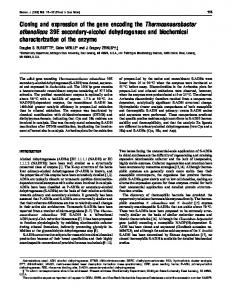
oBPR35 pBPR6 8
pBPR 114
=
S
S
'
'
200 bp
NB '
~
a
NB
S
lJ i
t
.N B
~
V
Fig. 1. Schematicrepresentation of the structure of bovine PACAP receptor cDNA. The restrictionendonucleasecleavagesites used for mapping are indicated (B, BamHI; N, NcoI; S, SmaI). The box indicates the protein coding region and the dotted area indicatesthe signal sequence. The alternativelyspliced exon of pBPRll4 is crosshatched.
Y. Miyamoto et al. / Biochimica et Biophysica Acta 1218 (1994) 297-307
300
I
T~GC~TG~A~c6CAC~CC~AG~TGC~AA~Ac~GA~6~T~T~TCG~TC~CT~CT~C~G~CT~GCTTC~T~GAG~C~T~6CT~T~
104 208 312 I 416
CGGCAAGGCAGACC•GGCTGGGCGG•CGCGCGGCGcGGGGCGGGCTAGGGAAGGCCGGGGGCCTCGCGC•CGGGCCCCGGG•GGCGACTGAC•GCGG•GGCGGC GGCGGC•GCG••TCCAAGGCGAG•GTGGTCCCCGCGTGCGCACAAGCTCGCCGCCGCGCAGGGACCCACGGACACCGGCGCCGGGCGGACACACAGACGCGGAG ATCGGG~T~T~C~G~CT~TCAGCG~ACG~G~T~C~ATCC~TGGG~GGAG~GGGG~G~GGA~TCG~c~TG~G~G~cT~CG~GGAGT~TG~C~GGGC Met Arg GIy GIy Arg AGACCCGCAGCC~GCGG~CCCGCCGCGAGGCCCCTGGGTGAGCAGCCTGTAGA~A~CTGGGGTTGAGCAGTGG~GGCTGTGA ATG AGA GGC GGG CGG
103 207 311 415 5 512
6 613
His Trp Pro Glu Pro Pro Cys Arg Leu Arg Ser Val Met Ala Set l i e Ala Gin Val Ser Leu Ala Ala Lcu Leu Lcu CAC TGG CCC GAG CCG CCT TGC AGG CTG AGA AGC GTC ATG GCC AGC ATC GCG CAG GTC TCC CTG GCT GCT CTC CTC CTG
31 590
32 591
Leu Pro Met Ala Thr AlaVMet His Ser Asp Cys lle Phe Lys Lys Glu Gin Ala Met Cys Leu Glu Lys llc Gln Arg CTG CCT ATG GCC ACC GCC ATG CAT TCC GAC TGC ATC TTC AAG AAG GAG CAA GCC ATG TGC CTG GAG AAG ATC CAG AGG
57 668
669
58 Val Asn Asp Leu Met GIy Leu ~sn Asp Ser Ser Pro GIy Cys Pro GIy Met Trp Asp ~sn lie ThF Cys Trp Lys Pro GTG AAT GAC CTG ATG GGC TTG AAT GAC TCC TCC CCA GGG TGC CCT GGG ATG TGG GAC AAC ATC ACG TGT TGG AAG CCC
83 746
84 Ala His Val GIy Glu Met Val Leu Va] Ser Cys Pro Glu Leu Phc Arg lie Phe Asn Pro Asp Gin Val Trp Glu Thr 747 GCC CAC GTG GGT GAG ATG GTC CTG GTC AGT TGC CCT GAA CTC TTC CGA ATC TTC AAC CCA GAG CAA GTC TGG GAG ACG
109 824
110 825
Glu Thr lie GIy Glu Phe GIy Phe Ala Asp Set Lys Ser Leu Asp Leu Ser Asp Met Arg Val Val $er Arg ~sn Cys GAA ACC ATC GGA GAG TTC GGT TTT GCA GAC AGT AAA TCC TTG GAT CTC TCA GAC ATG AGG GTG GTG AGC CGG AAT TGC
135 902
136 903
Thr Glu Asp Gly Trp Ser Glu Pro Phe Pro His Tyr Phe Asp Ala Cys GIy Phe Glu Glu Tyr Glu Ser Glu Thr GIy ACG GAG GAT GGA TGG TCA GAG CCA TTC CCT CAT TAT TTC GAT GCC TGT GGG TTT GAG GAG TAC GAA TCT GAG ACT GGG
161 980
162 981
Asp Gin Asp Tyr Tyr Tyr Leu Ser Val Lys Ala Leu Tyr Thr Val G]y Tyr Set Thr Ser Leu Val Thr Leu Thr Thr GAC CAG GAT TAC TAC TAC CTG TCA GTG AAG GCC CTG TAC ACA GTT GGC TAC AGC ACG TCC CTC GTC ACC CTC ACC ACT
187 1058
188 Ala Met Val lie Leu Cys Arg Phe Arg Lys Leu His Cys Thr Arg Asn Phe lie His Met Ash Leu Phe Val Ser Phe 1069 GCC ATG GTC ATC CTG TGT CGT TTC CGG AAG CTG CAC TGC ACC CGC AAC TTC ATC CAC ATG AAC CTC TTC GTG TCG TTT
213 1136
214 Wet Leu Arg Ala lle Ser Val Phe lie Lys Asp Trp lie Leu Tyr Ala Glu Gin Asp Ser Asn His Cys Phe Val Ser I137 ATG CTG AGG GCC ATC TCC GTC TTC ATC AAA GAC TGG ATC CTC TAT GCT GAG CAG GAC AGC AAT CAC TGC TTT GTC TCC
239 1214
240 Thr Val Glu Cys Lys Ala Val Met Val Phe Phe His Tyr Cys Val Val Ser Asn Tyr Phe Trp Leu Phe l i e Glu GIy 1215 ACT GTG GAA TGC AAG GCT GTG ATG GTT TTC TTC CAC TAC TGT GTT GTA TCC AAC TAC TTC TGG CTG TTC ATC GAG GGC
265 1292
266 Leu Tyr Leu Phe Thr Leu Leu Val Glu Thr Phe Phe Pro Glu Arg Arg Tyr Phe Tyr Trp Tyr I{e lie lie GIy Trp 1293 CTG TAT CTC TTC ACC CTG CTG GTG GAG ACC TTC TTC CCC GAG AGG AGA TAT TTC TAC TGG TAC ATC ATC ATT GGC TGG
291 1370
292 GIy Thr Pro Thr Val Cys Va{ Ser Val Trp AIa Met Leu Arg Leu Tyr Phe Asp Asp Thr GIy Cys Trp Asp Met Asn 1371 GGG ACA CCA ACT GTG TGT GTG TCT GTG TGG GGT ATG CTG AGG CTC TAG TTG GAT GAG ACA GGC TGC TGG GAT ATG AAT
317 1448
318 Asp Asn Thr Ala Lea Trp Trp Val lie Lys GIy Pro Val Val GIy Ser lle Met Val Asn Phe Val Leu Phe l i e GIy 1449 GAC AAC ACG GCT CTG TGG TGG GTG ATC AAA GGC COT GTA GTT GGC TOG ATA ATG GTT AAT ?TT GTG CTC TTC ATC GGC
343 1526
344 lle lle Val lle Leu Val Gin Lys Leu Gin Set Pro Asp Met GIy Gly Ash Glu Ser Ser Ilc Tyr Phe Ser Cys Val 1527 ATC ATT GTC ATC CTT GTG CAG AAA CTT CAG TCT CCA GAC ATG GGA GGC AAC GAG TCC AGC ATC TAC TTC AGC TGC GTG
369 1604
370 Gln Lys Cys Tyr Cys Lys Pro Gin Arg Ala Gin Gin His Ser Cys Lys Met Ser GIu Leu Set Thr lie Thr Leu Arg 1606 CAG AAA TGC TAC TGC AAG CCA CAG CGG GCT CAG GAG CAC TCT TGC AAG ATG TCA GAA CTG TOG ACC ATT ACT CTA CGG
395 1682
i
396 Leu Ala Arg Set Thr Leu Leu Leu l i e Pro Leu Phe Giy l i e His Tyr Thr Val Phe Ala Phe Set Pro Glu Asn Va] 1683 CTC GCC AGG TCC ACC TTG CTG CTC ATC CCA CTC TTT GGA ATC CAC TAC ACT GTC TTT GCT TTC TCC CCG GAG AAC GTC
421 1760
Ser Lys Arg Glu Arg Leu Val Phe Glu Leu GIy Leu GIy Ser Phe Gin GIy Phe Val Val Ala Val Leu Tyr Cys Phe AGC AAG AGG GAG AGA CTG GTG TTT GAG CTG GGT CTG GGC TCC TTC CAG GGC TTT GTG GTG GCT GTT CTC TAT TGC TTT
447 1838
448 Leu Asn Gly Glu Val Gin Ala Glu l i e Lys Arg Lys Trp Arg Ser Trp Lys Val Ash Arg Tyr Phe Thr Met Asp Phe 1839 CTG AAT GGA GAG GTG CAG GCG GAG ATC AAG AGG AAG TGG CGG AGC TGG AAG GTG AA6 666 TAC TTC ACC ATG GAG TTC
473 1916
474 Lys His Arg His Pro Ser Leu Ala Ser Ser GIy Val Ash Gly Gly Thr Gin Leu Ser lie Leu Ser Lys Ser Ser Ser 1917 AAG CAC CGG CAC CCA TCC CTG GCC AGC AGC GGG GTG AAC GGG GGC ACC CAG CTC TCC ATC CTG AGC AAG AGC AGC TCC
499 1994
600 1995
Gin lle Arg Met Set Gly Leu Pro Ala Asp Asn Leu Ala Thr ,** CAG ATC CGC ATG TCT GGG CTT CCG GCC GAC AAC CTG GCC ACC TGA GCCCACCCTGCCCCCTCCTCTCCTCTGTACGCAGGCTGGGGCTG
513 2083
2084 2188 2292 2396 2500 2604 2708 2812
TGGTGGGGCGCCGGCCCACGCATGTTGTGCCTCTTCTCGCCTTCGGGCAGGCCC•GGGCTGGGCGCCTGGCC••CGAGGTTGGAGAAGGATGCGGGACAGGCAG CTGTTTAGCCTTC•TGTTTTGGCG•TGGCCCA••CACCGTGGGTCCCTGGGCCTGCACCCAGACAT•TAAT•CTCCTTAATTGGGAA•TCATCCATTCTTTCCC TTTCCC•AGTCCTTGCTTATTAAGAGGTTC•AGTCACCTACC•AATTCAG•AGCTTAAGTAACC••TA•CCACCGTGACTGCGTGGG•GGCCTCCCATGGGCTG AGCT•CTGACTTGGCTTTGGGGGCCTTGGGCTGGGGCCCTCCTT•AAGCCCCCCCTG•A•TTGTCGG•CCTC•A•GTGTGACTCCTTTG•GTCTACTCGCC•CC CCCGTGGCC•TTTGC•GCCCTGGTCCAGTC•CCG•GGTTACTGGAAGTCCAGCTTGG•TGGCCAGAC•GCTTTTTGGC•CAGGC•GACCCATGCTCACCC•ACA TTTTAGTGT•CAGGTGCCCAGGTGCCC•GGTGCCCAGCTCCTGGGCATCAG•CAGTGGGAAAGCTCC•GGG•TCTACC•TTCAGAGACTTC•GTTTGG•TGTAG GG•TAAGGCCAGAG••A•GTTCTGGAGCTTTT•ATTTGGC•C•AGAAAAA••TGCCA•GATCCAGAAAAGTGGATCTGAGTG•AATTT•GATGCAA•G•GCTTG GAG
2187 2291 2385 2499 2603 2707 2811 2814
422 1761
Fig. 2. Nucleotide and predicted amino acid sequences of the bovine P A C A P receptor cDNAs. The numbers on both sides correspond to the amino acid and nucleotide positions of the c D N A encoding the bovine P A C A P receptor. A non-sense codon in the 5' non-coding region i~ double-underlined. An arrowhead indicates the cleavage site of the signal peptide. The deleted sequence which was observed in p B P R l l 4 L, underlined. The potential N-glycosylation sites in the putative extracellular domains are marked by stars.
Y Miyamoto et aL/Biochimica et Biophysica Acta 1218 (1994) 297-307
min in a Hitachi RP42 rotor. Pellet was then suspended in a buffer containing 20 mM Tris-HCl (pH.7.4), 0.25 M sucrose, 2 mM EDTA, 0.5 mM PMSF, 20 /xg/ml of leupeptin, and 1 /zg/ml of pepstatin, and used as a membrane fraction.
Receptor binding and affinity labeling experiments Competitive binding and saturation binding experiments were performed as described by Ohtaki et al. [23]. For saturation binding experiments, the membrane fractions prepared from the transformed CHO cells were incubated in a buffer containing 20 mM Tris-HC1 (pH 7.4), 5 mM magnesium acetate, 2 mM EGTA, 0.5 mM PMSF, 20 /zg/ml of leupeptin, 4 /zg/ml of E-64,and 1 /xg/ml of pepstatin with 100 pM I25I-PACAP27 at 25°C for 75 min. Bound and free ligands were separated by filtration through glass fiber filters (Whatman, G F / F , UK) treated with polyethyleneimine. Non-specific binding was determined in the presence of 1/~M PACAP27. For affinity labeling experiments, the membrane fractions were incubated with I25I-PACAP27 as described above, and then washed twice with phosphatebuffered saline. Cross-linking was carried out in the presence of 0.1 mM bis(2-(succinimidooxycarbonyloxy) ethyl)sulfone (BSOCOES) at 4°C for 30 min. Excess of BSOCOES was quenched with 0.1 M Tris. After the cross-linking, the membrane fraction was centrifuged and dissolved in a sample buffer for SDS-polyacrylamide gel electrophoresis (PAGE). Samples were subjected to SDS-PAGE in 8% polyacrylamide gels. The gels were autoradiographied at -80°C for 4 days.
Assay of intracellular cAMP in transfected CHO cells The transformed CHO cells were grown in 24-well multiwell plates for 3 days after plating. The cells were washed twice with an assay solution (Hank's balanced salt solution containing 0.05% BSA), and pre-incubated in an assay solution containing 0.2 mM 3-isobutyl-l-methylxanthine for 1 h. Various concentrations of PACAP27,PACAP38 or VIP were then added to the medium and incubated at 37°C for 30 min. The cells were washed with the assay solution, and the intracellular cAMP was extracted with a solution (500/xl of the assay solution and 100/zl of 20% perchloric acid) on crushed-ice for 30 min. The extract was neutralized with 1.5 M KOH, and subjected to enzyme immunoassay. The amount of cAMP was estimated with a cAMP enzyme immunoassay system, BIOTRAK (Amersham, UK).
Quantification of inositol phosphates Inositol lipid turnover was assayed essentially as described by Berridge et al. [30]. The transformed CHO cells were grown in 24-well multiwell plates for 4 days after plating. The cells were labeled with 5 ~Ci of myo-[3H]inositol (19.1 Ci/mmol, Amersham) per well
301
in the growth medium overnight at 37°C. The cells were washed with an assay buffer (20 mM Hepes, 140 mM NaCI, 4 mM KC1, 1 mM Na2HPO4, 1 mM MgCI2, 1.25 mM CaC12, 10 mM LiC1, 10 mM glucose, and 0.1% BSA) [31], and pre-incubated with the assay buffer at 37°C for 20 min. The cells were incubated with 500 ~1 of the assay buffer containing various concentrations of PACAP27,PACAP38 and VIP at 37°C for 20 min, and 100 ~1 of 20% perchloric acid was added to each well to terminate the reactions. After treatment for 10 min at room termperature, the extract was kept on ice for 20 min. The extract was then centrifuged and the supernatant was neutralized with 1.5 M KOH. Total inositol phosphates were separated from free inositol by ion-exchange chromatography (AG1-X8, Bio-Rad, USA), and eluted with 1 M ammonium formate/0.1 M formic acid, followed by scintillation counting. 3. Results
Cloning of a cDNA encoding the PACAP receptor We screened the bovine brain cDNA library with a chemically synthesized probe corresponding to the partial N-terminal amino acid sequence of the natural PACAP receptor. From this screening, we obtained 124 candidate clones. Furthermore, 20 clones were selected from this group on the basis of signal intensity on the autoradiographic films. Nucleotide sequencing of these clones revealed that only one clone (pBPR35) encoded an amino acid sequence corresponding to the N-terminus of the purified receptor. However, this cDNA was devoid of a C-terminal coding region. Therefore, we subsequently screened a bovine brain cDNA library and a hippocampal cDNA library with this cDNA as a probe. As a consequence, we obtained two positive clones, pBPR68 and pBPRll4. A restriction enzyme map of these cloned cDNAs is shown in Fig. 1. Nucleotide sequencing of pBPR68 and pBPR114 revealed that pBPR68 encoded the C-terminal coding region and that pBPRII4 encoded a putative isoform of the PACAP receptor, which was generated by deletion of an alternative exon area represented by the cross-hatched portion in Fig. 1. The 3' non-coding region of pBPR68 was so long that the region is not illustrated in Fig. 1. The length of the unillustrated region, which did not include a poly(A) tail, was about 5.5 kbp (data not shown). Fig. 2 shows the nucleotide sequence encoding the entire PACAP receptor, based on pBPR35 and pBPR68,together with the deduced amino acid sequence. The initiation codon ATG was found at position 485 (Fig. 2). According to this position of the initiation codon, the nucleotide sequence of the PACAP receptor cDNA contained a maximum open reading frame encoding a polypeptide of 513 amino
Y. Miyamoto et al. /Biochimica et Biophysica Acta 1218 (1994) 297-307
302 Index
+/
4 3 2 1 0 -1 2 -3 -4.
¸
0
120
240
360
480
600
Residues
-< 9 . 4 9 - , 7.46
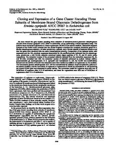
Fig. 3. Hydropathy plot of the bovine PACAP receptor. The hydropathy plot of the deduced amino acid sequence of the PACAP receptor was obtained by the method of Kyte and Doolittle [29]. The numbers along the x-axis indicate the positions of the amino acid residues of the precursor protein. The large numbers 1-7 mean the seven putative transmembrane domains. The deleted sequence which was observed in pBPRI14 is marked by an arrowhead.
acid residues. The first 37 amino acids of the Nterminus were assumed to be a signal sequence because the following 29 amino acids corresponded exactly to the N-terminal sequence of the purified natural PACAP receptor. The two undetermined residues in the N-terminal sequence of the purified natural PACAP receptor were found to be cysteine. Therefore, the mature PACAP receptor was thought to consist of 476-amino acid residues with a calculated molecular mass of 54.2 kDa. Four potential N-glycosylation sites (Asn-X-Thr/Ser) were found in the putative extracellular domains as described below (Fig. 2). Analysis of hydrophobicity revealed that eight clusters of hydrophobic amino acid residues existed in the amino acid sequence (Fig. 3). The first N-terminal cluster corresponded to the signal peptide sequence. The remaining hydrophobic clusters were thought to form seven transmembrane domains. A hydrophilic region between the first and second hydrophobic clusters appears to be the N-terminal exracellular domain. This region contained three potential N-glycosylation sites (Fig. 2). One more potential N-glycosylation site was found in the putative third extracellular loop. The 20
ii i~ii
i
i
..,
4.40
42.37 -< 1 . 3 5
-< 0.24
Fig. 4. Northern blot analysis of the bovine PACAP receptor. Poly(A) + RNA was prepared from the bovine cerebral-cortex and hippocampus. 5/zg of poly(A) + RNA from each source per lane was probed under conditions of high stringency with a 32p-labeled probe of the bovine PACAP receptor cDNA fragment. Size marker locations are shown at right of panel.
nucleotide sequence of pBPRll4 corresponded almost exactly to that of pBPR68 except for the deletion of 84 bp at nucleotide positions between 1595 and 1678,which is underlined in Fig. 2. This region was hydrophilic and located in the putative third intracellular loop (Fig. 3).
Expression of PACAP receptor mRNA Northern blot analysis was performed using poly(A) + RNA from the hippocampus and cerebral-cortex. A major band hybridized with the cDNA was observed at about 7 kb, and two additional minor bands were detected at about 2.2 and 2.7 kb in both tissues (Fig. 4). 0.2
A
15
:i LL "O t-
10
"O t-
O
0.1
rn
,
i
100
,
i
200
,
i
300
,
i
400
Total Llgand (pM)
,
i
500
,
0.(1 600
10
2 0
Bound (pM)
Fig. 5. Scatchard plot analysis of 125I-PACAP27 binding. (A) The specific saturation binding of 12SI-PACAP27. Membrane fractions from CHO cells transfected with the PACAP receptor cDNA were incubated with various concentrations of 125I-PACAP27 at 25°C for 75 min, followed by separation of the bound and free ligand. Non-specific binding was determined in the presence of 1 /xM PACAP27. (B) Scatchard plot analysis of ~2SI-PACAP27. Data are expressed as the mean + S.E. of triplicate samples. Where no error bars are visible, they lie within the symbol.
Y.. Miyamoto et al. / Biochimica et Biophysica Acta 1218 (1994) 297-307
Characterization of the PACAP receptor expressed on CHO cells The molecular characteristics of the PACAP receptor encoded by the cloned cDNA were compared with the natural PACAP receptor. An expression plasmid, pBPR-T, which contained the entire open reading frame (nucleotide positions 409-2136, Fig. 2) under the control of the cytomegalo virus promoter was introduced into CHO-K1 cells. A membrane fraction prepared from the transfected cells apparently bound 1zSI-PACAP27. The saturation binding experiments (Fig. 5A) and following Scatchard plot analysis indicated that the membranes have a single class of binding sites with a dissociation constant (K d) of 102 _+7.9 pM, and that the content of the receptor per total proteins was 0.13 pmol/mg (Fig. 5B). The K d value was similar to that of the PACAP receptor on the bovine brain membranes [23]. Competitive binding experiments revealed that PACAP38 (IC50 = 0.13 nM) displaced the 1251PACAP27 binding more potently than PACAP27 (IC50 = 0.30 nM) while VIP was not effective (Fig. 6). This result was similar to that obtained from the natural PACAP receptors except for the lower affinity of VIP (data not shown). The inhibitory constant (K i) and Hill coefficient were derived from the equation of Chang [32]. The K i value was 150 + 7.8 pM for PACAP27 and 60.9 + 3.8 pM for PACAP38. The Hill coefficient was 1.01 + 0.04 pM for PACAP27 and 1.06 _+0.05 pM for PACAP38. A coefficient of approximately one means that there is only a single class of receptors. The molecular mass of the PACAP receptor expressed on CHO cells was compared to that of the PACAP receptor from bovine brain by affinity-labeling techniques. A single band with a molecular mass of
01
120
._= m
10o
.R
o o D.. O E E
80 60
M
1 2
3 4
2 0 0 I"
92.5 " " 69
46
Fig. 7. Affinity-labeling of 12>I-PACAP27 to the PACAP receptors. Membrane fractions from the bovine brain and the transformed CHO cells were incubated with 110 pM tzsI-PACAP27 at 4°C overnight in the presence and absence of unlabeled PACAP27. The receptor-ligand complexes were cross-linked with BSOCOES and analyzed by SDS-PAGE. The gel was autoradiographed. Lane M, 14C-labeled protein molecular weight markers; lane 1, membrane fractions of the CHO cells in the absence of unlabeled PACAP27; lane 2, membrane fractions of the CHO cells in the presence of unlabeled PACAP27; lane 3, membrane fractions of the bovine brain in the absence of unlabeled PACAP27; lane 4,membrane fractions of the bovine brain in the presence of unlabeled PACAP27.
65-75 kDa was labeled in the membrane fractions from transfected CHO cells (Fig. 7, lane 1). On the other hand, a major band with a molecular mass of 56-66 kDa was densely labeled, and a minor band with a molecular mass of 73 kDa was lightly labeled, in the membrane fractions from the bovine brain (Fig. 7, lane 3). These molecular masses include the 3 kDa crosslinked PACAP27. Therefore, the real molecular masses were 62-72 kDa for the cloned receptor, 53-63 kDa for the major band of the natural receptor, and 69 kDa for the minor band of the natural receptor. None of these bands were seen when crosslinking was done in the presence of 1 /~M unlabeled PACAP27 (Fig. 7, lanes 2 and 4). The calculated molecular mass of the 476-amino acid peptide deduced from the bovine PACAP receptor cDNA was 54.2 kDa (see above). The discrepancy between these molecular masses might result from differences in the extent of glycosylation.
40 20 o
c o.
(kDa)
303
-20 -12
L -11
.
t -10
.
, -9
Concentration
.
i
~
-8
i -7
,
-6
(IogM)
Fig. 6. Competitive binding of 125I-PACAP27 to the cloned P A C A P receptor. The binding of 125I-PACAP27 to m e m b r a n e s from the transformed C H O cells was determined in the presence of the indicated concentrations of PACAP27 (o), PACAP38 ([]) and VIP (zx). Data are expressed as the m e a n + S.E. of triplicate samples. W h e r e no error bars are visible, they lie within the symbol.
Intracellular accumulation of cAMP mediated by the cloned PACAP receptors To examine whether the cloned PACAP receptor expressed in CHO cells could transduce the signal for activation of adenylate cyclase, we examined the stimulation of adenylate cyclase activity by PACAP27, PACAP38 and VIP (Fig. 8). PACAP38 stimulated the accumulation of cAMP in the CHO cells more potently than PACAP27, whereas VIP was less effective. This result was almost parallel with that of the competitive binding experiments.
Y.. Miyamoto et al. / Biochimica et Biophysica Acta 1218 (1994) 297-307
304
single class of PACAP receptor can stimulate dual signaling cascades.
71
¢L =E o
~
s
u
4. D i s c u s s i o n
To
4
O0
3
e0 0 ._>
2
0
J
-13
-12
i
i
-11
-10
,
A
,
-9
i
i
-8
-7
-6
Concentration (IogM) Fig. 8. Stimulation of c A M P content by PACAP27, PACAP38 and VIP. The transformed C H O cells were incubated for 30 min at 37°C with various concentrations of PACAP27 ( 1 /xM) in PACAP-Rhopl,which was characterized in our work. This result
306
Y. Miyamoto et al. / Biochimica et Biophysica Acta 1218 (1994) 297-307
differs from our result. It is unclear what causes this discrepancy. The discrepancy may be derived from the properties of the used cell line, because the cell line transfected with the PACAP receptor was different each other. We used CHO cells, and they used porcine renal epithelial LLC PK1 cells. In this paper, we succeeded in cloning a bovine cDNA encoding the PACAP receptor. The binding properties of the PACAP receptor expressed on CHO cells were identical to the natural PACAP receptor in the bovine brain. The PACAP receptors simultaneously stimulated dual signaling pathways, activation of adenylate cyclase and phospholipase C. We believe that our study will facilitate the study of signal transduction through PACAP receptors.
Acknowledgments We are grateful to Mihoko Sato for her excellent technical assistance. We thank to Dr. Hisayoshi Okazaki for valuable discussions and encouragement throughout this work, Dr. Shuji Hinuma for critical reading of this manuscript and indispensable advice, and Dr. Chiharu Kimura for synthesis of the oligonucleotides used for screening.
References [1] Miyata, A., Arimura, A., Dahl, R.R., Minamino, N., Uehara, A., Jiang, L., Culler, M.D. and Coy, D.H. (1989) Biochem. Biophys. Res. Commun. 164, 567-574. [2] Miyata, A., Jiang, L., Dahl, R.R., Kitada, C., Kubo, K., Fujino, M., Minamino, N. and Arimura, A. (1990) Biochem. Biophys. Res. Commun. 170, 643-648. [3] Culler, M.D. and Paschall, C.S. (1991) Endocrinology 129, 2260-2262. [4] Tatsuno, I., Somogyvari-Vigh, A., Mizuno, K., Gottshall, P.E., Hidaka, H. and Arimura, A. (1991) Endocrinology 129, 17971804. [5] Watanabe, T., Masuo, Y., Matsumoto, H., Suzuki, N., Ohtaki, T., Masuda, Y., Kitada, C., Tsuda, M. and Fujino, M. (1992) Biochem. Biophys. Res. Commun. 182, 403-411. [6] Kawai, K., Ohse, C., Watanabe, Y., Suzuki, S., Yamashita, K. and Ohashi, S. (1992) Life Sci. 50, 257-261. [7] Kimura, C., Ohkubo, S., Ogi, K., Hosoya, M., Ito, Y., Onda, H., Miyata, A., Jiang, L., Dahl, R.R., Stibbs, H.H., Arimura, A. and Fuiino, M. (1990) Biochem. Biophys. Res. Commun. 166, 81-89. [8] Ogi, K., Kimura, C., Onda, H., Arimura, A. and Fujino, M. (1990) Biochem. Biophys. Res. Commun. 173, 1271-1279. [9] Ohkubo, S., Kimura, C., Ogi, K., Okazaki, K., Hosoya, M., Onda, H., Miyata, A., Arimura, A. and Fujino, M. (1992) DNA Cell Biol. 11, 21-30. [10] Hosoya, M., Kimura, C., Ogi, K., Ohkubo, S., Miyamoto, Y., Kugoh, H., Shimizu, M., Onda, H., Oshimura, M., Arimura., A. and Fujino, M. (1992) Biochim. Biophys. Acta 1129, 199-206. [11] Lain, H.-C., Takahashi, K., Ghatei, M.A., Kanse, S.M., Polak, J.M. and Bloom, S.R. (1990) Eur. J. Biochem. 193, 725-729. [12] Gottschall, P.E., Tatsuno, I., Miyata, A. and Arimura, A. (1990) Endocrinology 127, 272-277.
[13] Ohtaki, T., Watanabe, T., Ishibashi, Y., Kitada, C., Tsuda, M., Gottschall, P.E., Arimura, A. and Fujino, M. (1990) Biochem. Biophys. Res. Commun. 171,838-844. [14] Gourlet, P., Woussen-Colle, M.-C., Robberecht, P., De Neef, P., Cauvin, A., Vandermeers-Piret, M.-C., Vandermeers, A. and Christophe, J. (1991) Eur. J. Biochem. 195, 535-541. [15] Shivers, B.D., G6rcs, T.J., Gottschall, P.E. and Arimura, A. (1991) Endocrinology 128, 3055-3065. [16] Ishihara, T., Shigemoto, R., Mori, K., Takahashi, K. and Nagata, S. (1992) Neuron 8, 811-819. [17] Cauvin, A. Buscail, L., Gourlet, P., De Neef, P., Gossen, D., Arimura, A., Miyata, A., Coy, D.H., Robberecht, P. and Christophe, J. (1990) Peptides 11,773-777. [18] Buscail, L., Gourlet, P., Cauvin, A., De Neef, P., Gossen, D., Arimura, A., Miyata, A., Coy, D.H., Robberecht, P. and Christophe, J. (1990) FEBS Lett. 262, 77-81. [19] Watanabe, T., Ohtaki, T., Kitada, C., Tsuda, M. and Fujino, M. (1990) Biochem. Biophys. Res. Commun. 173, 252-258. [20] Robberecht, P., Gourlet, P., Cauvin, A., Buscail, L., De Neef, P., Arimura, A. and Christophe, J. (1991) Am. J. Physiol. 260, G97-GI02. [21] Deutsch, P.J. and Sun, Y. (1992)J. Biol. Chem. 267, 5108-5113. [22] Masuo, Y., Ohtaki, T., Masuda, Y., Tsuda, M. and Fujino, M. (1992) Brain Res. 575, 113-123. [23] Ohtaki, T., Masuda, Y., Ishibashi, Y., Kitada, C., Arimura, A. and Fujino, M. (1993) J. Biol. Chem. 268, 26650-26657. [24] Chirgwin, J,M., Przybyla, A.E., MacDonald, R.J. and Rutter, W.J. (1979) Biochemistry 18, 5294-5299. [25] Okayama, H., Kawaichi, M., Brownstein, M., Lee, F., Yokota, T. and Arai, K. (1987) Methods Enzymol. 154, 3-28. [26] Sambrook, J., Fritsch, E.F. and Maniatis, T. (1989) Molecular Cloning: A Laboratory Manual, 2nd Edn., Cold Spring Harbor Laboratory, Cold Spring Harbor. [27] Church, G.M. and Gilbert, W. (1984) Proc. Natl. Acad. Sci. USA 81, 1991-1995. [28] Brumberg, B., Wright, C.V.E., De Roberfis, E.M. and Cho, K.W.Y. (1991) Science 253, 194-196. [29] Kyte, J. and Doolittle, R.F. (1982) J. Mol. Biol. 157, 105-132. [30] Berridge, M.J., Dawson, R.M.C., Downes, P., Heslop, J.P. and lrvine, R.F. (1983) Biochem. J. 212, 473-482. [31] Sakurai, T., Yanagisawa, M., Takuwa, Y., Miyazaki, H., Kimura, S., Goto, K. and Masaki, T. (1990) Nature 348, 732-735. [32] Chang, K.-J., Jacobs, S. and Cuatrecasas, P. (1975) Biochim. Biophys. Acta 406, 294-303. [33] Ishihara, T., Nakamura, S., Kaziro, Y., Takahashi, T., Takahashi, K. and Nagata, S. (1991) EMBO J. 10, 1635-1641. [34] Mayo, K.E. (1992) Mol. Endocrinol. 6, 1734-1744. [35] Sexton, P.M., Houssami, S., Hilton, J.M., O'Keeffe, L.M., Center, R.J., Gillespie, M.T., Darcy, P. and Findlay, D.M. (1993) Mol. Endocrinol. 7, 815-821. [36] Jelinek, L.J., Lok, S., Rosenberg, G.B., Smith, R.A., Grant, F.J., Biggs, S., Bensch, P.A., Kuijper, J.L., Sheppard, P.O., Sprecher, C.A., O'Hara, P.J., Foster, D., Walker, K.M., Chen, L.H.J., McKernan, P.A. and Kindsvogel, W. (1993) Science 259, 16141616. [37] Kozak, M. (1984) Nucleic Acid Res. 12, 857-872. [38] Lin, S.-C., Lin, C.R., Gukovsky, I., Lusis, A.J., Sawchenko, P.E. and Rosenfeld, M.G. (1993) Nature 364, 208-213. [39] Hosoya, M., Onda, H., Ogi, K., Masuda, Y., Miyamoto, Y., Ohtaki, T., Okazaki, H., Arimura, A. and Fujino, M. (1993) Biochem. Biophys. Res. Commun. 194, 133-143. [40] Pisegna, J.R. and Wank, S.A. (1993) Proc. Natl. Acad. Sci. USA 90, 6345-6349. [41] Morrow, J.A., Lutz, E.M., West, K.M., Fink, G. and Harmar, A.J. (1993) FEBS Lett. 329, 99-105. [42] Hashimoto, H., Ishihara, T., Shigemoto, R., Mori, K. and Nagata, S. (1993) Neuron 11,333-342.
Y. Miyamoto et al. / Biochimica et Biophysica Acta 1218 (1994) 297-307
[43] Smrcka, A.V., Hepler, J.R., Brown, K.O. and Sternweis, P.C. (1991) Science 251, 804-807. [44] Taylor, S.J., Chae, H.Z. Rhee, S.G. and Exton, J.H. (1991) Nature 350, 516-518. [45] Van Sande, J., RaspS, E., Perret, J., Lejeune, C., Maenhaut, C., Vassart, G. and Dumont, J.E. (1990) Mol. Cell. Endocrinol. 74, R1-R6.
307
[46] Chabre, O., Conklin, B.R., Lin, H.Y., Lodish, H.F., Wilson, E., Ives, H.E., Catanzariti, L., Hemmings, B.A. and Bourne, H.R. (1992) Mol. Endocrinol. 6, 551-556. [47] Yada, T., Vigh, S. and Arimura, A. (1993) Peptides 14, 235-239. [48] Splengler, D., Waeber, C., Pantaloni, C., Holsboer, F., Bockaert, J., Seeburg, P.H. and Journot, L. (1993) Nature 365, 170-175.