Combination of Wavelet Transform and Morphological Filtering for Enhancement of Magnetic Resonance Images Akansha Srivastava1, Alankrita1, Abhishek Raj1 and Vikrant Bhateja1 1 Department of Electronics and Communication Engineering, Shri Ramswaroop Memorial Group of Professional Colleges, Lucknow-227105 (U.P.), India {
[email protected],
[email protected],
[email protected],
[email protected]}
Abstract. Brain tumor is an abnormal mass of tissue with uncoordinated growth inside the skull which may invade and damage nerves and other healthy tissues. Limitations posed by the image acquisition systems leads to the inaccurate analysis of magnetic resonance images (MRI) even by the skilled neurologists. This paper presents an improved framework for enhancement of cerebral MRI features by incorporating enhancement approaches of both the frequency and spatial domain. The proposed method requires de-noising, enhancement using a non-linear enhancement function in wavelet domain and then iterative enhancement algorithm using the morphological filter for further enhancing the edges is applied. A good enhancement of Region Of Interest(ROI) is obtained with the proposed method which is well portrayed by estimates of three quality metrics. Contrast improvement index (CII), peak signal to noise ratio (PSNR) and average signal to noise ratio (ASNR). Keywords: brain tumor; contrast improvement index; daubechies wavelet; discrete wavelet transform; logistic function; magnetic resonance imaging; morphological filter.
1 Introduction Human beings have battled cancer since their existence. In the United States, every year more than 200,000 people are diagnosed with a primary or metastatic brain tumor. The full repertoire of numbers reported till date reflects the enormous complexity and diversity of cancer. Malignant solid brain tumors are masses of tissues formed inside skull as a result of abnormal and uncoordinated growth due to proliferation of atypical cells. The grown mass may evolve in a more complex manner, once it starts affecting the neighboring healthy tissues and nerves. Neuroradiologists classify brain tumors into two groups, namely: glial tumors (gliomas) and non-glial tumors. There are more than 120 different types of brain tumors, which further increase the complexity of the effective treatment [1]. MR images are noisy and suffer from poor contrast. Due to these miscellanea in tumor characteristics for
different patients, coupled with the limitations posed by the various image acquisition devices, analysis of MR images, becomes a complex task, even for the skilled neurologists. The comprehensive survey indicates the exponential increase in the number of research work going on in the medical world for brain cancer indicating the fatal traits of brain tumor. An efficient image contrast enhancement module is the primary pre-requisite of any computer aided detection system employed for medical diagnosis. Common methods for image contrast enhancement include techniques such as histogram equalization [2], mathematical morphology [3], and transform domain techniques such curvelet transform [4]. However, these conventional enhancement techniques pose limitations such as inappropriate enhancement of the desired ROI, thereby complicating the process of image segmentation. The present work proposes a combination of these traditional contrast enhancement algorithms to enhance the targeted lesion with respect to its background without enhancement of noise and other artifacts. Wavelet analysis serves as an important tool for the practical implementation of image de-noising and enhancement in the frequency domain. Significant amount of design flexibility can be obtained using wavelet transform. Choice of requisite level of decomposition, selection of wavelet family, spatial frequency tiling coupled with various thresholding techniques can be well optimized for enhancement as well as background noise suppression in the region of interest. Kim et al. used partially overlapped sub-block histogram equalization (POSHE) technique [2] for image enhancement. The technique used a blocking effect reduction filter (BERF) for suppression of the blurring effect. But, this in turn leads to a tradeoff problem between visual quality and computational speed-up. Kharrat et al. proposed a morphological filter [3] for contrast enhancement of magnetic resonance images using structuring element of radius 30 pixels. However, usage of large sized structuring elements poses the problem of image blurring leading to loss of useful information. Also, the hardware implementation of large sized structuring element is cumbersome. Starck et al. proposed an algorithm [4] for enhancement of gray and color images using curvelet transform. This transform based approach works better than the wavelet transform in case of noisy images, however for near noiseless images this transform is not well suited. In case of noise free images, the results of curvelet enhancement are not much better than wavelet based enhancement as the enhancement function tends towards Velde’s approach in cases of weak noise. The edges and contours can be detected effectively using wavelets in such cases. Yang et al. proposed an algorithm [5] for medical image de-noising by soft thresholding using wavelet transform followed by enhancement using non linear histogram equalization. Besides enhancing the ROI, histogram equalization also enhances the noise contents of the image which is undesirable. Sardy et al. proposed a robust wavelet based estimator for image de-noising [6]. However, the authors have used a universal threshold parameter for image de-noising. Universal threshold computes a global threshold value for the image without emphasizing on local details. As a consequence, undesired blurring is observed. Chen et al. proposed image denoising using neighborhood wavelet coefficients [7]. This approach is based on the assumption that adjacent wavelet coefficients have similar value. Hence, this algorithm will not produce optimal results for images with sharp edges and variations. Arulmozhi et al. proposed three spatial domain filters [8] for contrast enhancement. These algorithms were application specific, but the author has not mentioned in particular that, which
technique works best for which specific application. In addition to this, the author suggests to apply histogram equalization on the final enhanced output, which could lead to over enhancement. With the discussed literature review, it can be inferred that combination of non-linear filtering techniques in the transform domain, known as multiscale analysis techniques, can catalyze the performance of image contrast enhancement algorithms by providing remarkably improved results for image denoising along with feature enhancement. This paper presents an improved framework for contrast enhancement of cerebral MRI features by incorporating multiscale analysis, thereby assisting neurologists in early detection of cerebral cancer. The proposed method requires denoising and enhancement using a non-linear enhancement function in wavelet domain followed by the application of an iterative enhancement algorithm using the morphological filter to further enhance the edges and filter out the residual noise. The proposed morphological filter uses a variable sized structuring elements (not exceeding, a symmetric size of 5x5), thereby providing a consistent enhancement of ROI along with suppression of background noise.
2 Wavelet Transform and Multi-Scale Analysis The primary aim of wavelet analysis is to decompose a given input signal on a set of ‘basis functions’. To capture the frequency evolution of a non-static signal, it is necessary that the basis function should have a compact support in both time and frequency domain [9]. A continuous wavelet transform decomposes an input signal over dilated and translated wavelet function. The wavelet transform of a signal at time u and scale s is performed as
1 t u ( )dt 0 s s . (1) Assuming that the energy of Ψ*(ω) is concentrated in a positive frequency interval centered at η, the time-frequency support of a wavelet atom Ψu,s(t) is symbolically represented by a Heisenberg rectangle centered at (u, η/s) with time and frequency supports spread proportional to s and 1/s respectively. An orthogonal (non-redundant wavelet transform) can be constructed by dicretising the dilation parameters on an exponential sampling using fixed dilation steps, on the other hand, the translation parameter can be varied in integral multiples of a dilation dependent step [10]. For the purpose of medical imaging one has to generally deal with the discrete pixel intensities. Therefore, the focus centers on discrete wavelet transform. Wavelets find extensive applications in image processing. Since an image is two dimensional, so the dimensionality of wavelet transform for images is also increased by one. Hence, wavelet transform for an image is given by: Wf (u, s ) f , u , s
f (t )
Wt p; q1, q2 f y1 , y2 , a;q1,q2 y1 , y2
y q y q 1 f y1 , y2 1 1 , 2 2 dy1dy2 . ∬ p p p
(2)
where: translation in two dimensions is given by q1 and q2. Similarly, the inverse wavelet transform can be computed as:
f y1 , y2
y q y q 1 dp W p; q1, q2 1 1 , 2 2 dq1dq2 . 3∬ t r 0 p p p
| 1 , 2 |2 where: r 1 d1d2 , 2∬ 4 12 22
a;q q y1 , y2 1, 2
1 y1 q1 y2 q2 . ( , ) p p p
(3)
(4)
(5)
and φa;q1,q2(y1,y2) is basic wavelet function in two dimension [11].
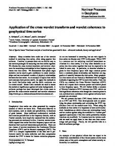
3 Proposed Enhancement Framework This paper proposes an improved framework for enhancing the contrast of cerebral MRI features by incorporating enhancement approaches of both the frequency and spatial domain. Multiscale analysis techniques are employed, which decomposes an image signal using wavelets followed by enhancement and de-noising of only those sub images which contain the necessary information. Morphological filter is then applied to the reconstructed image to further enhance the features along with filtering of residual noise. The block diagram of the proposed framework for enhancement of cerebral MRI features is shown in fig.1. 3.1 Wavelet Based Sub-band Coding In wavelet transform, an image is represented at different resolution levels. It decomposes any image into low frequency approximation coefficients and high frequency detail coefficients, viz. horizontal, vertical and diagonal coefficients, at any resolution. Discrete Wavelet Transform (DWT) transforms the image into an array of wavelet coefficients. These coefficients are then modified as per the requirement and the image is reconstructed from these modified coefficients by using Inverse Discrete Wavelet Transform (IDWT). In the proposed method, a two-level decomposition of the input signal is performed using daubechies wavelet family. In the higher frequency spectrum, with the increase in the information content, noise also increases. Therefore, de-noising is performed by doing soft thresholding of the high frequency coefficients. Soft Thresholding [5] can be mathematically defined as: t Th ; t Th . f (t ) t Th ; t Th 0; t T h
(6)
MRI Database
Pre-processing
Decomposition using Discrete Wavelet Transform (DWT)
Low frequency Components
High frequency Components
(Approximation Coefficients)
(Detail Coefficients)
Tuning of function parameters Non-Linear Enhancement Operator
Soft Thresholding
Reconstruction using Inverse Discrete Wavelet Transform (IDWT)
Reconstructed Image [I]
-
Opening
Closing
3x3 structuring element
3x3 structuring element
+
Image [I]
1st Iteration of proposed morphological filter
Image [II]
-
Opening
Closing
5x5 structuring element
Image [II]
5x5 structuring element
+
2nd Iteration of proposed morphological filter
Final Enhanced Image
Fig. 1. Block diagram of the proposed framework for contrast enhancement of Cerebral MRI Features
As in different high frequency sub-images, the noise properties are different, different threshold levels are used for each sub-image. The threshold Th is defined as:
Th 2log Ni .
(7)
where: σ is the noise standard deviation and Ni is the size of sub-image. After accessing the coefficients at the fine level, IDWT is applied for reconstructing the image back into the spatial domain. The non-linear enhancement operator is then applied to the approximation coefficients.
3.2 Non Linear Enhancement Operator The fundamental problem posed in the enhancement of brain tumor images is the inability to emphasize the desired features without the enhancement of noise. This drawback is overcome by the global enhancement techniques equipped with multistate Adaptive Gain [12]. A logistic function is real-valued, differentiable, and monotonically increasing function given by:
logistic( x)
1 . 1 e- x
(8)
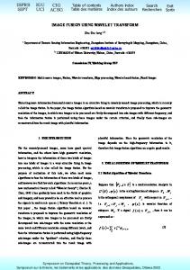
This is also the requirement of the non-linear transformation function for conventional histogram equalization. The graphical variation for logistic(x) is shown in fig. 2(a). Linear combination of logistic functions with an adaptive gain factor is used for the preparation of non-linear mask for contrast enhancement of cerebral MRI features. This mask is spatially moved over the ROI to produce the enhanced image. The function modifies the gray levels by suppression of pixel values of very small amplitude, and enhancement of only those pixels larger than a certain threshold within each level of the transform space. The non-linear enhancement operator [13] (as shown in fig. 2(b)) used to accomplish the above operation is given by:
y x a[logistic{k ( x b)} logistic{ k( x b)}] .
(9)
where: x denotes the gray level value of the original ROI at co-ordinate (i, j). k and b are the parameters for control of enhancement and the threshold respectively and a is given by: a
1 . logistic{k (1 b)} logistic{k 1 b }
where: the parameters b Є R while k Є N.
(10)
The enhancement operator y(x) is differentiable as well as continuous, hence it is monotonically increasing within the interval [-1, 1]. This further ensures that the enhancement using y(x) does not include any discontinuities into the enhanced image. In this approach, the threshold parameter b controls the noise level of the ROI during the process of enhancement. All the pixel intensity values above the threshold level are enhanced while the ones below it are suppressed. This threshold level can be calculated by solving the equation y(x)-x=0 or by finding the standard deviation of pixel values.
(a)
(b)
Fig. 2 (a). Graphical variation of Logistic Function [logistic(x)], (b) Graphical variation of Non-Linear Enhancement operator y(x).
However, with the above function (9), the threshold is controlled through the parameter b (where 0