Further, the cell lines' susceptibility to cell disruption methods was investigated by employing three different animal cell lines (HeLa. HH, Vero, and HeLa S3 ...
BIOTECHNOLOGY
TECHNIQUES
Volume 8 No.6 (June 1994) pp.425-430 Received 20th April
COMPARISON OF CELL DISRUPTION METHODS FOR DETERMINING P-GALACTOSIDASE ACTIVITY EXPRESSED IN ANIMAL CELLS Jung Hyun Shin, Gyun Min Lee*, and Jung Hoe Kim
Departmentof Biotechnology, Korea AdvancedInstitute of Scienceand Technology 373-1, Kusong-Dong, Yusong-Gu, Taejon 30.5-701,Korea ABSTRACT To determine P-galactosidase activity expressed in animal cells, the cells are often disrupted. In this study, four different cell disruption methods (freezing and thawing, sonication, homogenization, and lysis buffer) were compared with regard to their efficiency in the determination of p-galactosidase activity. Further, the cell lines’ susceptibility to cell disruption methods was investigated by employing three different animal cell lines (HeLa HH, Vero, and HeLa S3 cells) infected by recombinant vaccinia virus (vSC8) expressing fi-galactosidase gene. Regardless of cell lines used, three different cell disruption methods except homogenization did not show any significant difference in the final p-galactosidase activity recovered from the cells. Homogenization was inefficient, and required up to 300 strokes to recover /3-galactosidaseactivity fully from HeLa S3 cells. The use of lysis buffer was recommendable becauseof its convenience. However, if there was no need for an immediate assay, it was convenient to keep cell samples collected during the culture frozen at -2OOCuntil the assay. Since thawing of the cells was enough to recover the P-galactosidase, further treatment on the frozen cells after thawing was unnecessary. INTRODUCTION
P-Galactosidaseis one of the most widely studied glycosidasesbecauseof its universal occurrencecoupled with the simple enzyme assay and the availability of numerous substrates(Wallenfels, 1962, Miao et al., 1993). In a number of genetic studies using recombinant DNA technology, p-galactosidasehasbeen widely used as an internal reference of gene, marker enzyme of fusion protein or chimeric receptor (Miao et al., 1993a; Gu et al., 1993; King et at., 1992; Utsumi et al., 1989; Tsui et al., 1988; Yokota et al., 1986, Casadabanet al., 1983). The introduction of o-nitrophenyl P-D-galactoside (ONPG) as a substratefor a sensitive assayby Pardeeet al. (1959) expedited the general choice of P-galactosidasefor genetic studies (Shimizu et al., 1987). Since p-galactosidaseis an intracellular enzyme, cell disruption is often required to determine its activity in host cells. Although cell disruption methods to reduce the loss of enzyme activity have been extensively studied in bacteria and fungi (Miller, 1992;Foster, 1992; Szoke et al., 1988; Gunsalus, 1955), limited information is available for animal cell disruption. Animal cells, which are enclosed solely by plasma membrane and relatively big, are easier to be disrupted than bacteria and fungi (Dignam, 1990; Salusbury, 1989). The susceptibility of cells should be considered when choosing a method of cell disruption. Further, one should pay attention to conditions used in cell disruption which may alter the activity or native structure of /3-galactosidase. This nonspecific inactivation can result in poor reproducibility which makes interpretation of such studies difficult. Accordingly, the cell disruption methods for bacteria and fungi need to be modified for animal cells to ensurea consistent P-galactosidase aSSay. In this study, we employ four different cell disruption methodsfrequently used in the laboratory (freezing and thawing, sonication, homogenization,and lysis buffer) in determining P-galactosidaseactivity in animal cells infected by recombinant vaccinia virus (vSC8) carrying P-galactosidasegene(Chakrabarti et al., 1985), and thereby, seek to find the efficient animal cell disruption method for a consistent /3galactosidaseassay.
425
Cell lines, media, and culture maintenance Two anchorage-dependent and one anchorageindependent cell lines were used in this study. One anchorage-dependentcell line was human cervical carcinoma (HeLa) HH cell provided by Dr. J. Choi at KAIST. The other anchoragedependentcell line was At&au green monkey kidney (Vero) cell obtained from American Type Culture Collection (ATCC, Rockville, MD). HeLa S3 cell adapted to grow in suspension was provided by Dr. B. Ann at Korea University. The HeLa HH cell was cultivated in Dulbecco’s modified Eagle’s medium (DMEM, Gibco Laboratories, Grand Island, NY) supplementedwith 10% fetal bovine serum (FBS, Gibco Laboratories). The Vero cell was cultivated in DMEM supplementedwith 10% horse serum (Gibco Laboratories). The HeLa S3 cell was cultivated in DMEM/F12 (Gibco Laboratories) supplemented with 10% FBS. No antibiotics were usedin the media. All cell lines used were grown as monolayers in polystyrene tissue culture flasks and were incubated at 37OCin a humidified, 5% CO2 incubator (Forma Scientific, Marietta, OH). The cells were transferredevery 3 or 4 days. Virus strain Both original vaccinia virus (strain WR) and recombinant vaccinia virus (vSC8) containing b-gal inserted into TK locus (Chakrabarti et al., 1985) were kindly provided by Dr. B. Ann (Korea University). Vaccinia virus titers were determinedfrom plaque assayson Vero cells as describedpreviously (Burleson et al., 1992). Cell culture, virus infection, and virus recovery For HeLa HH and Vero cells, cell cultures were carried out in T-25 cm2 flasks. Two extra cultures were set up to estimate cell concentrations for determining the timing of viral infection. When the cells were at the exponential phase of growth, the medium was completely removed from the flask. After washing the cells twice with Dulbecco’sphosphatebuffered saline A (PBSA Ca2+- and Mg2+-free), the cells were infected with 1 ml of vaccinia virus solution, adjusted for a multiplicity of infection (MOI) of 5. The viral infection was carried out by incubating the culture at 37OCin a CO2 incubator. After oue hour incubation, 8 ml of fresh medium was added to the culture. After another 36 h incubation, the medium was removed from the flask. The infected cells were recoveredby trypsiuization, and suspendedin a fresh medium,and then chilled on ice. For HeLa S3 cells, cell cultures were carried out in spinner flasks containing 50 ml of medium. HeLa S3 cells which had been maintained as a monolayer were used as an inoculum and did not require adaptation to grow in suspensionin spinner flasks. HeLa S3 cells in the exponential growth phase were harvestedfrom the spinner flask and centrifuged at 1000rpm for 10 min. After discarding the supernatant, the cells were resuspendedin 10 ml of fresh medium, and transferred to the spinner flask. The cells were infected with 1 ml of vaccinia virus suspension,adjustedfor a MO1 of 5. The infection was carried out for an hour in the spinner flask with intermittent stirring which was placed in a CO2 incubator. After one hour incubation, the culture volume was brought up to 50 ml by adding a fresh medium. The further procedureof virus recovery for HeLa S3 cells was identical to that of HeLa HH cells. Cell disruption methods Four different cell disruption methodswere applied for disrupting the infected cells. Freezing and thawina; One cycle of freezing and thawing consistsof freezing the cells in a ethanol/dry ice bath,. followed by thawing the cells in a water bath at 37oC. One cycle takesapproximately 7 minutes. . o catron; The frequency of 2OkHzwas applied to 1 ml of cell suspensionin a sonicator with a small tip (Mzel W-385, Heat Systems-Ultrasonics,New York). One secondpulse cycling was used to minimize the inactivation of 8-galactosidase. Homogenization: The cells were disrupted by gentle strokes with a tight-fitting pestle in a hand-held Wheaton Dounce homogenizer (size; lml, Wheaton,NJ). The clearancebetween mortar and pestle was 44 pm to 69 pm. Lvsis buffet After adding 15 ltl chloroform and 15 ttl 0.1% sodium dodecyl sulfate (SDS) solution to 1 ml of cell suspension,the cell suspensionwas agitatedvigorously for 10 seconds(Miller, 1992). Analytical methods Cell concentration was determined by counting cells with a hemocytometer under an inverted microscope. Viability was determined by trypan blue dye-exclusion method. After applying various cell disruption methodsto the cells, the degreeof cell disruption was observedmicroscopically. The activity of B-galactosidasewas determinedon the basisof the rate of cleavageof o-nitrophenyl B-D-galactosidase(ONPG) as describedpreviously (Sambrooket al., 1989). The optical density (OD) of reactions was simultaneously measuredat 420 and 540 nm. The following equation was used to calculate the P-galactosidaseactivity in International Units (IU) per milliliter using an extinction coefficient of
426
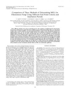
4.5 ml/pm01 for o-nitrophenol (Wickham et al., 1992) and a correction factor of 1.1. The correction factor defined as a OD420/OD540 was usedto minimize the interferenceof light scatteringby cell debris and was determinedexperimentally (refer to Fig. 1). ITJ/ml=:3D420 - l.lxOD~O)(dilution factor)/(4Sml/pmol)(min of incubation) RESULTS
AND DISCUSSION
Correction
for interference by cell debris in determining
P-galactosidase activity
Before
various cell disruption methodsam comparedwith regard to their efficiency in recovery of P-galactosidase, the proper method for the determination of fi-galactosidaseactivity needsto be established.The activity of P-galactosidase is often determined on the basis of the rate of cleavage of ONPG by measuring me absorbance at 420 nm (King er af., 1992, Shimizu ec al., 1987; Wickham et a!., 1992). However, the reading at 420 nm (OD420) is actually a combination of absorbanceby o-nitrophenol and light scattering by the cell debris. Contribution from light scatteringonly by cell debris to OD420, which was determined by measuring the OD420 of the lysate of the cells infected with wild type virus, was more thau 10% (dam not shown). Accordingly, the light scattering by cell debris should be minimized for an accurate quantification of p-galactosidase.The interference by the cell debris was not eliminated by centrifugation even at 13,000rpm for 4 minutes (data not shown). Alternatively, the light scatteringby cell debris at 420 nm can be corrected for by obtaining the absorbanceat 540 nm as long as the ratio of OD420 to OD540 is constant. The light scattering at 540 nm is causedmainly by cell debris. Figure 1 shows that the correction factor can be used for correcting the light scattering by
1.5
cell debris at 420 mn. For HeLa HH cells, the 1.2 -....--..f..r.... ......*
correction factor was approximately 1.14 ti.04 (standard deviation), which is slightly
0 g 0.9 -
lower than that (=1.75) of E. coli
%
(Miller,
.
...._.d...& ......-.......-I?_____, .
q 0.6 -
1992). The correction factors of Vero cells and
8
HeLa S3 cells, which were determined in the
0.3
same manner, were 1.05 kO.03 and 1.10 kO.05, respectively. Using these correction 0
factors, a true absorbanceof o-nitrotrophenol
2x10³
4x10³
6x10'
8x10'
1x10⁶
Cells/ml
was estimated as described in Materials and methods. Figure 1. Estimation of a correction factor of HeLa HH cells. The dotted line represents an average value of a cmrection factor.
Effect of cell disruption
methods on recovery of P-galactosidase activity
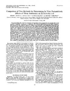
The intracellular
p-galactosidaseexpressedin various cell lines was recoveredusing different cell disruption methods. Figure 2 shows that the intracellular P-galactosidaseexpressedin HeLa HH cells was recovered using various cell disruption methods.When the P-galactosidasewas releasedfrom the cells by freezing and thawing, one cycle of freezing and thawing was enough to recover the P-galactosidase (Fig. 2(A)).
427
The enzyme activity recoveredper 106 cells significantly increasedfrom 0.35 to 1.55 1~/106 cells after one cycle of freezing and thawing. The initial enzyme activity indicates that of the intact cells. When the pgalactosidasewas releasedfrom the cells by sonication, lo-20 secondsof sonication was enough to release the fi-galactosidase(Fig. 2(B)). Sonication longer than 70 secondsgradually deactivatedp-galactosidase.No viable cells were observed microscopically after ca. 40 second-sonication.When the /3-galactosidasewas releasedfrom the cells by homogenization, the recovery of p-galactosidasewas very inefficient (Fig. 2(C)). Although the recovery of p-galactosidaseincreasedby repeatedstrokesin a homogenizer,the strokesover 100 times was not enough to fully recover the P-galactosidase.Microscopic observation showed that a significant portion of the cells were intact even after 100 strokes.When the p-galactosidasewas released from the cells using a lysis buffer, the enzyme activity recovered was 1.35 III/lo6 cells which was comparableto that obtainedby either freezing and thawing or sonic&ion.
0
Time
Number
(SW)
of Stroke
Time
Number
Isal
of Stroke
* Numbe:
lime
Number
of Cycle6
lsec)
of Stroke
Figure 2. Effect of various cell disruption methods on recovery of fi-galactosidaseexpressed in HeLa HH cells. Figure 3. Effect of various cell disruption methods on recovery of P-galactosidaseexpressed in Vero cells. Figure 4. Effect of various cell disruption methods on recovery of p-galactosidase expressed in HeLa S3 cells. (A) freezing and thawing, (B) sonication, (C) homogenization.
428
6
Figure 3 shows that the intracellular p-galactosidaseexpressedin Vero cells was recovered using various cell disruption methods.The effect of various cell disruptiou methodson recovery of p-galactosidase from Vero cells was similar to that from HeLa HH cells. The homogenization was most inefficient in disrupting the Vero cells. Even after 160 strokes, the enzyme activity recovered was 0.02 III/lo6 cells which was still significantly lower than that obtained by other cell disruption methodsused. When the pgalactosidase was released from the cells using a lysis buffer, the enzyme activity recovered was 0.03 III/lo6 cells. Figure 4 shows that the intracellular p-galactosidaseexpressedin HeLa S3 cells was recovered using various cell disruption methods.Since HeLa HH and Vero cells am anchorage-dependent cells, trypsin treatment was necessaryto recover the cells from the T-flasks. The trypsin treatment may influence the cells’ susceptibility to cell disruption methods. This hypothesis was tested by employing HeLa S3 cells which grow in suspension.As shown in. Fig.4, the effect of various cell disruption methodson recovery of p-galactosidase from HeLa S3 cells was similar to that from other cell lines used. Accordingly, trypsin treatment doesnot appearto influence the cells susceptibility to cell disruption methods.In addition, it was found that most of p-galactosidaseactivity could be also recoveredby homogenization. Strokesup to 300 were ueeded,though. Although no significant difference in the cells’ susceptibility to cell disruption methods except homogenization was observedamong the cell lines used, the enzyme activity recoveredper cell was very different depending on the cell lines used.The P-galactosidaseactivity recoveredper cell in HeLa HH cells was 2-5 times higher than that in other cell lines used (refer to Figures 2, 3, and 4). This difference in the enzyme activity recoveredper cell are probably due to the difference in infectivity and/or expression level among the cell lines used. Since the biochemical assay using ONPG yields an average value of Bgalactosidaseactivity in the whole cell population, cell to cell heterogeneitywas unnoticed. Accordingly, the infectivity and the expressionlevel per infected cell could not be evaluated Storage stability of P-galactosidase Regardlessof cell lines used, one cycle of freezing and thawing was enough to recover the p-galactosidase.Accordingly, if the j/l-grdactosidaseactivity of frozen cells is preserved during the storage at -20°C, no further treatment on the frozen cells after thawing may be necessaryfor P-galactosidaseassay.When there is no immediateneedfor fi-galactosidaseassay,various cell samplescollected during the culture can be kept frozen at -2OOCfor later assay. The stability of P-galactosidaseof frozen cells at -2flOC was tested. As shown in Fig& the j3galactosidaseof frozen cells at -2OOCwas stableover a month, suggestingthat cell samplescollected during the culture can be kept frozen at -2OOCfor the later assay. In addition, since thawing of the cells was enough to recover the P-galactosidase,further treatmenton the frozen cells except thawing was unnecessary (data not shown). In conclusion, three different cell disruption methods,except homogenization, did not show any significant difference in the final P-galactosidaseactivity recovered from the cells. Homogenization was inefficient and required up to 300 strokesto recover P-galactosidaseactivity fully from HeLa S3 cells. Based on the results obtained here, we makethe following suggestionsfor a covenient /3-galactosidaseassayin the
429
laboratory. When the fi-galactosidase activity of the cells needs to be measured immediately during the
culture, the use of lysis buffer is recommendablebecauseof its convenience.Otherwise, it is convenient to keep cell samplescollected during the culture frozen at -20°C until the assay.When the assayis needed,the P-galactosidaseactivity of the frozen cells is measuredafter thawing. Since thawing of the cells is enough to recover the p-galactosidase,further treatmenton the frozen cells after thawiug is unnecessary. Acknowledgement The authors thank Dr. J. Choi and Dr. B. Ann for providing cell lines and virus. We also thank Mr. M.K. Kim for technical assistance.This work was supported by the KOSBF researchgrant to the BERC in KAIST.
.$’
.
‘~l.oOu
.
lv
l
.
l
. % 0.75
-
0.25
-
0 zi 2 0.50 5 ci z ‘G $
0.00 0
20
40
60
80
Time (day) Figure 5. Storage stability of p-galactosidase in frozen HeLa HH cells at -2OOC.The activity of & galactosidasein frozen cells was measured after thawing at 37%.
References Burleson, F. G., Chambers,T. M. and Wiedbrauk D. L. (1992). Virology: a Laboratory Manual. pp. 7284,ls.t ed. San Diego: Academic Press. Casadaban,M. J., Martinez-Arias, A., Shapira, S. K., and Chou, J. (1983). Methods Enzymol. 100,293308.
Chakrabarti, S., Brechling, K. and Moss, B. (1985). Mol. Cell. Biol. 5, 3403-3409. Dignam, J. D. (1990). Methods Enzymol. 182, 194-203. Foster, D. (1992). BiolTechnology 10, 1539-1541. Gu, M. B., Todd, P., and Kampala, D. S. (1993). Biotechnol. Bioeng. 42, 1113-1123. Guns&s, I. C. (1955). Methods Enzymol. 1, 51-62. King, G. A., Daugulis, A. J., Faulkner, P., and Goosen,M. F. A. (1992). Biotechnol. Prog. 8, 567-571. Miao, F., Todd, P., and Kompala, D. S. (1993). Biotechnol. Bioeng. 42, 708-715. Miao, F., Drake, S. K., and Kompala, D. S. (1993a).Biotechnol. Prog. ,Y, 153-159. Miller, J. H. (1992). A Short Course in Bacterial Genetics, pp. 71-80, Cold Spring Harbor: Cold Spring Harbor
Laboratory.
Pardee,A. B., Jacob, F. and Monod, J. (1959). Mol. Biol. 1, 165-178. Salusbury, T. (1989). Disruption. In: Protein Purification Methods, E. L. V. Harris and S. Angal, eds. pp. 87-97, Oxford: IRL Press. Sambrook,J., Fritsch, E. F. and Man&is, T. (1989). Molecular Cloning: a Laboratory Manual. 2nd ed. Cold Spring Harbor: Cold Spring Harbor Laboratory. Shimizu, N., Fukuzono, S., Nishimura N., Odawara,Y., and Fujiwara, K. (1987). J. Ferment. Technol. 65, 7-10.
Szoke, A., Campagna,R., Kroner, K. H. and Hustedt, H. (1988). Biotechnol. Biotechn. 2, 3540. Tsui, P., Helu, V. and Freundlich, M. (1988). J. Bacterial. 170, 4950-4953. Utsumi, R., Brissette, R.E., Rampersaud,A., Foist, S.A., Oosawa,K. and Inouye, M. (1989). Science 245, 1246-1249. Wallenfels, K. (1962). Methods Enzymol. ,5, 212-219. Wickham, T. I., Davis, T., Granados,R. R., Shuler, M. L. and Wood, H. A. (1992). Biotechnol. Prog. 8, 391-396. Yokota, H., Yokoo, K. and Nagata Y. (1986). Biochim. Biophys. Acta 868,4S-SO.
430