Am J Physiol Lung Cell Mol Physiol 305: L747–L755, 2013. First published September 13, 2013; doi:10.1152/ajplung.00023.2013.
Cycloheximide and lipopolysaccharide downregulate ␣ENaC mRNA via different mechanisms in alveolar epithelial cells Francis Migneault, Émilie Boncoeur, Frédéric Morneau, Mihai Pascariu, André Dagenais, and Yves Berthiaume Institut de recherches cliniques de Montréal, Centre de recherche, Centre hospitalier de l’Université de Montréal (CRCHUM) and Département de médecine, Université de Montréal, Montréal, Québec, Canada Submitted 22 January 2013; accepted in final form 9 September 2013
Migneault F, Boncoeur É, Morneau F, Pascariu M, Dagenais A, Berthiaume Y. Cycloheximide and lipopolysaccharide downregulate ␣ENaC mRNA via different mechanisms in alveolar epithelial cells. Am J Physiol Lung Cell Mol Physiol 305: L747–L755, 2013. First published September 13, 2013; doi:10.1152/ajplung.00023.2013.— Active Na⫹ transport mediated by epithelial Na⫹ channel (ENaC) is vital for fetal lung fluid reabsorption at birth and pulmonary edema resolution. Previously, we demonstrated that ␣ENaC expression and activity are downregulated in alveolar epithelial cells by cycloheximide (Chx) and Pseudomonas aeruginosa. The regulatory mechanisms of ␣ENaC mRNA expression by Chx and lipopolysaccharide (LPS) from P. aeruginosa were further studied in the present work. Both agents decreased ␣ENaC mRNA expression to 50% of control values after 4 h. Chx repressed ␣ENaC expression in a dose-dependent manner independently of protein synthesis. Although extracellular signal-regulated kinases 1 and 2 (ERK1/2) and p38 mitogenactivated protein kinase (MAPK) pathways were activated by the two treatments, their mechanisms of ENaC mRNA modulation were different. First, activation of the signaling pathways was sustained by Chx but only transiently by LPS. Second, ERK1/2 or p38 MAPK inhibition attenuated the effects of Chx on ␣ENaC mRNA, whereas suppression of both signaling pathways was necessary to alleviate the outcome of LPS on ␣ENaC mRNA. The molecular mechanisms involved in the decrease of ␣ENaC expression were investigated in both conditions. LPS, but not Chx, significantly reduced ␣ENaC promoter activity via the ERK1/2 and p38 MAPK pathways. These results suggest that LPS attenuates ␣ENaC mRNA expression via diminution of transcription, whereas Chx could trigger some posttranscriptional mechanisms. Although LPS and Chx downregulate ␣ENaC mRNA expression similarly and with similar signaling pathways, the mechanisms modulating ENaC expression are different depending on the nature of the cellular stress. epithelial sodium channel; inflammation; cellular stress; signal transduction; messenger ribonucleic acid
revealed by the lung edema susceptibility of mice in which ENaC expression and activity are decreased (16, 41). Multiple agents have been found to modulate ENaC activity and expression in AEC. Glucocorticoids and cAMP (9, 10, 21, 25) increase ENaC expression and activity, whereas factors involved in lung inflammation and lesions, such as interleukin (IL)-1 (45), IL-4 (19), tumor necrosis factor-␣ (TNF-␣) (10, 11), and transforming growth factor- (TGF-) (17), downregulate them. The complex control of ␣ENaC gene expression is mediated via transcriptional activity of its promoter (7, 9, 10, 36, 45) but could also entail posttranscriptional regulation of transcript stability (11). We reported previously that ␣ENaC expression (9, 12) and activity (5) are downregulated by cycloheximide (Chx) in AEC and in the lungs infected with Pseudomonas aeruginosa. Chx and lipopolysaccharide (LPS) from P. aeruginosa have been shown to inhibit ENaC mRNA expression in AEC (9, 38). Although it is a translation inhibitor, Chx is also known to induce cellular stress (37) and to modulate ENaC gene expression (9, 26). LPS, a glycolipid that constitutes a major portion of the outermost membrane of gramnegative bacteria, is a potent proinflammatory molecule that can evoke lung inflammation. Although LPS has been reported to decrease ENaC activity and expression in AEC (5, 14), little is known about its signal transduction pathways and mechanisms. In the present work, we investigated the hypothesis that Chx, an inducer of cellular stress, and LPS could modulate ␣ENaC mRNA expression in AEC isolated from rat lungs. The signaling pathways and mechanisms associated with ␣ENaC mRNA regulation were studied under these two conditions. We observed that Chx and LPS downregulated ␣ENaC mRNA expression in the same way after activation of the same signaling pathways. However, ␣ENaC mRNA modulation occurred via different mechanisms, depending on the cell stress induced. MATERIALS AND METHODS
vital for fluid movement across the alveolar epithelium in the lungs to reabsorb fetal lung fluid at birth (40) and for pulmonary edema resolution (4, 34). Two essential components of this active transport system are amiloride-sensitive epithelial Na⫹ channel (ENaC) at the apical surface of alveolar epithelial cells (AEC) and Na⫹-K⫹-ATPase at the basolateral surface (4, 34). To date, four homologous ENaC subunits ␣, , ␥, and ␦, have been identified in humans (2, 6, 33). The importance of ␣ENaC function has been demonstrated in the ␣ENaC knockout mouse model. These mice develop respiratory distress and die shortly after birth because of their inability to clear fluid from alveolar spaces (24). The crucial role of ENaC in alveolar liquid clearance is further ACTIVE Na⫹ TRANSPORT IS
Address for reprint requests and other correspondence: Y. Berthiaume, IRCM, 110 Ave. des Pins Ouest, Montreal (Quebec) Canada H2W 1R7 (e-mail:
[email protected]). http://www.ajplung.org
Materials. Minimum essential medium (MEM) and FBS were purchased from Life Technologies (Burlington, ON). Porcine pancreatic elastase was obtained from Worthington Biochemical (Lakewood, NJ). LPS from P. aeruginosa (serotype 10, strain ATCC 27316), Chx, and emetine were procured from Sigma-Aldrich (Oakville, ON). Primary antibodies against extracellular signal-regulated kinases 1 and 2 (ERK1/ 2), phosphorylated-ERK (p-ERK), p38 mitogen-activated protein kinase (MAPK), and phosphorylated-p38 (p-p38) were sourced from Cell Signaling Technology (Beverly, MA), and MAPK inhibitors (PD-98059, SB-203580) were from Calbiochem (Gibbstown, NJ). PD-184352 came from LC Laboratories (Woburn, MA), and Birb-796 was from Selleck Chemicals (Houston, TX). AEC isolation. AEC were isolated from male Sprague-Dawley rats, as described previously (9) and according to a procedure approved by the institutional animal care committee of Institut de recherches cliniques de Montréal in accordance with the Canadian Council of Animal Care
1040-0605/13 Copyright © 2013 the American Physiological Society
L747
L748
CYCLOHEXIMIDE AND LIPOPOLYSACCHARIDE DECREASE ␣ENaC mRNA EXPRESSION IN AEC
standards. Perfused lungs were digested with elastase, and cells were purified by a differential adherence technique on bacteriological plastic plates coated with rat immunoglobulin G (IgG). They were maintained in MEM containing 10% FBS, 0.08 mg/l gentamicin, 0.2% NaHCO3, 0.01 M HEPES, and 2 mM L-glutamine. They were then plated at 1 ⫻ 106 cells/cm2 densities in plastic dishes and cultured at 37°C with 5% CO2 in a humidified incubator. The medium was supplemented with Septra (3 g/ml trimethoprim and 17 g/ml sulfamethoxazole) for the first 3 days. After this time period, the medium was replaced, and the cells were cultured without Septra. Semiquantitative reverse transcription-polymerase chain reaction. Total RNA from 24-mm wells was purified with TRIzol reagent according to the manufacturer’s instructions (Life Technologies). Total RNA (3 g) was reverse-transcribed to cDNA with MMLV-RT (Life Technologies) in the presence of oligo(dT) primers (Roche Diagnostics, Laval, QC). cDNA was amplified with Taq polymerase (Life Technologies), using specific primers designed for rat ␣ENaC or -actin. The PCR primers for ␣ENaC amplification generated a 501-bp amplicon from exon 5 to 11 (forward: 5=-GAG CCT GCC TTT ATG GAT GA-3=, and reverse: 5=-GAG CTT TGC AAC TCC GTT TC-3=; 1 M final concentration of each), whereas -actin primers amplified a 349-bp amplicon from exon 3 to 4 (forward: 5=-CTA AGG CCA ACC GTG AAA AG-3=, and reverse: 5=-GCC ATC TCT TGC TCG AAG TC-3=; 0.25 M final concentration of each). Semiquantitative reverse transcription-polymerase chain reaction (RT-PCR) amplification was undertaken according to a well-established laboratory protocol (10). To remain in the linear phase of amplification, the ␣ENaC product was amplified for 19 PCR cycles, whereas -actin amplification was stopped after 15 cycles. The amplification products were separated on agarose gels, stained with SyBr Safe (Life Technologies), and analyzed by a Typhoon Gel Imager. For the semiquantitative evaluation of ␣ENaC cDNA in rat AEC, the signal was normalized to -actin amplification of the same cDNA sample. RT-quantitative PCR. Total RNA was isolated from 24-mm wells by directly lysing the cells with TRIzol Reagent according to the manufacturer’s protocol (Life Technologies). ␣ENaC mRNA expression was estimated by quantitative PCR (qPCR). Total RNA (1 g) was reverse transcribed to cDNA with MMLV-RT (Life Technologies) in the presence of random primers (Roche Diagnostics). For qPCR amplification, 15 ng of cDNA were amplified with forward and reverse primers at 225 nM each with Platinum SYBR Green qPCR surperMix-UDG (Life Technologies) in a final volume of 15 l. For ␣ENaC, forward 5=-CGT CAC TGT CTG CAC CCT TA-3 and reverse 5=-CCT GGC GAG TGT AGG AAG AG-3 primers amplified a 128-bp amplicon between exon 1 and 2 of the rat Scnn1a gene (between nt 830 and 957 of NM_031548). The -actin signal was used for normalization. Forward 5=-ACC GTG AAA AGA TGA CCC AGA T-3= and reverse 5=-CAC AGC CTG GAT GGC TAC GT-3 primers amplified a 78-bp amplicon between exon 3 and 4 of Actb gene (between nt 421 and 498 of NM_031144.2). PCRs were amplified for 40 cycles in a Rotor-Gene Q 5 Plex thermal cycler (Qiagen, Toronto, ON). After 10 min incubation at 95°C to activate Taq polymerase, the samples were amplified for 40 cycles with a 10-s denaturation step at 95°C, a 15-s annealing step at 58°C, and a 20-s elongation step at 72°C. A high-resolution melting curve was generated at the end of amplification to assess the nature of the amplicon. Standard cDNA was run in parallel to the samples tested in duplicate to normalize the amplification signal from run to run. For quantitative analysis, the fluorescence signal of dsDNA was converted to gene copy number, calculated from the dilution curve of a standard cDNA from untreated cells. The expression of a given signal was normalized for the fluorescence of -actin and reported as percentage of expression compared with untreated cells from the same animal. Immunoblotting. AEC treated or not with 1.0 M Chx or 15 g/ml LPS were washed two times with PBS and lysed for 15 min under agitation at 4°C in lysis buffer (1% Triton X-100, 150 mM NaCl, 5 mM EDTA, and 50 mM Tris, pH 7.5) supplemented with protease inhibitor and phosphatase inhibitor cocktails (Life Technologies). The cells were subsequently scraped with a rubber policeman, collected, and centrifuged
at 12,000 g for 10 min. Protein concentration of the supernatants was evaluated with the Bradford method (Bio-Rad Laboratories, Mississauga, ON). Total proteins (50 g) were solubilized in sample buffer (62.5 mM Tris·HCl, pH 6.8, 2% SDS, 0.2% bromophenol blue, 10% glycerol, and 7.7% -mercaptoethanol), subjected to SDS-polyacrylamide gel electrophoresis, and transferred electrophoretically onto polyvinylidene difluoride membranes. The membranes were blocked with 5% dried fat-free milk {or 5% wt/vol BSA in Tris-buffered saline pH 7.4 with 0.05% Tween 20 (TBST) [or PBS with 0.05% Tween 20 (PBST)]} for 1 h at room temperature and then incubated overnight at 4°C with primary antibody [anti-ERK1/2 (1:1,000 42- and 44-kDa proteins; Cell Signaling Technology), anti-p-ERK1/2 (1:1,000; Cell Signaling Technology), antip38 (1:1,000, 38 kDa; Cell Signaling Technology), or anti-p-p38 (1: 1,000; Cell Signaling Technology)] in TBST (or PBST) plus 5% milk or 5% BSA. After being washed with TBST, the membranes were incubated with goat anti-rabbit (for p-ERK1/2, p38, and p-p38, 1:4,000; Santa Cruz Biotechnologies, Santa Cruz, CA) or goat anti-mouse (for ERK1/2, 1:4,000; ThermoFisher Scientific, Ottawa, ON) IgG linked to horseradish peroxidase for 1 h. After TBST washes, the membranes were incubated with ECL⫹ (GE Healthcare Life Sciences, Baie d’Urfé, QC) for 5 min before the luminescent signals were recorded in a ChemiDoc XRS⫹ system (Bio-Rad Laboratories). The intensity of each specific band was quantified with Image Lab software. Transient transfection of AEC and luciferase assay. The modulation of ␣ENaC promoter activity and the implication of the 3=-untranslated region (3=-UTR) by Chx or LPS was tested in AEC. For the promoter activity, cells were transiently transfected with a 2.9-kb BamHI-MscI fragment of the mouse ␣ENaC promoter (9 –11) (␣ENaC-Luc) cloned upstream of the firefly luciferase (Luc) reporter gene. For the 3=-UTR, cells were transiently transfected with a pLENTI vector containing an 894-bp fragment (Luc-3=-UTR complete) or a 374-bp fragment (Luc-3=UTR deletion) of the rat 3=-UTR ␣ENaC cloned downstream of the firefly Luc reporter gene. pRL-SV40 (Promega, Madison, WI), a plasmid expressing Renilla reniformis luciferase (RL), was cotransfected for normalization of the Luc response. Over the years, several approaches were tested, including liposomes and viral vectors, for transfection of the difficult to transfect AEC in primary culture. The NEON electroporation transfection system was the only technique that could achieve a 25–30% transfection efficiency as monitored by recording GFP-positive clones. On day 1, 14 ⫻ 106 cells were plated in 100-mm dishes after AEC isolation. On day 2, the cells were trypsinized, washed with PBS, and resuspended in Resuspension Buffer R. In each well, 400,000 cells were mixed with 2 g ␣ENaC-Luc and 0.4 g pRL-SV40 for transfection with the NEON Transfection System (Life Technologies) and cultured without antibiotics in MEM ⫹ 10% FBS. On day 4, the cells were rinsed, and fresh medium (MEM ⫹ antibiotics) was added. Chx or LPS was included for 24-h treatment after prior exposure (or not) to the MAPK inhibitors (PD-98059 or SB-203580). On day 5, Chx or LPS was added to untreated cells for 6-h treatment. Firefly and RL assays were undertaken with the Dual-Luciferase Reporter Assay System, as specified by the manufacturer (Promega). Luminometry measurements were undertaken in an EnVision Multilabel reader (PerkinElmer, Montreal, QC). Statistical analysis. All data are presented as means ⫾ SE. Groups were compared by the Mann-Whitney test, one-phase decay nonlinear regression, ANOVA, the Kruskal-Wallis test, and post hoc Bonferroni and Dunn’s analysis with GraphPad Prism 5 software (GraphPad software, San Diego, CA). P ⬍ 0.05 probability was considered to be significant (P values and statistical tests are reported for each experiment.). RESULTS
Chx and LPS decrease ␣ENaC mRNA expression time dependently. AEC were treated from 15 min to 24 h with 1.0 M Chx or 15 g/ml LPS. Both treatments progressively reduced steady-state mRNA levels of the ␣ENaC subunit. The mRNA abundance following Chx treatment (1.0 M) presented a significant time-dependent variation with a 50% de-
AJP-Lung Cell Mol Physiol • doi:10.1152/ajplung.00023.2013 • www.ajplung.org
CYCLOHEXIMIDE AND LIPOPOLYSACCHARIDE DECREASE ␣ENaC mRNA EXPRESSION IN AEC
A
Chx 1.0 µM Ctrl
15’
30’
1hr
2hrs
4hrs
A
CHX 6hrs Ctrl 0.1 µM 0.5 µM 1.0 µM 5.0 µM 8.8 µM
αENaC mRNA
β-actin mRNA
B
αENaC mRNA expression (% of control)
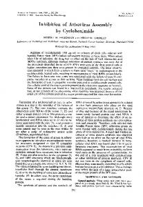
crease after 4 h (Fig. 1A). The changes in mRNA abundance by LPS treatment (15 g/ml) were very similar, with a significant time-dependent correlation and a 50% diminution after 4 h (Fig. 1B). In the presence of LPS or Chx, ␣ENaC mRNA levels reached a plateau (⬃35% of untreated cells) after 6 h. Lactate dehydrogenase release and caspase-3/7 activity were measured to evaluate if the downregulation of ␣ENaC mRNA expression by Chx and LPS was caused by cell death. No apoptosis or necrosis of AEC cells was detected (4 or 6 h) at the Chx and LPS concentrations tested in Fig. 1 (data not shown). Chx decreases ␣ENaC mRNA expression in a dose-dependent manner. To investigate the effect on ␣ENaC mRNA expression, AEC were treated for 6 h with different concentrations of Chx (0.1– 8.8 M). As reported previously (9), 5.0 and 8.8 M Chx reduced ␣ENaC mRNA expression to ⬃30% of untreated cell values at 6 h (Fig. 2). Chx concentrations (0.1, 0.5, and 1.0 M), reported to not inhibit protein synthesis in
100
6hrs 24hrs
80
*
60
*
40
*
75
*
50
*
*
*
0
Ctrl
0.1 µM 0.5 µM 1.0 µM 5.0 µM 8.8 µM
CHX 6hrs
25 0
0
2
4
6 Hours
8 12
24
LPS 15 µg/mL Ctrl
15’
30’
1hr
2hrs
4hrs
6hrs 24hrs
αENaC mRNA β-actin mRNA αENaC mRNA expression (% of control)
*
Fig. 2. Dose-dependent modulation of ␣ENaC mRNA expression by Chx in AEC. AEC were treated with 0.1– 8.8 M Chx for 6 h. A: representative agarose gel showing a dose-dependent decrease of ␣ENaC mRNA expression after RT-PCR. B: the percentages of ␣ENaC mRNA expression determined by RT-qPCR are depicted for each concentration. The data are expressed as percentages ⫾ SE compared with untreated cells after normalization with -actin. Chx significantly decreased ␣ENaC mRNA expression at each concentration. *P ⬍ 0.05 by the Kruskal-Wallis test and Dunn’s post hoc test; n ⱖ 3 for each experimental condition.
100
*
75
*
50
*
*
25 0
0
2
4
6 Hours ⫹
8 12
24
Fig. 1. Modulation of epithelial Na channel (␣ENaC) mRNA expression by cycloheximide (Chx) and lipopolysaccharide (LPS) in alveolar epithelial cells (AEC). AEC were treated for 15 min to 24 h with 1.0 M Chx (A) or 15 g/ml LPS (B). The time-dependent decrease of ␣ENaC mRNA expression determined by reverse transcription (RT)-quantitative PCR (qPCR) is shown along with representative agarose gel from classical RT-polymerase chain reaction (RT-PCR) after Chx (A) and LPS (B) treatment. ␣ENaC mRNA level decreased significantly in a time-dependent manner: R ⫽ 0.8414 for Chx (A) and R ⫽ 0.7779 for LPS (B). The data are expressed as percentages of the ␣ENaC signal compared with untreated cells ⫾ SE after normalization with -actin. *P ⬍ 0.0001 by the Kruskal-Wallis test. Time-dependent correlation was determined by 1-phase decay nonlinear regression. Cells from at least 3 different animals (n ⱖ 3) were studied in each experimental condition.
collecting duct epithelial cells (26), also decreased ␣ENaC mRNA expression (P ⬍ 0.05, Fig. 2). Emetine does not reduce ␣ENaC mRNA expression. To assess the importance of de novo protein synthesis on ␣ENaC mRNA expression, AEC were treated with 20 M emetine, a well-known translation inhibitor, which was unable to suppress ␣ENaC mRNA expression within 6 h (Fig. 3). Chx and LPS activate ERK1/2 and p38 MAPK signaling pathways. Because Chx and LPS decreased ␣ENaC mRNA levels, the intracellular signaling pathways involved were inαENaC mRNA expression (% of control)
αENaC mRNA expression (% of control)
B
100
*
20
αENaC mRNA β-actin mRNA
L749
150 125 100 75 50 25 0
Ctrl
2h
3h 4h Time
5h
6h
Fig. 3. Modulation of ␣ENaC mRNA expression by emetine in AEC. AEC were treated with 20 M emetine, an inhibitor of protein synthesis, for 2– 6 h. ␣ENaC mRNA expression was quantified by RT-qPCR and expressed as percentages ⫾ SE of untreated cells after normalization with -actin. Emetine did not inhibit ␣ENaC mRNA expression (1-way ANOVA and Bonferroni’s post hoc test). Cells from at least 5 different rats (n ⱖ 5) were studied in each experimental condition.
AJP-Lung Cell Mol Physiol • doi:10.1152/ajplung.00023.2013 • www.ajplung.org
CYCLOHEXIMIDE AND LIPOPOLYSACCHARIDE DECREASE ␣ENaC mRNA EXPRESSION IN AEC
L750
vestigated. Chx and LPS are known to modulate ERK1/2 and p38 MAPK, two pathways that regulate ENaC expression (1, 17, 26, 28, 32, 39, 45, 51). The levels and activation of these MAPKs were examined by Western blotting after different times (5 min to 24 h) of treatment. Cells exposed to 1.0 M Chx showed a sustained increase of activated p-ERK1/2 with 15 min to 24 h of treatment (Fig. 4A). p38 MAPK was also significantly triggered within 5 min of treatment with peak activation at 15–30 min, followed by lower, sustained stimulation detected until 24 h (Fig. 4B). Cells exposed to 15 g/ml LPS showed a different activation pattern, with transient ERK1/2 and p38 MAPK phosphorylation at 15 min, but returning to baseline after 2 h (Fig. 4, C and D). In all cases, total MAPK levels remained constant. Chx and LPS decrease ␣ENaC mRNA expression via ERK1/2 and p38 MAPK signaling pathways. To assess the role of ERK1/2 and p38 MAPK in the modulation of ␣ENaC mRNA expression by Chx and LPS, AEC were pretreated for 1 h with PD-98059 (20 M) or PD-184352 (10 M), two distinct ERK1/2 kinase inhibitors, and SB-203580 (10 M) or
A
*
1000
Birb-796 (100 nM), two distinct p38 MAPK inhibitors. Inhibition of ERK1/2 or p38 MAPK was sufficient to reduce the effect of Chx on ␣ENaC mRNA expression (Fig. 5A), whereas concomitant inhibition of both signaling pathways was necessary to alleviate the outcome of LPS on ␣ENaC mRNA (Fig. 5B). Jun NH2–terminal kinase (SAPK/JNK), a stress activated protein kinase, was also investigated. Both Chx and LPS induced transient SAPK/JNK phosphorylation after 15–30 min. However, suppression with JNK inhibitor II (5 M) was unable to block the effect of Chx and LPS (data not included). Chx and LPS differentially inhibit ␣ENaC promoter activity. To further understand the mechanism of ␣ENaC gene downregulation, its promoter activity was measured after exposure to 1.0 M Chx or 15 g/ml LPS for 6 or 24 h. Rat AEC were transiently cotransfected with the ␣ENaC-Luc construct containing the murine ␣ENaC promoter upstream of firefly Luc gene (9 –11) and with pRL-SV40 plasmid expressing RL for normalization of Luc activity. LPS significantly decreased ␣ENaC promoter activity to 48 and 32% of control values at 6 and 24 h, respectively (Fig. 6). Chx had no significant impact
B P-P38/T-P38 (% of control)
P-ERK/T-ERK (% of control)
600 400
600 400 200
200
C
*
800
800
0
1000
0
Ctrl
5'
15' 30' 60' 90' 2hrs 3hrs 4hrs 5hrs 6hrs 24hrs
Ctrl
5'
15' 30' 60' 90' 2hrs 3hrs 4hrs 5hrs 6hrs 24hrs
p-p44 p-p4 p-p42 p-p4
p-p38
p4 p44 p4 p42
p38
D
800
*
* 1000
P-P38/T-P38 (% of control)
P-ERK/T-ERK (% of control)
600
400
500
200
0
Ctrl
5'
0
15' 30' 60' 90' 2hrs 3hrs 4hrs 5hrs 6hrs 24hrs
Ctrl
5'
15' 30' 60' 90' 2hrs 3hrs 4hrs 5hrs 6hrs 24hrs
p-p4 p-p44 p-p42 p-p4
p-p38
p44 p4 p42 p4
p38
Fig. 4. Activation of mitogen-activated protein kinase (MAPK) signaling pathways by Chx and LPS in AEC. AEC were treated for 5 min to 24 h with 1.0 M Chx (A and B) or 15 g/ml LPS (C and D) before protein extraction. Representative immunoblots of phosphorylated (p)-extracellular signal-regulated kinase (ERK), ERK, p-p38, and p38 MAPK are presented. ERK and p38 MAPK activation was quantified by the phospho-to-total ratio for each time point and expressed as percentages of untreated cell values. Chx sustained ERK (A) and p38 MAPK (B) activation, whereas LPS transiently activated ERK (C) and p38 MAPK (D). *P ⬍ 0.05 compared with untreated cells by the Kruskal-Wallis test and Dunn’s post hoc test; n ⱖ 3 for each experimental condition. AJP-Lung Cell Mol Physiol • doi:10.1152/ajplung.00023.2013 • www.ajplung.org
A
α ENaC mRNA expression (% of control)
CYCLOHEXIMIDE AND LIPOPOLYSACCHARIDE DECREASE ␣ENaC mRNA EXPRESSION IN AEC
150 § §
100
50
α ENaC mRNA expression (% of control)
*
0
CHX (1 µM) PD98059 (20 µM) PD184352 (10 µM) SB203580 (10 µM) Birb796 (100 nM)
B
§
§
-
+
-
-
-
-
+
+
+
+
-
-
+
-
-
-
+
-
-
-
-
-
-
+
-
-
-
+
-
-
-
-
-
-
-
- + -
+
-
-
-
-
-
+
- - -
-
+
150 § §
100
50
LPS (15µg/mL) PD98059 (20 µM) PD184352 (10 µM) SB203580 (10 µM) Birb796 (100 nM)
*
*
*
*
*
0 -
+
-
-
-
-
+
+
+
+
+
-
-
+
-
-
-
+
-
-
-
+
+ -
-
-
-
+
-
-
-
+
-
-
-
+
-
-
-
-
+
-
-
-
+
-
+
-
-
-
-
-
-
+
-
-
-
+
-
+
L751
Chx but not LPS inhibits Luc activity via ␣ENaC 3=-UTR. The 3=-UTR of a transcript plays an important role in the modulation of mRNA stability. To study if ␣ENaC 3=-UTR sequences could modulate the Chx or LPS response, we developed chimeric constructs where different portions of these sequences were cloned downstream of the firefly Luc reporter gene. The role of ␣ENaC 3=-UTR on transcript stability was estimated by measuring Luc activity of these constructs in cells treated or not for 24 h with 1.0 M Chx or 15 g/ml LPS. Chx, but not LPS, significantly decreased Luc activity to 59% of untreated cells expressing the construct bearing the complete (894-bp) ␣ENaC 3=-UTR (Fig. 8A). However, Chx and LPS were unable to inhibit Luc activity in cells expressing a Luc chimera bearing a shorter (374-bp) ␣ENaC 3=-UTR domain deleted of its 3=-distal portion (Fig. 8B). DISCUSSION
We reported previously that proinflammatory cytokines and Chx can decrease steady-state ␣ENaC mRNA levels (9, 11). In the present study, we evaluated the mechanisms involved in Chx and LPS effects on ␣ENaC mRNA expression in AEC. We found that both treatments decreased ␣ENaC mRNA via ERK and p38 MAPK activation but acted on ENaC mRNA expression via different mechanisms. Modulation of ␣ENaC mRNA was tested after Chx and LPS treatment. In both conditions, the steady-state ␣ENaC mRNA levels were decreased similarly, indicating that common pathways could be activated by LPS and Chx (Fig. 1). Although we established earlier that LPS can modulate ENaC current via protein kinase C activation (5), Chx is known mostly as a protein synthesis inhibitor. For this reason, we conducted experiments to determine if translation inhibition was involved in Chx modulation of ␣ENaC mRNA. First, the impact of Chx on ␣ENaC mRNA expression was measured over a range of
Fig. 5. Effect of different MAPK inhibitors on ␣ENaC mRNA modulation in AEC treated with 1.0 M Chx (A) or 15 g/ml LPS (B). AEC were pretreated for 1 h with PD-98059 (ERK inhibitor, 20 M, bars 3 and 7), PD-184352 (ERK inhibitor, 10 M, bars 4 and 8), SB-203580 (p38 MAPK inhibitor, 10 M, bars 5 and 9), or Birb-796 (p38 MAPK inhibitor, 100 nM, bars 6 and 10) before 4 h Chx (1.0 M) or LPS treatment (bars 2 and 7–10). The percentages of ␣ENaC mRNA expression determined by RT-qPCR are depicted for each experimental condition. The data are expressed as percentages ⫾ SE compared with untreated cells after normalization with -actin. Whereas ERK and p38 MAPK inhibitors were individually able to block the effect of Chx on ␣ENaC mRNA expression, concomitant treatment of both MAPKs was needed to inhibit ␣ENaC downregulation by LPS. *P ⬍ 0.05 compared with untreated cells, and §P ⬍ 0.05 compared with Chx- or LPS-treated cells (bar 2) by the Kruskal-Wallis test and Dunn’s post hoc test; n ⱖ 5 for each experimental condition.
on ␣ENaC promoter activity at 6 h but reduced it to 65% of untreated cells after 24 h of treatment. Combined treatments after 24 h significantly decreased ␣ENaC promoter activity to 34% of untreated cells. Because ␣ENaC mRNA expression is regulated by ERK and p38 MAPK, we assessed their roles in the regulation of ␣ENaC promoter activity. Transfected cells were pretreated with PD-98059 (20 M) or SB-203580 (10 M), followed by Chx or LPS for 24 h. ERK or p38 MAPK inhibition could not block the effect of Chx on ␣ENaC promoter activity (Fig. 7A). However, combined incubation with the two inhibitors significantly reduced the effect of LPS, with promoter activity rising with treatment from 32 to 65% of control values (Fig. 7B).
Fig. 6. Modulation of ␣ENaC promoter activity by Chx and LPS in AEC. AEC were transfected with the ␣ENaC-luciferase (Luc) construct where firefly Luc gene was regulated by a 3-kb portion of the ␣ENaC promoter. Cells were cotransfected with pRL-SV40 coding for Renilla reniformis luciferase (RL) for normalization. Posttransfection (48 h), cells were treated with 1.0 M Chx or 15 g/ml LPS for 6 or 24 h. Luc and Renilla signals were measured by Dual-Luciferase Reporter Assay (Promega) in an EnVision Multilabel reader. ␣ENaC promoter activity is expressed as percentages ⫾ SE of Luc activity compared with untreated cells after normalization with RL. LPS significantly inhibited ␣ENaC promoter activity at 6 and 24 h. Chx significantly inhibited ␣ENaC promoter activity at 24 h. *P ⬍ 0.05 by the Kruskal-Wallis test and Dunn’s post hoc test compared with untreated cells; n ⱖ 6 from different animals were tested in duplicate for each experimental condition.
AJP-Lung Cell Mol Physiol • doi:10.1152/ajplung.00023.2013 • www.ajplung.org
CYCLOHEXIMIDE AND LIPOPOLYSACCHARIDE DECREASE ␣ENaC mRNA EXPRESSION IN AEC
A
% αENaC promoter activity (Luc/RL)
L752 100
* 50
0 -
+
-
PD98059 (20 µM)
-
-
SB203580 (10 µM)
-
-
B
% αENaC promoter activity (Luc/RL)
CHX (1 µM)
-
+
+
-
+
-
-
+
-
+
+
100 §
50
*
0
LPS (15 µg/mL)
-
+
-
PD98059 (20 µM)
-
-
SB203580 (10 µM)
-
-
-
+
+
-
+
-
+
+
Fig. 7. Effect of MAPK inhibitors on Chx- and LPS-mediated ␣ENaC promoter activity in AEC. AEC were transiently cotransfected with ␣ENaCLuc construct and pRL-SV40 coding for RL for normalization of the Luc signal. Later (48 h), the cells were pretreated with PD-98059 (20 M) or SB-203580 (10 M) for 1 h followed by Chx (1.0 M) (A) or LPS (15 g/ml) (B) for 24 h. The Luc signal was measured in an EnVision Multilabel reader by Dual-Luciferase Reporter Assay (Promega). ␣ENaC promoter activity is expressed as percentages ⫾ SE of Luc activity compared with untreated cells after normalization with RL. A: individual ERK and p38 MAPK inhibitors could not block the effect of Chx on ␣ENaC promoter activity, whereas combination of ERK and p38 MAPK inhibitors suppressed the outcome of LPS on ␣ENaC promoter activity (B). *P ⬍ 0.01 compared with untreated cells (bar 1), and §P ⬍ 0.05 compared with Chx- or LPS-treated cells (bar 2) by the Mann-Whitney test; n ⫽ 4 from different animals were tested in duplicate for each condition.
concentrations. We noted that Chx, whatever the concentrations tested (0.1– 8.8 M), significantly reduced the ␣ENaC mRNA level (Fig. 2), even at concentrations (0.1 and 0.5 M) that are not considered to interfere with protein synthesis, as the one reported in Madin-Darby canine kidney (MDCK) cells (26). To further study the implication of protein synthesis on the modulation of ␣ENaC mRNA in AEC, we tested the effect of emetine, a protein synthesis inhibitor acting similarly to Chx on translation (20). Whereas Chx decreased the ␣ENaC mRNA level, emetine had no influence (Fig. 3). These results suggest that the suppression of ␣ENaC expression by Chx is not dependent on protein synthesis inhibition. Our findings are in agreement with those of Itani et al. (26) who reported that modulation of ␣ENaC expression in kidney epithelial cells by Chx was not reliant on protein synthesis inhibition.
Because ␣ENaC mRNA modulation by Chx is not caused by translation inhibition per se, we tested if this response could be secondary to activation of a stress signal cascade in cells. To determine how Chx and LPS could trigger such cell responses, the intracellular signaling pathways known to modulate ENaC expression were investigated by immunoblotting. The results reveal that active phosphorylated isoforms of ERK and p38 MAPK were detected after both treatments (Fig. 4). Although Chx elicited sustained ERK and p38 MAPK activation for 24 h, the cell response to LPS was transient. Similar transient activation of p38 MAPK has been reported in response to LPS in fetal distal lung epithelial cells from rats (22). ERK1/2 and p38 MAPK have also been found to be activated in A549 AEC after LPS treatment (52). The evanescent nature of ERK and p38 MAPK activation by LPS could be linked to modulation of the proinflammatory response after TLR4 receptor activation by LPS (49). In contrast, Chx at the concentration used here is a stress agent with no specific receptor identified so far. However, its sustained activation of MAPK has been reported in other systems (8, 23, 31). Chx seems to elicit a nonspecific response that persists until the stress is removed. Treatment did not increase reactive oxygen species in cells (data not included) but, as with LPS-treated cells, it augmented the mRNA expression of proinflammatory cytokines (IL-1␣, IL-1, TNF) (data not presented), suggesting that Chx could induce a proinflammatory stress in these cells. Chx (27, 29, 44) and LPS (43, 46) have been shown to induce the expression of proinflammatory cytokines in numerous epithelial and immune cells. Because several of these cytokines have been reported to decrease ENaC expression (11, 19, 45), further work is needed to determine if they could be involved in the modulation of ␣ENaC expression reported here. Altogether, our results show that LPS and Chx activate signaling pathways in AEC that are known to impact ENaC expression. The influence of ERK and p38 inhibitors was studied to determine if these pathways were involved in the modulation of ␣ENaC mRNA by Chx and LPS. Because MAPK inhibitors may show some cross-specificity (13) and inhibit other targets in addition to ERK and p38, two inhibitors, with different chemical structure and mode of action, were tested for each MAPK investigated. PD-98059 (15) and PD-184352 (47) were used to inhibit ERK, whereas SB-203580 (18) and Birb-796 (42) were employed to suppress p38 MAPK. The results in Fig. 5 disclose that both ERK and p38 MAPK are involved in ␣ENaC mRNA downregulation by Chx and LPS. However, ERK or p38 MAPK inhibition was sufficient to abrogate ␣ENaC mRNA downregulation by Chx, whereas concomitant inhibition of both pathways was needed to attenuate the effect of LPS on the transcript. The results presented here are concordant with several reports showing that ERK and p38 MAPK can modulate ␣ENaC expression. However, the impact of p38 MAPK activation might be tissue-specific. p38 MAPK is also involved in ␣ENaC mRNA downregulation in AEC after IL-1 activation (45). However, in MDCK cells, p38 MAPK mediates Chx superinduction of ␣ENaC mRNA in the presence of glucocorticoids (26). Several reports have stated that ERK activation can decrease ␣ENaC expression in AEC. TGF- (17) and K⫹ channel inhibitors (3) downregulate ␣ENaC mRNA expression via ERK activation in AEC. Our data confirm that, as in human H441 pulmonary epithelial cells (1), ERK is involved in ␣ENaC mRNA downregulation by LPS.
AJP-Lung Cell Mol Physiol • doi:10.1152/ajplung.00023.2013 • www.ajplung.org
CYCLOHEXIMIDE AND LIPOPOLYSACCHARIDE DECREASE ␣ENaC mRNA EXPRESSION IN AEC
L753
Fig. 8. Effect of Chx and LPS on the Luc activity of Luc chimeras bearing different portions of ␣ENaC 3=-untranslated region (3=-UTR). AEC were transfected by electroporation with a pLENTI vector coding for the Luc reporter gene linked upstream of the complete (894-bp) ␣ENaC 3=-UTR sequences (A) or bearing a shorter ␣ENaC 3=-UTR (374 bp) deleted of its 3=-distal portion (B). pRL-SV40 plasmid coding for RL was cotransfected for normalization of the Luc signal. Posttransfection (48 h), the cells were treated with 1.0 M Chx or 15 g/ml LPS, and the Luc/RL activity was tested after 24 h of treatment. The Chx and LPS Luc-to-RL activity ratio is expressed as percentage ⫾ SE compared with untreated cells. Chx significantly inhibited Luc activity of the construct bearing the complete ␣ENaC 3=-UTR sequences. This inhibition was abrogated in the Luc chimera bearing the shorter ␣ENaC 3=-UTR sequence. LPS had no effect on the Luc-to-RL ratio for the two constructs tested. *P ⬍ 0.01 by Kruskal-Wallis and Dunn’s post hoc test compared with untreated cells; n ⫽ 5 for each experimental condition.
We explored the possibility that other signaling pathways besides ERK and p38 MAPK could be implicated in ENaC mRNA modulation after Chx and LPS treatments. Although transient JNK activation was detected in Chx- and LPS-treated AEC (data not included), ␣ENaC mRNA modulation was not, however, affected by JNK inhibitors (data not presented), as in A549 AEC (51). These results demonstrate that ENaC mRNA expression is influenced by a complex signaling pathway that might be stress specific. The steady-state level of a transcript is the combined result of gene transcription and mRNA stability. We previously found that these two processes were involved in ␣ENaC mRNA downregulation by TNF (10, 11). Because ERK (3, 17, 30, 53) and p38 (26, 45) MAPK have been shown to modulate ␣ENaC promoter activity, we studied the effect of Chx and LPS on the mouse ␣ENaC promoter (9, 10). LPS, but not Chx, decreased Luc activity significantly at 6 h (Fig. 6). Furthermore, ERK and p38 MAPK inhibitors partially prevented LPS-mediated suppression of ␣ENaC promoter activity. Because the reduction brought by LPS on ␣ENaC promoter activity is very similar to the decrease of the transcript levels (Fig. 1B), we propose that the decline of ␣ENaC mRNA detected in LPS-treated cells could be mainly explained by inhibition of ␣ENaC promoter activity. Few studies in the literature have addressed the repression of ␣ENaC transcription in AEC. In H441 cells, it has been shown by electromobility shift assays that NF-B can bind to a NF-B consensusbinding sequence present in ␣ENaC promoter and that LPS treatment increased NF-B binding activity in the treated cells (1). This suggests that NF-B could be a good candidate for the modulation of ENaC expression by LPS. In other cell systems, the Ras/ERK pathway has been shown to inhibit transcription of the ␣ENaC gene in salivary epithelial cells (30). ERK signaling also plays a role in the downregulation of ␣ENaC transcription by TGF- in AEC (17). Because inhibition of ERK and p38 is needed to inhibit ␣ENaC expression by LPS, it suggests that several transcription factors acting in synergy are needed to inhibit ␣ENaC promoter activity. These questions should be studied further in the modulation of ␣ENaC expression by proinflammatory conditions in AEC.
Chx had a small but statistically nonsignificant effect on ENaC promoter activity at 6 h but a significant impact at 24 h. In addition, ERK and p38 MAPK inhibition, sufficient to repress the effect of Chx on ␣ENaC mRNA expression, was able to inhibit the fall of promoter activity at 24 h (Fig. 7). These data suggest that the rapid fall of transcript level observed at 6 h cannot be explained by inhibition of promoter activity. Posttranscriptional mechanisms involving mRNA stability might therefore explain ␣ENaC mRNA downregulation by Chx at 6 h. The 3=-UTR of a transcript plays an important role in the modulation of mRNA stability. For this reason, the involvement of ␣ENaC mRNA 3=-UTR was investigated in the modulation by Chx and LPS. Luc activity of constructs bearing different lengths of ␣ENaC 3=-UTR was tested in the presence of Chx or LPS. Chx, but not LPS, decreased Luc activity significantly at 24 h in the construct bearing the complete ␣ENaC 3=-UTR sequence (Fig. 8A). Furthermore, in the construct where the 3= part of 3=-UTR was deleted, the effect of Chx was abrogated, showing that the distal part of ␣ENaC 3=-UTR is required to modulate Luc activity by Chx. Because the 3=-UTR is largely responsible for the mRNA stability modulation (50), these results suggest that the stability of the mRNA is likely involved in the mechanism of modulation by Chx. The data presented in this paper show that Chx and LPS downregulate ␣ENaC mRNA abundance similarly via the ERK and p38 MAPK pathways. However, the mechanisms of ENaC inhibition by these two agents are different, since LPS impedes ␣ENaC promoter activity, whereas at 6 h the Chx effect involves posttranscriptional mechanisms. A reduction of ENaC activity and expression could slow the resolution process of acute lung injury (ALI) and respiratory distress symptom (ARDS) (4). Because various pathological conditions ranging from bacterial or viral pneumonia to major trauma and lung transplantation can lead to ALI/ARDS (35, 48), the results presented here indicate that ␣ENaC mRNA modulation during cellular stress in AEC is complex and implicates mechanisms that are specific to the nature of the injury process. ACKNOWLEDGMENTS Manuscript editing by Ovid Da Silva is acknowledged.
AJP-Lung Cell Mol Physiol • doi:10.1152/ajplung.00023.2013 • www.ajplung.org
L754
CYCLOHEXIMIDE AND LIPOPOLYSACCHARIDE DECREASE ␣ENaC mRNA EXPRESSION IN AEC
GRANTS F. Migneault was supported by studentships from the Quebec Respiratory health training program of the Canadian Institutes of Health Research and the respiratory health network of Fonds de la recherche en santé du Québec (FRSQ), a studentship from FRSQ, and a studenship from the Faculté des Études Supérieures et Postdoctorales, Université de Montréal. É. Boncoeur was awarded a fellowship from the Canadian Thoracic Society and the Canadian Cystic Fibrosis Foundation. This work was funded in part by the Canadian Cystic Fibrosis Foundation and the Canadian Institutes of Health Research. DISCLOSURES No conflicts of interest, financial or otherwise are declared by the authors. AUTHOR CONTRIBUTIONS Author contributions: F. Migneault, É.B., F. Morneau, M.P., A.D., and Y.B. conception and design of research; F. Migneault, É.B., F. Morneau, and M.P. performed experiments; F. Migneault, É.B., F. Morneau, M.P., and A.D. analyzed data; F. Migneault, É.B., A.D., and Y.B. interpreted results of experiments; F. Migneault prepared figures; F. Migneault, A.D., and Y.B. drafted manuscript; F. Migneault, A.D., and Y.B. edited and revised manuscript; F. Migneault, É.B., F. Morneau, M.P., A.D., and Y.B. approved final version of manuscript. REFERENCES 1. Baines DL, Albert AP, Hazell MJ, Gambling L, Woollhead AM, Dockrell ME. Lipopolysaccharide modifies amiloride-sensitive Na⫹ transport processes across human airway cells: role of mitogen-activated protein kinases ERK 1/2 and 5. Pflugers Arch 459: 451–463, 2010. 2. Bangel-Ruland N, Sobczak K, Christmann T, Kentrup D, Langhorst H, Kusche-Vihrog K, Weber WM. Characterization of the epithelial sodium channel delta-subunit in human nasal epithelium. Am J Respir Cell Mol Biol 42: 498 –505, 2010. 3. Bardou O, Prive A, Migneault F, Roy-Camille K, Dagenais A, Berthiaume Y, Brochiero E. K(⫹) channels regulate ENaC expression via changes in promoter activity and control fluid clearance in alveolar epithelial cells. Biochim Biophys Acta 1818: 1682–1690, 2012. 4. Berthiaume Y, Matthay MA. Alveolar edema fluid clearance and acute lung injury. Respir Physiol Neurobiol 159: 350 –359, 2007. 5. Boncoeur E, Tardif V, Tessier MC, Morneau F, Lavoie J, GendreauBerthiaume E, Grygorczyk R, Dagenais A, Berthiaume Y. Modulation of epithelial sodium channel activity by lipopolysaccharide in alveolar type II cells: Involvement of purinergic signaling. Am J Physiol Lung Cell Mol Physiol 298: L417–L426, 2010. 6. Canessa CM, Schild L, Buell G, Thorens B, Gautschi I, Horisberger JD, Rossier BC. Amiloride-sensitive epithelial Na⫹ channel is made of three homologous subunits. Nature 367: 463–467, 1994. 7. Chang CT, Hung CC, Chen YC, Yen TH, Wu MS, Yang CW, Phillips A, Tian YC. Transforming growth factor-beta1 decreases epithelial sodium channel functionality in renal collecting duct cells via a Smad4dependent pathway. Nephrol Dial Transplant 23: 1126 –1134, 2008. 8. Croons V, Martinet W, Herman AG, Timmermans JP, De Meyer GR. Selective clearance of macrophages in atherosclerotic plaques by the protein synthesis inhibitor cycloheximide. J Pharmacol Exp Ther 320: 986 –993, 2007. 9. Dagenais A, Denis C, Vives MF, Girouard S, Masse C, Nguyen T, Yamagata T, Grygorczyk C, Kothary R, Berthiaume Y. Modulation of ␣-ENaC and ␣1-Na⫹-K⫹-ATPase by cAMP and dexamethasone in alveolar epithelial cells. Am J Physiol Lung Cell Mol Physiol 281: L217–L230, 2001. 10. Dagenais A, Frechette R, Clermont ME, Masse C, Prive A, Brochiero E, Berthiaume Y. Dexamethasone inhibits the action of TNF on ENaC expression and activity. Am J Physiol Lung Cell Mol Physiol 291: L1220 –L1231, 2006. 11. Dagenais A, Frechette R, Yamagata Y, Yamagata T, Carmel JF, Clermont ME, Brochiero E, Masse C, Berthiaume Y. Downregulation of ENaC activity and expression by TNF-alpha in alveolar epithelial cells. Am J Physiol Lung Cell Mol Physiol 286: L301–L311, 2004. 12. Dagenais A, Gosselin D, Guilbault C, Radzioch D, Berthiaume Y. Modulation of epithelial sodium channel (ENaC) expression in mouse lung infected with Pseudomonas aeruginosa (Abstract). Respir Res 6: 2, 2005.
13. Davies SP, Reddy H, Caivano M, Cohen P. Specificity and mechanism of action of some commonly used protein kinase inhibitors. Biochem J 351: 95–105, 2000. 14. Deng W, Wang DX, Zhang W, Li CY. Regulation of epithelial sodium channel alpha-subunit expression by adenosine receptor Aa in alveolar epithelial cells. Chin Med J (Engl) 124: 1551–1555, 2011. 15. Dudley DT, Pang L, Decker SJ, Bridges AJ, Saltiel AR. A synthetic inhibitor of the mitogen-activated protein kinase cascade. Proc Natl Acad Sci USA 92: 7686 –7689, 1995. 16. Egli M, Duplain H, Lepori M, Cook S, Nicod P, Hummler E, Sartori C, Scherrer U. Defective respiratory amiloride-sensitive sodium transport predisposes to pulmonary oedema and delays its resolution in mice. J Physiol 560: 857–865, 2004. 17. Frank J, Roux J, Kawakatsu H, Su G, Dagenais A, Berthiaume Y, Howard M, Canessa CM, Fang X, Sheppard D, Matthay MA, Pittet JF. Transforming growth factor-beta1 decreases expression of the epithelial sodium channel alphaENaC and alveolar epithelial vectorial sodium and fluid transport via an ERK1/2-dependent mechanism. J Biol Chem 278: 43939 –43950, 2003. 18. Frantz B, Klatt T, Pang M, Parsons J, Rolando A, Williams H, Tocci MJ, O’Keefe SJ, O’Neill EA. The activation state of p38 mitogenactivated protein kinase determines the efficiency of ATP competition for pyridinylimidazole inhibitor binding. Biochemistry 37: 13846 –13853, 1998. 19. Galietta LJ, Pagesy P, Folli C, Caci E, Romio L, Costes B, Nicolis E, Cabrini G, Goossens M, Ravazzolo R, Zegarra-Moran O. IL-4 is a potent modulator of ion transport in the human bronchial epithelium in vitro. J Immunol 168: 839 –845, 2002. 20. Grollman AP. Structural basis for inhibition of protein synthesis by emetine and cycloheximide based on an analogy between ipecac alkaloids and glutarimide antibiotics. Proc Natl Acad Sci USA 56: 1867–1874, 1966. 21. Guney S, Schuler A, Ott A, Hoschele S, Zugel S, Baloglu E, Bartsch P, Mairbaurl H. Dexamethasone prevents transport inhibition by hypoxia in rat lung and alveolar epithelial cells by stimulating activity and expression of Na⫹-K⫹-ATPase and epithelial Na⫹ channels. Am J Physiol Lung Cell Mol Physiol 293: L1332–L1338, 2007. 22. Haddad JJ, Land SC. Redox/ROS regulation of lipopolysaccharideinduced mitogen-activated protein kinase (MAPK) activation and MAPKmediated TNF-alpha biosynthesis. Br J Pharmacol 135: 520 –536, 2002. 23. Hershko DD, Robb BW, Wray CJ, Luo GJ, Hasselgren PO. Superinduction of IL-6 by cycloheximide is associated with mRNA stabilization and sustained activation of p38 map kinase and NF-kappaB in cultured caco-2 cells. J Cell Biochem 91: 951–961, 2004. 24. Hummler E, Barker P, Gatzy J, Beermann F, Verdumo C, Schmidt A, Boucher R, Rossier BC. Early death due to defective neonatal lung liquid clearance in alpha-ENaC-deficient mice. Nat Genet 12: 325–328, 1996. 25. Itani OA, Auerbach SD, Husted RF, Volk KA, Ageloff S, Knepper MA, Stokes JB, Thomas CP. Glucocorticoid-stimulated lung epithelial Na⫹ transport is associated with regulated ENaC and sgk1 expression. Am J Physiol Lung Cell Mol Physiol 282: L631–L641, 2002. 26. Itani OA, Cornish KL, Liu KZ, Thomas CP. Cycloheximide increases glucocorticoid-stimulated ␣ -ENaC mRNA in collecting duct cells by p38 MAPK-dependent pathway. Am J Physiol Renal Physiol 284: F778 –F787, 2003. 27. Ito K, Kiyosawa N, Kumagai K, Manabe S, Matsunuma N, Yamoto T. Molecular mechanism investigation of cycloheximide-induced hepatocyte apoptosis in rat livers by morphological and microarray analysis. Toxicology 219: 175–186, 2006. 28. Lee JW, Fang X, Dolganov G, Fremont RD, Bastarache JA, Ware LB, Matthay MA. Acute lung injury edema fluid decreases net fluid transport across human alveolar epithelial type II cells. J Biol Chem 282: 24109 – 24119, 2007. 29. Li K, Guo D, Zhu H, Hering-Smith KS, Hamm LL, Ouyang J, Dong Y. Interleukin-6 stimulates epithelial sodium channels in mouse cortical collecting duct cells. Am J Physiol Regul Integr Comp Physiol 299: R590 –R595, 2010. 30. Lin HH, Zentner MD, Ho HL, Kim KJ, Ann DK. The gene expression of the amiloride-sensitive epithelial sodium channel alpha-subunit is regulated by antagonistic effects between glucocorticoid hormone and ras pathways in salivary epithelial cells. J Biol Chem 274: 21544 –21554, 1999. 31. Lin WW, Hsu YW. Cycloheximide-induced cPLA(2) activation is via the MKP-1 down-regulation and ERK activation. Cell Signal 12: 457–461, 2000.
AJP-Lung Cell Mol Physiol • doi:10.1152/ajplung.00023.2013 • www.ajplung.org
CYCLOHEXIMIDE AND LIPOPOLYSACCHARIDE DECREASE ␣ENaC mRNA EXPRESSION IN AEC 32. Liu L, Duke BJ, Malik B, Yue Q, Eaton DC. Biphasic regulation of ENaC by TGF-␣ and EGF in renal epithelial cells. Am J Physiol Renal Physiol 296: F1417–F1427, 2009. 33. Matalon S, Benos DJ, Jackson RM. Biophysical and molecular properties of amiloride-inhibitable Na⫹ channels in alveolar epithelial cells. Am J Physiol Lung Cell Mol Physiol 271: L1–L22, 1996. 34. Matthay MA, Folkesson HG, Verkman AS. Salt and water transport across alveolar and distal airway epithelia in the adult lung. Am J Physiol Lung Cell Mol Physiol 270: L487–L503, 1996. 35. Matthay MA, Ware LB, Zimmerman GA. The acute respiratory distress syndrome. J Clin Invest 122: 2731–2740, 2012. 36. McTavish N, Getty J, Burchell A, Wilson SM. Glucocorticoids can activate the alpha-ENaC gene promoter independently of SGK1. Biochem J 423: 189 –197, 2009. 37. Michels AA, Kanon B, Konings AW, Bensaude O, Kampinga HH. Cycloheximide- and puromycin-induced heat resistance: different effects on cytoplasmic and nuclear luciferases. Cell Stress Chaperones 5: 181– 187, 2000. 38. Morneau F, Dagenais A, Berthiaume Y. LPS down regulation of á-ENaC mRNA is mediated by phospho-Inositol 3-Kinase in alveolar epithelial cells. Am J Respir Crit Care Med 163: 2001. 39. Mustafa SB, Castro R, Falck AJ, Petershack JA, Henson BM, Mendoza YM, Choudary A, Seidner SR. Protein kinase A and mitogenactivated protein kinase pathways mediate cAMP induction of alphaepithelial Na⫹ channels (alpha-ENaC). J Cell Physiol 215: 101–110, 2008. 40. O’Brodovich H. Epithelial ion transport in the fetal and perinatal lung. Am J Physiol Cell Physiol 261: C555–C564, 1991. 41. Olivier R, Scherrer U, Horisberger JD, Rossier BC, Hummler E. Selected contribution: limiting Na⫹ transport rate in airway epithelia from ␣-ENaC transgenic mice: a model for pulmonary edema. J Appl Physiol 93: 1881–1887, 2002. 42. Pargellis C, Tong L, Churchill L, Cirillo PF, Gilmore T, Graham AG, Grob PM, Hickey ER, Moss N, Pav S, Regan J. Inhibition of p38 MAP kinase by utilizing a novel allosteric binding site. Nat Struct Biol 9: 268 –272, 2002. 43. Raoust E, Balloy V, Garcia-Verdugo I, Touqui L, Ramphal R, Chignard M. Pseudomonas aeruginosa LPS or flagellin are sufficient to activate TLR-dependent signaling in murine alveolar macrophages and airway epithelial cells. PLoS One 4: e7259, 2009.
L755
44. Roger T, Out TA, Jansen HM, Lutter R. Superinduction of interleukin-6 mRNA in lung epithelial H292 cells depends on transiently increased C/EBP activity and durable increased mRNA stability. Biochim Biophys Acta 1398: 275–284, 1998. 45. Roux J, Kawakatsu H, Gartland B, Pespeni M, Sheppard D, Matthay MA, Canessa CM, Pittet JF. Interleukin-1beta decreases expression of the epithelial sodium channel alpha-subunit in alveolar epithelial cells via a p38 MAPK-dependent signaling pathway. J Biol Chem 280: 18579 – 18589, 2005. 46. Schulz C, Farkas L, Wolf K, Kratzel K, Eissner G, Pfeifer M. Differences in LPS-induced activation of bronchial epithelial cells (BEAS2B) and type II-like pneumocytes (A-549). Scand J Immunol 56: 294 –302, 2002. 47. Sebolt-Leopold JS, Dudley DT, Herrera R, Van BK, Wiland A, Gowan RC, Tecle H, Barrett SD, Bridges A, Przybranowski S, Leopold WR, Saltiel AR. Blockade of the MAP kinase pathway suppresses growth of colon tumors in vivo. Nat Med 5: 810 –816, 1999. 48. Sugita M, Ferraro P, Dagenais A, Clermont ME, Barbry P, Michel RP, Berthiaume Y. Alveolar liquid clearance and sodium channel expression are decreased in transplanted canine lungs. Am J Respir Crit Care Med 167: 1440 –1450, 2003. 49. Thorley AJ, Grandolfo D, Lim E, Goldstraw P, Young A, Tetley TD. Innate immune responses to bacterial ligands in the peripheral human lung–role of alveolar epithelial TLR expression and signalling. PLoS One 6: e21827, 2011. 50. Tourriere H, Chebli K, Tazi J. mRNA degradation machines in eukaryotic cells. Biochimie 84: 821–837, 2002. 51. Wang HC, Zentner MD, Deng HT, Kim KJ, Wu R, Yang PC, Ann DK. Oxidative stress disrupts glucocorticoid hormone-dependent transcription of the amiloride-sensitive epithelial sodium channel alpha-subunit in lung epithelial cells through ERK-dependent and thioredoxinsensitive pathways. J Biol Chem 275: 8600 –8609, 2000. 52. Wu TT, Chen TL, Loon WS, Tai YT, Cherng YG, Chen RM. Lipopolysaccharide stimulates syntheses of toll-like receptor 2 and surfactant protein-A in human alveolar epithelial A549 cells through upregulating phosphorylation of MEK1 and ERK1/2 and sequential activation of NF-kappaB. Cytokine 55: 40 –47, 2011. 53. Zentner MD, Lin HH, Wen X, Kim KJ, Ann DK. The amiloridesensitive epithelial sodium channel alpha-subunit is transcriptionally down-regulated in rat parotid cells by the extracellular signal-regulated protein kinase pathway. J Biol Chem 273: 30770 –30776, 1998.
AJP-Lung Cell Mol Physiol • doi:10.1152/ajplung.00023.2013 • www.ajplung.org