VECTOR QUANTIZATION. SEGMENTATION. Dr. H. B. Kekre. Computer Engineering. MPSTME, NMIMS University,. Vileparle(w). Mumbai 400â056, India.
Dr. H. B. Kekre et. al. / International Journal of Engineering Science and Technology Vol. 2(8), 2010, 3753-3757
DETECTION OF TUMOR IN MRI USING VECTOR QUANTIZATION SEGMENTATION Dr. H. B. Kekre Computer Engineering MPSTME, NMIMS University, Vileparle(w) Mumbai 400–056, India
Dr.Tanuja Sarode Assistant Professor TSEC, Bandra(W), Mumbai–50, India
Ms.Kavita Raut Lecturer Thadomal Shahani Engineering College, Bandra(W), Mumbai – 50, India Abstract: Magnetic Resonance Imaging (MRI) is the state-of-the-art medical imaging technology which allows cross sectional view of the body with unprecedented tissue contrast. MRI plays an important role in assessing pathological conditions of the ankle, foot and brain. It has rapidly evolved into an accepted modality for medical imaging of disease processes in the musculoskeletal system, especially the foot and brain due to the use of non-ionizing radiation. MRI provides a digital representation of tissue characteristic that can be obtained in any tissue plane. The images produced by an MRI scanner are best described as slices through the brain. MRI has the added advantage of being able to produce images which slice through the brain in both horizontal and vertical planes. This paper presents a vector quantization segmentation method to detect cancerous mass from MRI images. In order to increase radiologist’s diagnostic performance, computer-aided diagnosis (CAD) scheme have been developed to improve the detection of primary signatures of this disease: masses and microcalcifications. Keywords: MRI; Segmentation; Vector Quantization; Clustering. 1. Introduction: Image Segmentation plays a crucial role in many medical imaging applications by automating or facilitating the delineation of anatomical structures. In the human brain imaging and diagnosis, Magnetic Resonance Imaging (MRI) can provide volumetric images of the brain with good soft tissue contrast – segmentation is then a postprocessing operation which abstracts quantitative description of anatomically relevant structures [1, 2]. The objective of segmenting different types of soft-tissue in MRI brain images is to label complex structures with complicated shapes, as white matter, grey matter, CSF and other types of tissues in neurological conditions. This leads to the development of quantitative algorithms to analyze the neuro anatomical structures. Furthermore, the correspondence between disease status and degree of shape deformations in clinical neurology necessitates the use of computational methods to improve the techniques. In this thesis, algorithms for two-dimensional and threedimensional segmentation and reconstruction of anatomical objects of MR brain images are presented [3]. Segmentation is an important process to extract information from complex medical images. Segmentation has wide application in medical field. The main objective of the image segmentation is to partition an image into mutually exclusive and exhausted regions such that each region of interest is spatially contiguous and the pixels within the region are homogeneous with respect to a predefined criterion. Widely used homogeneity criteria include values of intensity, texture, color, range, surface normal and surface curvatures. During the past many researchers in
ISSN: 0975-5462
3753
Dr. H. B. Kekre et. al. / International Journal of Engineering Science and Technology Vol. 2(8), 2010, 3753-3757 the field of medical imaging and soft computing have made significant survey in the field of image segmentation. Image segmentation techniques can be classified as based on edge detection, region or surface growing, threshold level, classifier such as Hierarchical Self Organizing Map (HSOM), and feature vector clustering or vector quantization. Vector quantization has proved to be a very effective model for image segmentation process. Vector quantization is a process of portioning an n-dimensional vector space into M regions so as to optimize a criterion function when all the points in each region are approximated by the representation vector associated with that region. There are two processes involved in the vector quantization: one is the training process which determines the set of codebook vector according to the probability of the input data, the other is the encoding process which assigns input vectors to the code book vectors [3]. Tumors or calcifications are embedded in an inhomogeneous background. In mammograms, background objects may even appear brighter. Therefore, global threshold methods suffer considerable drawback. Vector quantization segmentation algorithm attempts to overcome such drawbacks. Vector quantization is based on clustering algorithm. Clustering is the process of grouping a data set in a way that the similarity between data within a cluster is maximized while the similarity between data of different clusters is maximized and is used for pattern recognition in image processing. 1.1 Vector Quantization: Vector quantization is a classical quantization technique from signal processing which allows the modeling of probability density functions by the distribution of prototype vectors. It was originally used for data compression. It works by dividing a large set of points (vectors) into groups having approximately the same number of points closest to them. Each group is represented by its centroid point, as in k-means and some other clustering algorithms [4]. Vector Quantization (VQ) [6-14] is an efficient technique for data compression and has been successfully used in various applications such as index compression [15, 16]. VQ has been very popular in a variety of research fields such as speech recognition and face detection [17, 18]. VQ is also used in real time applications such as real time video-based event detection [19] and anomaly intrusion detection systems [20], image segmentation [21-24], speech data compression [25], content based image retrieval CBIR [26] and face recognition [27]. The rest of the paper is organized as follows. Section 2 describes Morphological Segmentation method and Kekre’s Median Codebook Generation (KMCG) algorithm used for image segmentation of MRI images. Followed by the experimental results for mammographic images for comparison in section 3 and section 4 concludes the work. 2.
Algorithms for Segmentation
In this section we explain segmentation by Morphological segmentation method [28], and Kekre’s Median Codebook Generation (KMCG) algorithm [10] which is used for comparative performance of tumor detection. 2.1 Morphological Segmentation Method [28 – 30]:In this segmentation method, basic morphological transformations are used to separate the tumor mass from MRI image. The algorithm is as given below. 1. Gaussian Filter: The Gaussian filter is used to blur images and to remove detail and noise. It smoothes out the high frequency components making some of the noisier areas more uniform and easier to segment. 2. Threshold: Threshold is the simplest method of image segmentation. From grayscale image, threshold can be used to create binary images. 3. Dilation: Border of zeros is added to the image. Starting at appoint on this border, all connected components in the image with a value of zero are extracted. If all other pixels are set to one, the holes are removed from the image. 4. Open: Morphological open operation is used to remove small peninsulas coming out of the tumor and to strip off any non-tumor areas. 5. Restore: The tumor details will be restored by dilating the transformed image and performing a logical and operation with the original untransformed image. 2.2 Proposed Method:2.2.1 Input: Original Image Given T = (X1, X2,…, XM} be the training sequence consisting of M code vectors. Assume that source is of length k, i.e. Xm= {Xm1,Xm2,…, Xmk} for m=1,2,…M. Let the code book size be ‘N’.
ISSN: 0975-5462
3754
Dr. H. B. Kekre et. al. / International Journal of Engineering Science and Technology Vol. 2(8), 2010, 3753-3757 2.2.2 Output: Labeled image 1. Sort the matrix T with respect to the first member of all the vectors i.e first Column. 2. Compute initial code vector by taking the median of the matrix T. Current_code_book_size =1. 3. Matrix T is divided into two equal parts and sorted with respect to the second member of all the vectors. 4. Compute the codevectors by taking median of both the sorted matrices. 5. Current_code_book_size = current_code_book_size * 2; 6. Repeat step 3 while the current_code_book_size is less than or equal to N. 7. After the code vectors are formed, for each training vector find corresponding pixel in the original image and label it with the code vector number. 8. Search the code vector with value 255 in code book. 9. Keep that code vector as 255 and make the remaining code vectors as 0 10. Reconstruct the image by replacing the code vector corresponding to the index. 11. Post processing is applied on reconstructed image to segment exact tumor. 3. Results:
(a)
(b)
(c)
(d)
(e)
(f)
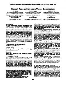
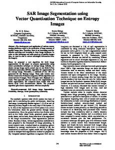
Fig.3.2: (a) Original Image, (b) Gaussian filtered Image, (c) Image after thresholding, (d) Image after morphological opening, (e) Image after Extracting boundary, (f) Superimposed Image.
(a)
(b)
(c)
(d)
(e)
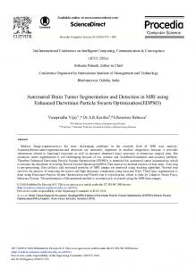
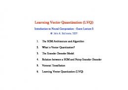
Fig.3.2: (a) Original Image, (b) Image after VQ segmentation, (c) Image after Extracting tumor Region, (d) Image after extracting boundary, (e) Superimposed image
(a)
(b)
(c)
(d)
(e)
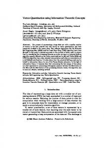
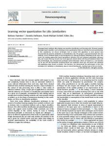
Fig.3.3: (a) Original Image, (b) Image after VQ segmentation, (c) Image after Extracting tumor region, (d) Image after Extracting boundary, (e) Superimposed image.
ISSN: 0975-5462
3755
Dr. H. B. Kekre et. al. / International Journal of Engineering Science and Technology Vol. 2(8), 2010, 3753-3757 4. Conclusion: In this paper morphological segmentation and proposed vector Quantization method are used for tumor detection in MRI images. From results it is observed that proposed method gives far better results compared to morphological segmentation. Morphological segmentation extracts other regions with tumor region. Thresholding is used to convert input image into binary image. Global threshold methods suffer from drawback as threshold value is given manually. The Algorithms are tested on twenty one MRI images. Identification rate for proposed method is 71.5% and for Morphological Segmentation Identification Rate is 66.7%. 5. References: [1]. H. B. Kekre, Tanuja K. Sarode ,Saylee Gharge, “Detection and Demarcation of Tumor using Vector Quantization in MRI Images” ,International Journal of Engineering Science and Technology(IJEST),Volume 2,No.2,pp:59-66,2009.
[2]. H. B. Kekre, Tanuja K. Sarode, Saylee Gharge, “Image Segmentation of MRI Images using Vector Quantization Techniques” International Conference on Contours of Computing Technology, 13th-14th March 2010, at Babasaheb Gawade Institute of Technology will be available on Springer LNCS Digital Library. [3]. Donald Mcrobbie. Medical Image Segmentation [Online]. Available: http:// encyclopedia.jrank.org [4]. Dr. H. B. Kekre , Saylee Gharge , “Image Segmentation of MRI using Texture Features,” International Conference on Managing Next Generation Software Applications, School of Science and Humanities, Karunya University, Coimbatore, Tamilnadu, 05-06 December, 2008. [5]. R. M. Gray, “Vector quantization”, IEEE ASSP Mag., pp.: 4-29, Apr. 1984 [6]. Y. Linde, A. Buzo, and R. M. Gray, “An algorithm for vector quantizer design,” IEEE Trans.Commun., vol. COM- 28, no. 1, pp.: 8495, 1980 [7]. H.B.Kekre, Tanuja K. Sarode, “New Fast Improved Clustering Algorithm for Codebook Generation for Vector Quantization”, International Conference on Engineering Technologies and Applications in Engineering, Technology and Sciences, Computer Science Department, Saurashtra University, Rajkot, Gujarat. (India), Amoghsiddhi Education Society, Sangli, Maharashtra (India) , 13th – 14th January 2008. [8]. H. B. Kekre, Tanuja K. Sarode, “New Fast Improved Codebook Generation Algorithm for Color Images using Vector Quantization,” International Journal of Engineering and Technology, vol.1, No.1, pp.: 67-77, September 2008. [9]. H. B. Kekre, Tanuja K. Sarode, “Fast Codebook Generation Algorithm for Color Images using Vector Quantization,” International Journal of Computer Science and Information Technology, Vol. 1, No. 1, pp.: 7-12, Jan 2009. [10]. H. B. Kekre, Tanuja K. Sarode, “An Efficient Fast Algorithm to Generate Codebook for Vector Quantization,” First International Conference on Emerging Trends in Engineering and Technology, ICETET-2008, held at Raisoni College of Engineering, Nagpur, India, pp.: 62- 67, 16-18 July 2008. Avaliable at IEEE Xplore. [11]. H. B. Kekre, Tanuja K. Sarode, “Fast Codebook Generation Algorithm for Color Images using Vector Quantization,” International Journal of Computer Science and Information Technology, Vol. 1, No. 1, pp.: 7-12, Jan 2009. [12]. H. B. Kekre, Tanuja K. Sarode, “Fast Codevector Search Algorithm for 3-D Vector Quantized Codebook”, WASET International Journal of cal Computer Information Science and Engineering (IJCISE), Volume 2, No. 4, pp.: 235-239, Fall 2008. Available: http://www.waset.org/ijcise. [13]. H. B. Kekre, Tanuja K. Sarode, “Fast Codebook Search Algorithm for Vector Quantization using Sorting Technique”, ACM International Conference on Advances in Computing, Communication and Control (ICAC3-2009), pp: 317-325, 23-24 Jan 2009, Fr. Conceicao Rodrigous College of Engg., Mumbai. Available on ACM portal. [14]. Jim Z.C. Lai, Yi-Ching Liaw, and Julie Liu, “A fast VQ codebook generation algorithm using codeword displacement”, Pattern Recogn. vol. 41, no. 1, pp.: 315–319,2008. [15]. C.H. Hsieh, J.C. Tsai, Lossless compression of VQ index with search order coding, IEEE Trans. Image Process. vol. 5, No. 11, pp.: 1579–1582, 1996. [16]. Chin-Chen Chang, Wen-Chuan Wu, “Fast Planar-Oriented Ripple Search Algorithm for Hyperspace VQ Codebook”, IEEE Transaction on image processing, vol 16, no. 6, pp.: 1538-1547, June 2007. [17]. C. Garcia and G. Tziritas, “Face detection using quantized skin color regions merging and wavelet packet analysis,”= IEEE Trans. Multimedia, vol. 1, no. 3, pp.: 264–277, Sep. 1999. [18]. H. Y. M. Liao, D. Y. Chen, C. W. Su, and H. R. Tyan, “Real-time event detection and its applications to surveillance systems,” in Proc. IEEE Int. Symp. Circuits and Systems, Kos, Greece, pp.: 509–512, May 2006. [19]. J. Zheng and M. Hu, “An anomaly intrusion detection system based on vector quantization,” IEICE Trans. Inf. Syst., vol. E89-D, no. 1, pp.: 201–210, Jan. 2006. [20]. H. B. Kekre, Tanuja K. Sarode, Bhakti Raul, ”Color Image Segmentation using Kekre’s Fast Codebook Generation Algorithm Based on Energy Ordering Concept”, ACM International Conference on Advances in Computing,Communication and Control (ICAC32009), pp.: 357-362, 23-24 Jan 2009, Fr. Conceicao Rodrigous College of Engg., Mumbai. Available on ACM portal. [21]. H. B. Kekre, Tanuja K. Sarode, Bhakti Raul, “Color Image Segmentation using Kekre’s Algorithm for Vector Quantization”, International Journal of Computer Science(IJCS), Vol. 3, No. 4, pp.: 287-292,Fall2008. Available:http://www.waset.org/ijcs. [22]. H. B. Kekre, Tanuja K. Sarode, Bhakti Raul, “Color Image Segmentation using Vector Quantization Techniques Based on Energy Ordering Concept” International Journal of Computing Science and Communication Technologies (IJCSCT) Volume 1, Issue 2, pp: 164-171, January 2009. [23]. H. B. Kekre, Tanuja K. Sarode, Bhakti Raul, “Color Image Segmentation Using Vector Quantization Techniques”,Advances in Engineering Science Sect. C (3), pp.: 35-42,July-September 2008.
ISSN: 0975-5462
3756
Dr. H. B. Kekre et. al. / International Journal of Engineering Science and Technology Vol. 2(8), 2010, 3753-3757 [24]. H. B. Kekre, Tanuja K. Sarode, “Speech Data Compression using Vector Quantization”, WASET International Journal of Computer and Information Science and Engineering (IJCISE), vol. 2, No. 4, pp.: 251-254, Fall 2008. Available: http://www.waset.org/ijcise.
[25]. H. B. Kekre, Ms. Tanuja K. Sarode, Sudeep D. Thepade,“Image Retrieval using Color-Texture Features from DCT on VQ Codevectors obtained by Kekre’s Fast Codebook Generation”, ICGST-International Journal on Graphics, Vision and Image Processing (GVIP), Volume 9, Issue 5, pp.: 1-8, September 2009. Available online.http://www.icgst.com/gvip/Volume9/Issue5/P1150921752. [26]. H. B. Kekre, Kamal Shah, Tanuja K. Sarode, Sudeep D. Thepade,”Performance Comparison of Vector Quantization Technique – KFCG with LBG, Existing Transforms and PCA for Face Recognition”, International Journal of Information Retrieval (IJIR), Vol. 02, Issue 1, pp.: 64-71,2009. [27]. Dr. H. B. Kekre , Saylee Gharge , “SAR Image Segmentation using co-occurrence matrix and slope magnitude,” ACM International Conference on Advances in Computing, Communication and Control (ICAC3-2009), pp.: 357-362, 23-24 Jan 2009, Fr. Conceicao Rodrigous College of Engg., Mumbai. Available on ACM portal. [28]. Parveen, N.R.S, “Segmenting Tumors in Ultrasound Images,” Proceeding of the 2008 International Conference on Computing, Communication and Networking, pp.: 1-5, 2008. [29]. Basim Alhadidi, Mohammad H. Zu`bi and Hussam N. Suleiman, “Mammogram Breast Cancer Image Detection Using Image Processing Functions,” Information Technology Journal, Volume: 6, Issue: 2, pp.: 217-221, 2007. [30]. S. Saheb Basha, Dr. K. Satya Prasad, “Automatic Detection Of Breast cancer mass in mammograms using morphological Operators and Fuzzy C-Means,” Journal of Theoretical and Applied Information Technology, pp.: 704-709, 2005.
AUTHOR BIOGRAPHIES Dr. H. B. Kekre has received B.E. (Hons.) in Telecomm. Engg. from Jabalpur University in 1958, M.Tech(Industrial Electronics) from IIT Bombay in 1960, M.S.Engg. (Electrical Engg.) from University of Ottawa in 1965 and Ph.D. (System Identification) from IIT Bombay in 1970. He has worked Over 35 years as Faculty of Electrical Engineering and then HOD Computer Science and Engg. at IIT Bombay. For last 13 years worked as a Professor in Department of Computer Engg. at Thadomal Shahani Engineering College, Mumbai. He is currently senior Professor working with Mukesh Patel School of Technology Management and Engineering, SVKM’s NMIMS University, Vile Parle(w), Mumbai, INDIA. His areas of interest are Digital Signal processing , Image Processing and computer networks. He has more than 250 papers in National / International Conferences / Journals to his credit. Recently six students working under his guidance have received best paper awards. Currently he is guiding ten Ph.D. students. Dr.Tanuja K. Sarode has Received Ph.D. from Mukesh Patel School of Technology, Management and Engg. SVKM’s NMIMS University, Vile-Parle (W), Mumbai, INDIA. She has more than 10 years of experience in teaching. She is currently working as Assistant Professor in Dept. of Computer Engineering at Thadomal Shahani Engineering College, Mumbai. She is life member of IETE, member of International Association of Engineers (IAENG) and International Association of Computer Science and Information Technology (IACSIT), Singapore. Her areas of interest are Image Processing, Signal Processing and Computer Graphics. She has 70 papers in National /International Conferences/journal to her credit. Ms. Kavita C.Raut has received B.E. (Computer Engg.) degree from Mumbai University in 2005, currently Pursuing M.E. from Thadomal Shahani Engineering College, Bandra (W), Mumbai. She has more than 4 years of experience in teaching. She is currently working as a lecturer in department of Computer Engineering at Thadomal Shahani Engineering College, Mumbai.
ISSN: 0975-5462
3757