Dr.H.B.Kekre. et al /International Journal of Engineering Science and Technology Vol.1(2), 2009, 59-66
Detection and Demarcation of Tumor using Vector Quantization in MRI images Dr. H. B. Kekre Ms. Tanuja K. Sarode Senior Professor, Ph.D. Scholar, MPSTME, SVKM’s Mukesh Patel School of Technology NMIMS University, Mumbai-56, Management and Engineering, India, SVKM’s NMIIMS University Assistant Professor, T.S.E.C., Mumbai-56, India. Mumbai-50, India.
[email protected] [email protected]
Ms. Saylee M. Gharge Ph.D. Scholar, MPSTME, SVKM’s NMIMS University, Mumbai-56, India, Lecturer, V.E.S.I.T, Mumbai-71 India.
[email protected]
ABSTRACT Segmenting a MRI images into homogeneous texture regions representing disparate tissue types is often a useful preprocessing step in the computer-assisted detection of breast cancer. That is why we proposed new algorithm to detect cancer in mammogram breast cancer images. In this paper we proposed segmentation using vector quantization technique. Here we used Linde Buzo-Gray algorithm (LBG) for segmentation of MRI images. Initially a codebook of size 128 was generated for MRI images. These code vectors were further clustered in 8 clusters using same LBG algorithm. These 8 images were displayed as a result. This approach does not leads to over segmentation or under segmentation. For the comparison purpose we displayed results of watershed segmentation and Entropy using Gray Level Co-occurrence Matrix along with this method.
Keywords - MRI, Texture features, Vector Quantization, Encoding. 1. INTRODUCTION Magnetic resonance imaging (MRI) is primarily a medical imaging technique most commonly used in Radiology to visualize the structure and function of the body. It provides detailed images of the body in any plane. MRI provides much greater contrast between the different soft tissues of the body than does computer tomography (CT), making it especially useful in neurological (brain), musculoskeletal, and oncological (cancer) imaging. Unlike CT it uses no ionizing radiation, but uses a powerful magnetic field to align the nuclear magnetization of (usually) hydrogen atoms in water in the body. Radiofrequency fields are used to systematically alter the alignment of this magnetization, causing the hydrogen nuclei to produce a rotating magnetic field detectable by the scanner. This signal can be manipulated by additional magnetic fields to build up enough information to reconstruct an image of the body.The advantages of magnetic resonance imaging (MRI) over other diagnostic imaging modalities are its high spatial resolution and excellent discrimination of soft tissues. MRI provides rich information about anatomical structure, enabling quantitative pathological or clinical studies [1]; the derivation of computerized anatomical atlases [2]; as well as pre and intra-operative guidance for therapeutic intervention [3, 4]. Such information is also valuable as an anatomical reference for functional modalities, such as PET [5], SPECT, and functional MRI [6]. Advanced applications that use the morphologic contents of MRI frequently require segmentation of the imaged volume into tissue types. This problem has received considerable attention. Such tissue segmentation is often achieved by applying statistical classification methods to the signal intensities [7, 8]. In the ideal case, differentiation between white and gray matter in the brain should be easy since these tissue types exhibit distinct signal intensities. In practice, spatial intensity inhomogeneities are often of sufficient magnitude to cause the distributions of signal intensities associated with these tissue classes to overlap significantly. In addition, the operating conditions and status of the MR equipment frequently affect the observed intensities, causing significant inter-scan intensity inhomogeneities that often necessitate manual training on a per-scan basis. While reported methods [9, 10, 11, 12, 13, 14] have had some success in correcting intra-scan inhomogeneities, such methods require supervision for the individual scan. The best approach to image segmentation may vary between different applications. The choice between manual, semiautomatic or fully automatic methods depends on the quality of the images, the number of objects needs to be segmented, the amount of available user time, and the required accuracy of the segmentation. The segmentation process is usually based on gray level intensity, color, shape or texture. Texture can be characterized by local variations of pixel values that repeat in a regular or random pattern on the object or image. It can also be defined as a repetitive arrangement of patterns over a region. A wide variety of texture segmentation techniques have been reported in the literature [15,16, 17-23,24]. We decided to choose a set of existing texture features [25-28] which can provide us good discriminating power and are easy to compute as compare to GLCM [29].
ISSN: 0975-5462
59
Dr.H.B.Kekre. et al /International Journal of Engineering Science and Technology Vol.1(2), 2009, 59-66 The work we have done is to propose a segmentation process which identifies on a MRI the opaque areas, suspect or not, present in the image using vector quantization which consumes moderate time but provide good accuracy with less complexity. Watershed algorithm has a drawback of over-segmenting the image making it obscure for identification of tumor. Segmentation using gray level co-occurrence matrix required huge time for tumor demarcation with less accuracy.
1.1 Vector Quantization Vector Quantization (VQ) [30-38] is an efficient technique for data compression and has been successfully used in various applications such as index compression [39, 40]. VQ has been very popular in a variety of research fields such as speech recognition and face detection [41, 42]. VQ is also used in real time applications such as real time video-based event detection [43] and anomaly intrusion detection systems [44], image segmentation [45-48], speech data compression [49], content based image retrieval CBIR [50] and face recognition [51]. Vector Quantization (VQ) techniques employ the process of clustering. Various VQ algorithms differ from one another on the basis of the approach employed for cluster formations. VQ is a technique in which a codebook is generated for each image. A codebook is a representation of the entire image containing a definite pixel pattern which is computed according to a specific VQ algorithm. The image is divided into fixed sized blocks that form the training vector. The generation of the training vector is the first step to cluster formation on these training vectors clustering methods is applied and codebook is generated. The method most commonly used to generate codebook is the Linde-Buzo-Gray (LBG) algorithm which is also called as Generalized Lloyd Algorithm (GLA). The rest of the paper is organized as follows. Section II describes Gray Level Co-occurrence Matrix(GLCM), Watershed algorithm and Linde Buzo Gray algorithm (LBG) algorithm used for image segmentation of MRI images. Followed by the experimental results for MRI images for comparison in section III and section IV concludes the work. 2. ALGORITHMS FOR SEGMENTATION In this section we explain segmentation by Gray level co-occurrence matrix [29], basic watershed algorithm [52-56] and Linde Buzo Gray algorithm (LBG) which are used for comparative performance of tumor detection. 2.1 Gray Level Co-occurrence Matrix Haralick suggested the use of gray level co-occurrence matrices (GLCM) for definition of textural features. The values of the co-occurrence matrix elements present relative frequencies with which two neighboring pixels separated by distance d appear on the image, where one of them has gray level i and other j. Such matrix is symmetric and also a function of the angular relationship between two neighboring pixels. The co-occurrences matrix can be calculated on the whole image, but by calculating it in a small window which scanning the image, the co-occurrence matrix can be associated with each pixel. By using gray level cooccurrence matrix we can extract different features like probability, entropy, energy, variance, inverse moment difference etc. Using co-occurrence matrix the major textural features are defined as: Maximum Probability: max(Pij)
(2.1)
Variance: (∑ (i − μ i ) 2 ∑ Pij )(∑ ( j − μ j ) 2 ∑ Pij )
(2.2)
Correlation:
∑∑ (i − μ i
x
)( j − μ y ) Pij / σ xσ y
(2.3)
j
where µx and µy are means and σx , σy are standard deviation (2.4) Entropy: ∑ ∑ Pij log( Pij ) i
j
Amongst all these features entropy has given us the best results. Hence in this paper we extracted entropy using gray level co-occurrence matrix and the results are displayed in Fig.6a alongwith that of watershed and LBG algorithms for comparison. 2.2 Watershed algorithm Watershed segmentation [52] classifies pixels into regions using gradient descent on image features and analysis of weak points along region boundaries. The image feature space is treated, using a suitable mapping, as a topological surface where higher values indicate the presence of boundaries in the original image
ISSN: 0975-5462
60
Dr.H.B.Kekre. et al /International Journal of Engineering Science and Technology Vol.1(2), 2009, 59-66 data. It uses analogy with water gradually filling low lying landscape basins. The size of the basins grows with increasing amount of water until they spill into one another. Small basins (regions) gradually merge together into larger basins. Regions are formed by using local geometric structure to associate the image domain features with local extremes measurement. Watershed techniques produce a hierarchy of segmentations, thus the resulting segmentation has to be selected using either some apriory knowledge or manually. These methods are well suited for different measurements fusion and they are less sensitive to user defined thresholds. We implemented watershed algorithm for MRI images as mentioned in [56].Results for MRI images are displayed in Fig 6(b).
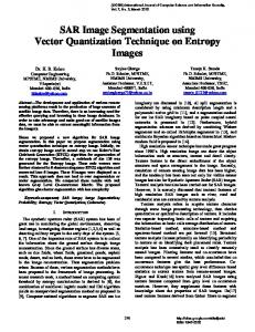
1.2 Linde Buzo and Gray (LBG)[29, 30] For the purpose of explaining this algorithm, we are considering two dimensional vector space as shown in Fig.1. This is obtained by considering two consecutive pixel values as x and y co-ordinates so that each pair is represented by point in x,y plane. In this algorithm centroid is computed as the first codevector C1 for the training set. In Fig. 1 two vectors v1 & v2 are generated by adding constant error to the codevector. Euclidean distances of all the training vectors are computed with vectors v1 & v2 and two clusters are formed based on nearest of v1 or v2. Procedure is repeated for these two clusters to generate four new clusters. This procedure is repeated for every new cluster until the required size of codebook is reached or specified MSE is reached.
Fig.1. LBG for 2 dimensional case
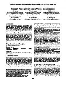
In this paper initially we have selected 128 as codebook size using 12 dimensional vector space. Thus the image is divided into 128 clusters which were further reduced to 8 by using requantization. The 8 clusters thus obtained were mapped onto the image generating 8 different images representing them. On all these images Canny’s operator was used to obtain the edge maps. These edge maps were superimposed on the original image giving clear demarcation of the tumor. The very first cluster gives the best results. However the other clusters also give comparatively better results as compare to watershed and GLCM algorithm. 3. RESULTS Defects in the metabolic system can lead to waste build-up that can cause altered levels of consciousness (ALC). Drug exposure is a common cause for ALC. Drug-induced ALC can result from an overdose of either over-the-counter or illegal drugs. Alcohol intoxication is probably the most common cause of drug-induced ALC. Structural abnormalities of the brain can lead to ALC [Figure 2a]. Tumors (benign or malignant) can form and crowd out the normal structures of the brain. As a result, weakness in the walls of the blood vessels in the brain (aneurysms) may begin to swell, or may even break, causing blood to pool inside the head and push the brain against the bony wall of the skull. The resulting damage can then cause ALC. For this image we generate codebook of size 128 using LBG algorithm and converting them to 8 segmented images are shown in Fig.2(b)-(i). After using Canny’s operator the results are displayed in Fig.3(a)-(h). Edge detected images Fig.3(a)-(h) were superimposed on original MRI tumor image Fig.2(a) and displayed as Fig.4(a)-(h) respectively which indicate textural variation as we move from one code-vector to the next. Fig.5(a) shows result for probability using GLCM and equalized probability is displayed in Fig 5(b).Extracted Entropy using GLCM as shown in Fig.5(c) with equalized entropy in Fig.5(d). Fig.6(a) shows superimposed edge map on original image for equalized Entropy using GLCM, Fig.6(b) displays similarly constructed image using watershed algorithm and Fig.6(c) indicates result for superimposed image for first code-vector amongst 8 codevectors using LBG algorithm.
ISSN: 0975-5462
61
Dr.H.B.Kekre. et al /International Journal of Engineering Science and Technology Vol.1(2), 2009, 59-66
(a)Original Image
(b)
(c)
(f)
(g)
(d)
(e)
(h)
(i)
Fig. 2: (a) Original brain tumor image,(b) Image for first code-vector, (c)-(i) Image for second -eighth code-vector,
(a)
(e)
(b)
(c)
(d)
(f)
(g)
(h)
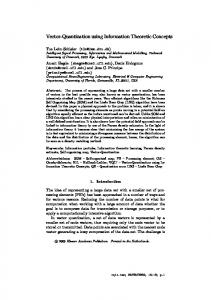
Fig.3: (a)-(h) Edge detected images for Fig.2(b)-(i) respectively.
(a)
ISSN: 0975-5462
(b)
(c)
(d)
62
Dr.H.B.Kekre. et al /International Journal of Engineering Science and Technology Vol.1(2), 2009, 59-66
(e)
(f)
(g)
(h)
Fig.4: (a)-(h) Superimposed images of Fig.3 (a)-(h) respectively on original image Fig.2(a)
(a)
(b)
(c)
(d)
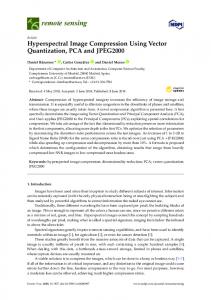
Fig. 5 : (a) Extracted Probability using GLCM of Fig 2a, (b) Equalized Probability for Fig.5a, (c) Entropy using GLCM for Fig.2a,(d) Equalized Entropy for Fig.5c.
(a)
(b)
(c)
Fig.6: (a) Segmented image for entropy using GLCM, (b) Segmented image using watershed algorithm, (c) Segmented image using proposed algorithm.
4. CONCLUSION Here we used Gray Level Co-occurrence Matrix, watershed algorithm and LBG algorithm for tumor detection and demarcation for MRI images. Initially a codebook of size 128 was generated for these images. These code vectors were further clustered in 8 clusters using same LBG algorithm. These 8 images were displayed as a results in Fig 2(b)-(i). From results (Fig.6) it is observed that GLCM, watershed gives over segmentation while LBG shows far better results for the same. This approach does not lead to over segmentation or under segmentation with less complexity. 5. ACKNOWLEDGEMENTS Authors sincerely would like to thank Dr. Manisha Mundhe for identifying tumor and approving the results. 6. REFERENCES [1] M. Shenton, R. Kikinis, F. Jolesz, et al. Abnormalities of the Left Temporal Lobe and Thought Disorder in Schizophrenia. N. Engl. J. Med: 604 to 612, 1992. [2] K. H¨ohne et al. A framework for the Generation of 3D Anatomical Atlases. In SPIE Vol. 1808, Visualization in Biomedical Computing 1992. [3] R. Kikinis, F.A. Jolesz, W.E. Lorensen, H.E. Cline, P.E Stieg, Black 3d Reconstruction of Skull Base Tumors from MRI Data for Neurosurgical Planning.In Proceedings of the Society of Magnetic Resonance in Medicine Conference, 1991. [4] R. Kikinis, D. Altobelli, W. Lorensen, W. Wells, and G. Ettinger. Pre- and intraoperative tumor localization using 3d renderings of mri's. 12th Annual Scientific Meeting of the Society of Magnetic Resonance in Medecine, 1993.
ISSN: 0975-5462
63
Dr.H.B.Kekre. et al /International Journal of Engineering Science and Technology Vol.1(2), 2009, 59-66 [5] D. Levin, X. Hu, K. Tan, et al. The Brain: Integrated Three-Dimensional Display of MR and PET Images. Radiology, 1989. [6] J. Belliveau, D. Kennedy, R. McKinstry, et al. Functional Mapping of the Human Visual Cortex by Magnetic Resonance Imaging. Science: 716 to 719, November 1991. [7] M. Vannier, D. Jordan, W. Murphy, Multi-Spectral Analysis of Magnetic Resonance Images. Radiology: 221 to 224, 1985. [8] M. Kohn, N. Tanna, G. Herman, et al. Analysis of Brain and Cerebrospinal Fluid Volumes with MR Imaging. Radiology, : 115 to 122, 1991. [9] R.B. Lufkin, T. Sharpless, B. Flannigan, and W. Hanafee. Dynamic-Range Compression in Surface-Coil MRI. AJR:379 to 382, 1986. [10] L. Axel, J. Costantini, and J. Listerud. Intensity Correction in Surface-Coil MR Imaging. AJR: 418 to 420, 1987. [11] K.O. Lim and A. P_erbaum. Segmentation of MR Brain Images into Cerebrospinal Fluid Spaces, White and Gray Matter. JCAT:588 to 593, 1989. [12] B. Dawant, A. Zijdenbos, and R. Margolin. Correction of Intensity Variations in MR Images for ComputerAided Tissue Classification. IEEE Trans. Med. Imaging: 770 to 781, 1993. [13] J. Gohagan, E. Spitznagel, W. Murphy, M. Vannier, Multispectral Analysis of MR Images of the Breast. Radiology:703 to 707, 1987. [14] S. Aylward and J. Coggins. Spatially Invariant Classification of Tissues in MR Images. In Proceedings of the Third Conference on Visualization in Biomedical Computing.SPIE, 1994. [15] P. Andrey, P. Tarroux, Unsupervised segmentation of Markov random field modeled textured images using selectionist relaxation, IEEE Transactions on Pattern Analysis and Machine intelligence 20 (3) (1998) 252–262 [16] U. Bhattacharya, B.B. Chaudhuri, S.K. Parui, An MLP-based texture segmentation method without selecting a feature set, Image Vision Computing 15 (1997) 937–948. [17] C. Bouman, B. Liu, Multiple resolution segmentation of textured images, IEEE Transactions on Pattern Analysis and Machine Intelligence 13 (2) (1991) 99–113. [18] J.L. Chen, A. Kundu, Unsupervised texture segmentation using multichannel decomposition and hidden Markov-models, IEEE Transactions on Image Processing 4 (5) (1995) 603–619. [19] G. Cross, A.K. Jain, Markov random field texture models, IEEE Transactions on Pattern Analysis and Machine Intelligence 5 (1982) . [20] C.S. Lu, P.C. Chung, C.F. Chen, Unsupervised texture segmentation via wavelet transform, Pattern Recognition 30 (5) (1997) . [21] J. Mao, A.K. Jain, Texture classification and segmentation using multiresolution simultaneous autoregressive models, Pattern Recognition 25 (2) (1992) 173–188. [22] D.K. Panjwani, G. Healey, Markov random field models for unsupervised segmentation of textured color images, IEEE Transactions on Pattern Analysis & Machine Intelligence (1995) [23] G. Rudolph, Convergence analysis of canonical genetic algorithms, IEEE Transactions on Neural Networks 5 (1) (1994) 96–101. [24]M. Unser, M. Eden, Multi resolution feature extraction and selection for texture segmentation, IEEE Transactions on Pattern Analysis and Machine Intelligence 11(1989). [25] Dr. H. B. Kekre , Saylee Gharge , “Selection of Window Size for Image Segmentation using Texture Features,” International Conference on Advanced Computing &Communication Technologies(ICACCT-2008) Asia Pacific Institute of Information Technology SD India, Panipat ,08-09 November,2008. [26] Dr. H. B. Kekre , Saylee Gharge , “Image Segmentation of MRI using Texture Features,” International Conference on Managing Next Generation Software Applications ,School of Science and Humanities, Karunya University, Coimbatore, Tamilnadu ,05-06 December,2008. [27] Dr. H. B. Kekre , Saylee Gharge , “Statistical Parameters like Probability and Entropy applied to SAR image segmentation,” International Journal of Engineering Research & Industry Applications (IJERIA), Vol.2,No.IV, pp.341-353. [28] Dr. H. B. Kekre , Saylee Gharge , “SAR Image Segmentation using co-occurrence matrix and slope magnitude,” ACM International Conference on Advances in Computing, Communication and Control (ICAC32009), pp.: 357-362, 23-24 Jan 2009, Fr. Conceicao Rodrigous College of Engg., Mumbai. Available on ACM portal. [29]Robert M. Hawlick, Statistical and Structural Approaches to Texture, IEEE Proceedings Of vol. 67, no. 5, May 1979. [30] R. M. Gray, “Vector quantization”, IEEE ASSP Mag., pp.: 4-29, Apr. 1984 [31] Y. Linde, A. Buzo, and R. M. Gray, “An algorithm for vector quantizer design,” IEEE Trans.Commun., vol. COM-28, no. 1, pp.: 84-95, 1980
ISSN: 0975-5462
64
Dr.H.B.Kekre. et al /International Journal of Engineering Science and Technology Vol.1(2), 2009, 59-66 [32] H.B.Kekre, Tanuja K. Sarode, “New Fast Improved Clustering Algorithm for Codebook Generation for Vector Quantization”, International Conference on Engineering Technologies and Applications in Engineering, Technology and Sciences, Computer Science Department, Saurashtra University, Rajkot, Gujarat. (India), Amoghsiddhi Education Society, Sangli, Maharashtra (India) , 13th – 14th January 2008. [33] H. B. Kekre, Tanuja K. Sarode, “New Fast Improved Codebook Generation Algorithm for Color Images using Vector Quantization,” International Journal of Engineering and Technology, vol.1, No.1, pp.: 67-77, September 2008. [34] H. B. Kekre, Tanuja K. Sarode, “Fast Codebook Generation Algorithm for Color Images using Vector Quantization,” International Journal of Computer Science and Information Technology, Vol. 1, No. 1, pp.: 7-12, Jan 2009. [35] H. B. Kekre, Tanuja K. Sarode, “An Efficient Fast Algorithm to Generate Codebook for Vector Quantization,” First International Conference on Emerging Trends in Engineering and Technology, ICETET2008, held at Raisoni College of Engineering, Nagpur, India, pp.: 62- 67, 16-18 July 2008. Avaliable at IEEE Xplore. [36] H. B. Kekre, Tanuja K. Sarode, “Fast Codebook Generation Algorithm for Color Images using Vector Quantization,” International Journal of Computer Science and Information Technology, Vol. 1, No. 1, pp.: 7-12, Jan 2009. [37] H. B. Kekre, Tanuja K. Sarode, “Fast Codevector Search Algorithm for 3-D Vector Quantized Codebook”, WASET International Journal of cal Computer Information Science and Engineering (IJCISE), Volume 2, No. 4, pp.: 235-239, Fall 2008. Available: http://www.waset.org/ijcise. [38] H. B. Kekre, Tanuja K. Sarode, “Fast Codebook Search Algorithm for Vector Quantization using Sorting Technique”, ACM International Conference on Advances in Computing, Communication and Control (ICAC32009), pp: 317-325, 23-24 Jan 2009, Fr. Conceicao Rodrigous College of Engg., Mumbai. Available on ACM portal [39] Jim Z.C. Lai, Yi-Ching Liaw, and Julie Liu, “A fast VQ codebook generation algorithm using codeword displacement”, Pattern Recogn. vol. 41, no. 1, pp.: 315–319, 2008. [40] C.H. Hsieh, J.C. Tsai, Lossless compression of VQ index with search order coding, IEEE Trans. Image Process. vol. 5, No. 11, pp.: 1579–1582, 1996. [41]Chin-Chen Chang, Wen-Chuan Wu, “Fast Planar-Oriented Ripple Search Algorithm for Hyperspace VQ Codebook”, IEEE Transaction on image processing, vol 16, no. 6, pp.: 1538-1547, June 2007. [42]C. Garcia and G. Tziritas, “Face detection using quantized skin color regions merging and wavelet packet analysis,” IEEE Trans. Multimedia, vol. 1, no. 3, pp.: 264–277, Sep. 1999. [43]H. Y. M. Liao, D. Y. Chen, C. W. Su, and H. R. Tyan, “Real-time event detection and its applications to surveillance systems,” in Proc. IEEE Int. Symp. Circuits and Systems, Kos, Greece, pp.: 509–512, May 2006. [44]J. Zheng and M. Hu, “An anomaly intrusion detection system based on vector quantization,” IEICE Trans. Inf. Syst., vol. E89-D, no. 1, pp.: 201–210, Jan. 2006. [45]H. B. Kekre, Tanuja K. Sarode, Bhakti Raul, ”Color Image Segmentation using Kekre’s Fast Codebook Generation Algorithm Based on Energy Ordering Concept”, ACM International Conference on Advances in Computing, Communication and Control (ICAC3-2009), pp.: 357-362, 23-24 Jan 2009, Fr. Conceicao Rodrigous College of Engg., Mumbai. Available on ACM portal. [46]H. B. Kekre, Tanuja K. Sarode, Bhakti Raul, “Color Image Segmentation using Kekre’s Algorithm for Vector Quantization”, International Journal of Computer Science (IJCS), Vol. 3, No. 4, pp.: 287-292, Fall 2008. Available: http://www.waset.org/ijcs. [47]H. B. Kekre, Tanuja K. Sarode, Bhakti Raul, “Color Image Segmentation using Vector Quantization Techniques Based on Energy Ordering Concept” International Journal of Computing Science and Communication Technologies (IJCSCT) Volume 1, Issue 2, pp: 164-171, January 2009. [48]H. B. Kekre, Tanuja K. Sarode, Bhakti Raul, “Color Image Segmentation Using Vector Quantization Techniques”, Advances in Engineering Science Sect. C (3), pp.: 35-42, July-September 2008. [49]H. B. Kekre, Tanuja K. Sarode, “Speech Data Compression using Vector Quantization”, WASET International Journal of Computer and Information Science and Engineering (IJCISE), vol. 2, No. 4, pp.: 251254, Fall 2008. available: http://www.waset.org/ijcise. [50]H. B. Kekre, Ms. Tanuja K. Sarode, Sudeep D. Thepade, “Image Retrieval using Color-Texture Features from DCT on VQ Codevectors obtained by Kekre’s Fast Codebook Generation”, ICGST-International Journal on Graphics, Vision and Image Processing (GVIP), Volume 9, Issue 5, pp.: 1-8, September 2009. Available online at http://www.icgst.com/gvip/Volume9/Issue5/P1150921752. [51]H. B. Kekre, Kamal Shah, Tanuja K. Sarode, Sudeep D. Thepade, ”Performance Comparison of Vector Quantization Technique – KFCG with LBG, Existing Transforms and PCA for Face Recognition”, International Journal of Information Retrieval (IJIR), Vol. 02, Issue 1, pp.: 64-71, 2009.
ISSN: 0975-5462
65
Dr.H.B.Kekre. et al /International Journal of Engineering Science and Technology Vol.1(2), 2009, 59-66 [51] L. Vincent, P. Soille, Watersheds in digital spaces: An efficient algorithm based on immersion Simulations , IEEE Trans. PAMI., 13 (6) (1991) 583–593. [52] F. Meyer, Topographic distance and watershed lines,Signal Processing, 38 (1) (1994) 113–125. [53] A. Bieniek, A. Moga, An efficient watershed algorithm based on connected components, Pattern Recognition, 33 (6) (2000) 907–916. [54] M. Frucci, Oversegmentation reduction by flooding regions and digging watershed lines, International Journal of Pattern Recognition and Artificial Intelligence, 20 (2006) 15–38. [55]L. E. Band, Topographic partition of watersheds with digital elevation models, Water Resources Res., 22 (1) (1986) 15–24. [56] Basim Alhadidi, Mohammad H. et al, “ Mammogram Breast Cancer Edge Detection Using Image Processing Function” Information Technology Journal 6(2):217-221,2007,ISSN-1812-5638. AUTHOR BIOGRAPHIES Dr. H. B. Kekre has received B.E. (Hons.) in Telecomm. Engg. from Jabalpur University in 1958, M.Tech (Industrial Electronics) from IIT Bombay in 1960, M.S.Engg. (Electrical Engg.) from University of Ottawa in 1965 and Ph.D. (System Identification) from IIT Bombay in 1970. He has worked Over 35 years as Faculty of Electrical Engineering and then HOD Computer Science and Engg. at IIT Bombay. For last 13 years worked as a Professor in Department of Computer Engg. at Thadomal Shahani Engineering College, Mumbai. He is currently Senior Professor working with Mukesh Patel School of Technology Management and Engineering, SVKM’s NMIMS University, Vile Parle(w), Mumbai, INDIA. His areas of interest are Digital Signal processing , Image Processing and computer networks. He has more than 250 papers in National / International Conferences / Journals to his credit. Recently six students working under his guidance have received best paper awards. Currently he is guiding ten Ph.D. students. Tanuja K. Sarode has Received M.E.(Computer Engineering) degree from Mumbai University in 2004, currently Pursuing Ph.D. from Mukesh Patel School of Technology, Management and Engg., SVKM’s NMIMS University, Vile-Parle (W), Mumbai, INDIA. She has more than 10 years of experience in teaching. Currently working as Assistant Professor in Dept. of Computer Engineering at Thadomal Shahani Engineering College, Mumbai. She is life member of IETE, member of International Association of Engineers (IAENG) and International Association of Computer Science and Information Technology (IACSIT), Singapore. Her areas of interest are Image Processing, Signal Processing and Computer Graphics. She has 35 papers in National /International Conferences/journal to her credit. Ms. Saylee M. Gharge has received M.E.(Electronics and telecomm.) degree from Mumbai University in 2007, currently Pursuing Ph.D. from Mukesh Patel School of Technology, Management and Engineering, NMIMS University, Vile-Parle (W), Mumbai. She has more than 9 years of experience in teaching. Currently working as a lecturer in department of electronics and telecommunication in Vivekanand Institute of Technology, Mumbai . Her areas of interest are Image Processing, Signal Processing. She has 19 papers in National /International Conferences/journal to her credit
ISSN: 0975-5462
66