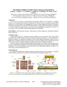
DIGITAL MICROFLUIDIC PLATFORM FOR HIGH-THROUGHPUT MONOCLONAL ANTIBODIES SCREENING AND ISOTYPING ANALYSIS Jie-Long He1*, Hsin-Yi Lu1, Yi-Han Lai1, Jie-Teng He2 and Shih-Kang Fan1 1 National Taiwan University, Taiwan and 2 Chiayi Chang Gung Memorial Hospital, Taiwan ABSTRACT Monoclonal antibody (mAb) technology is one of the most important scientific achievements in the twentieth century. However, the traditional mAbs screening encounters a bottleneck for the highthroughput process. Here we integrate the immunoassay methods on the digital microfluidic platform for the mAbs analysis that potentially holds high throughput. In our prototype devices, the nitrocellulose dots spotted on the Teflon surface let the isotyping antibodies be bound on the chip stably. Furthermore, the droplet of antibodies mixture and washing buffer were manipulated by EWOD (electrowetting-oncielectric) force. The preliminary results show that the isotyping analysis was executed on the digital microfluidic platform. KEYWORDS: Monoclonal antibody, Isotyping analysis, Electrowetting-on-dielectric INTRODUCTION The myeloma cells and the immunized mouse spleen cells were fused to obtain hybridoma cells, and then the cells were screened to recognize the specific antigens. Large amounts of the pure monoclonal antibodies (mAbs) were produced by this process. In our previous studies, hundreds of mAbs against various proteins of avian influenza virus had been established by the traditional strategy [1]. However, labor intensive and plenty reagent consuming make it difficult to become a high-throughput process. For that reason, this study had integrated the traditional immunoassay and the digital microfluidic platform. The mAbs produced by hybridoma cells are assorted by their isotype that differs in function. The major class and subclass have been identified in placental mammals (e.g. mouse), including IgA, IgG (IgG1, IgG2a, IgG2b or IgG3), and IgM. The mAbs isotyping analysis is one of the most important steps during hybridoma development to assure that the specific clones for further applications. The traditional determination methods are available in enzyme-linked immunosorbent assay (ELISA) and immune colloidal gold technique in the cassette. The goal of our study is developing lab-on-a-chip (LOC) modules which are based on the digital microfluidic techniques and mimic the traditional determination methods. The digital microfluidic platform which operates the droplets by electrowetting-on-dielectric (EWOD) between the two indiumtin oxide (ITO) glass plates has several advantages, including easy device process, simple structure, small sample demand and potential of modularity and automation. EXPERIMENTAL AND RESULTS The specific patterned single-layer electrodes were placed on the bottom ITO glass plate with the microfabrication processes [2]. The chip measures 4 cm × 4 cm (Figure 1A) with 19 square electrodes (1 mm × 1 mm) and 4 reservoir electrodes (Figure 1C). The spin-on glass (SOG, thickness 1 µm) was used as the dielectric layer, and then coated with a Teflon layer. The nitrocellulose dots were spotted on the Teflon surface stably to bind the isotyping antibodies (Figure 1B). The electrodes on the bottom plate of device were connected to a relay system. Alternating current of high electric potential was generated from a function generator and amplified with an amplifier. A data acquisition device controls the relay system with digital output signals programmed by LabVIEW software. The EWOD devices were placed on a microscope with CCD camera (Figure 1A). The 1 kHz and 40-76 VRMS AC signal was applied to manipulate the droplet.
(C).
(A).
Washing buffer Nitrocellulose dots
EB2+2nd Ab Blank
(B).
Isotyping Ab
Ab reserve
Upper ITO glass
Nitrocellulose
θ0
Teflon
V
θ(V)
SOG Electrodes (ITO)
Buffer reserve
Bottom ITO glass
Figure 1: Design of the digital microfluidic chip for mAbs isotyping. (A) The droplet manipulation in the chip was observed by microscope.(B) The cross section showing the structures of the patterned electrodes device. (C) The solution reservoirs and nitrocellulose dots arrangements on the digital microfluidic chip for mAbs isotyping. We used the immune colloidal gold technique in the cassette for rapid antibody isotyping (Thermo) (Figure 2A) and the commercialized mouse monoclonal antibody isotyping reagents (Sigma) for ELISA (Figure 2B) to identify the isotype of a specific influenza HA mAb, EB2 [3]. The isotyping analysis on the digital microfluidic chip used the isotyping antibodies (diluted 100X) for manual analysis (Figure 2C) and EWOD operations (Figure 2D). (D).
(A).
mAb+2nd Ab
TMB substrate
(B).
Nitrocellulose Teflon SOG Electrodes
(C).
Absorbance (405 nm)
0.45 0.40 0.30
2
0.25
3
0.20
IgG
IgA
IgM
(Anti-IgG, A or M)
1
0.35
Blank Isotyping Ab
TMB substrate
TMB stop solution
4
0.15
5
0.10 0.05
-
G1 +
0.00 -
+
G1
G2a
G2b
G3
A
M
G2b G2a
A G3
M
mAb isotype
Figure 2: The isotyping analysis of a specific influenza mAb, EB2. (A) Immune colloidal gold technique in the cassette. (B) Specific anti-immunoglobulin antibodies for the ELISA (n=16). (C) Manual analysis was operated on the digital microfluidic chip with the nitrocellulose dots, but without the electrodes (n=5). (D) The isotyping analysis on the prototype of the digital microfluidic platform. The EB2 and the 2nd Ab (goat anti-mouse horseradish peroxidase-conjugated secondary antibody) mixture was diluted 1000X for Ab incubation (Figure 3B). The PBST (PBS containing 0.1% Tween 20) was diluted 10X and used for washing (Figure 3C). Substrate 3, 3’, 5, 5’-tetramethylbenzidine (TMB, Sigma) was used and stopped by a stop solution. The reaction was measured quantitatively by the optical densities at 405 nm using a microtiter plate reader (Figure 2B) or qualitatively observed from the yellow color of the droplets (Figure 2C and 2D).
(B).
(A). Function generator (Agilent 33220A)
Microscopy with CCD
EB2+2nd Ab
EB2+2nd Ab
EB2+2nd Ab
Ab Incubating (C). Washing buffer
High voltage amplifier Relays system (A. A. Lab Systems A-304) (Fan-Tasy Lab)
Washing buffer
Washing buffer
Washing
Figure 3: Digital microfluidic platform for mAbs isotyping. (A) Digital microfluidic chip operating system. (B) The EB2 and the 2nd Ab mixture droplet covered over the nitrocellulose dots and was driven back and forth 3 times by EWOD for incubating. (C) The diluted PBST was used for washing of 5 times. The results of immune colloidal gold cassette for isotyping analysis shown that the EB2 mAb was IgG (Figure 2A). The ELISA results (Figure 2B) agreed with the colloidal gold cassette results. The digital microfluidic chip with the nitrocellulose layer performed by manual operations for isotyping analysis (Figure 2C) was strongly correlated with the previous traditional methods. The isotyping analysis operated by EWOD actuation brought the same conclusion (Figure 2D). In our device, the nitrocellulose dots on the Teflon surface let the isotyping antibodies could be bound on the chip stably. Although the current qualitative results based on the color of the droplets revealed the isotyping of IgG as the resuts from the cassette, the absorbance of the droplets will be further quantitatively evaluated by spectrometer at 405 nm and compared with the ELISA results. CONCLUSION We report for the first time the application of digital microfluidic platform to the monoclonal antibody isotyping analysis. With spotted nitrocellulose dots containing varied antibodies, the digital microfluidic platform is used for screening monoclonal antibodies with desired isotyping. ACKNOWLEDGEMENTS We are grateful to Prof. Rong-Huay Juang (National Taiwan University) for the mAb, EB2 supply. This study was funded by the Ministry of Science and Technology, Taiwan (NSC 101-2628-E-002-036). The funders had no role in the study design, analysis and preparation of the manuscript. REFERENCES [1] J. L. He, M. S. Hsieh, Y. C. Chiu, R. H. Juang, and C. H. Wang, “Preparation of monoclonal antibodies against poor immunogenic avian influenza virus proteins,” J. Immunol. Methods, 397, 43, 2013. [2] S.-K. Fan, Y.-W. Hsu, and C.-H. Chen, “Encapsulated droplets with metered and removable oil shells by electrowetting and dielectrophoresis,” Lab Chip, 11, 2500, 2011. [3] Y. T. Chen, R. H. Juang, J. L. He, W. Y. Chu, and C. H. Wang, “Detection of H6 influenza antibody by blocking enzyme-linked immunosorbent assay,” Vet. Microbiol., 142, 205, 2010. CONTACT * J.L. He; phone: +886-2-33664946;
[email protected]