Review
TRENDS in Neurosciences Vol.27 No.12 December 2004
Discrete synaptic states define a major mechanism of synapse plasticity Johanna M. Montgomery1 and Daniel V. Madison2 1 2
Department of Physiology, Faculty of Medical and Health Sciences, University of Auckland, New Zealand Department of Molecular and Cellular Physiology, Stanford University School of Medicine, Stanford, CA 94305, USA
Synapses can change their strength in response to afferent activity, a property that might underlie a variety of neural processes such as learning, network synaptic weighting, synapse formation and pruning. Recent work has shown that synapses change their strength by jumping between discrete mechanistic states, rather than by simply moving up and down in a continuum of efficacy. Coincident with this, studies have provided a framework for understanding the potential mechanistic underpinnings of synaptic plastic states. Synaptic plasticity states not only represent a new and fundamental property of CNS synapses, but also can provide a context for understanding outstanding issues in synaptic function, plasticity and development. The fact that certain excitatory synapses in the brain can change their strength in response to activity has long captured the imaginations of neuroscientists. These changes might underlie such diverse processes as learning and memory, alterations in coding of information in neural networks, and synaptic development and elimination [1]. Synaptic plasticity of the type discussed here has primarily been studied in the excitatory glutamatergic synapses of the brain, particularly of the hippocampus. This review of synaptic properties and plasticity is confined to the widely studied subset of excitatory glutamatergic synapses that are efferent from hippocampal CA3 pyramidal cells to CA3 and CA1 postsynaptic neurons. These synapses have multiple subtypes of glutamate receptors in their postsynaptic membranes, including AMPA receptors, NMDA receptors) and metabotropic glutamate (mGlu) receptors. Generally speaking, AMPA receptor subtypes mediate ion fluxes across the membrane during synaptic transmission at these synapses, whereas NMDA receptors and mGlu receptors are thought primarily to play a role in inducing or modulating plasticity of the AMPA-receptor-mediated transmission [2]. Persistent activity-dependent increases in synaptic transmission are referred to as long-term potentiation (LTP) [3] and decreases in synaptic transmission are termed long-term depression (LTD) [4,5]. Changes in synaptic strength can result from changes in glutamate receptor function [6], increased or decreased glutamate receptor expression in the postsynaptic density (PSD) [2], Corresponding author: Daniel V. Madison (
[email protected]). Available online 14 October 2004
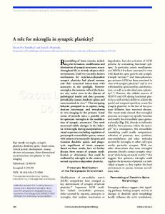
or changes in transmitter release, as at hippocampal mossy fiber terminals [7]. Blockade of postsynaptic exocytosis or endocytosis [by disrupting activity of N-ethylmaleimide-sensitive membrane fusion protein (NSF) [8] or inhibiting dynamin] prevents expression of LTP [9–11] and LTD, respectively [12–14]. Insertion of AMPA receptors into the synaptic membrane in an activity-dependent manner has been demonstrated using green fluorescent protein (GFP)-tagged AMPA receptor subunits [15,16]. Together with the finding that NMDAreceptor-mediated excitatory postsynaptic currents (EPSCs) do not change with increases in synaptic efficacy [17–19] (but see Refs [20,21]), these data show that potentiation or strengthening of excitatory synapses in the CA1 and CA3 regions of the hippocampus is associated with the specific recruitment and insertion of AMPA receptors from intracellular pools into the postsynaptic membrane [11,15]. Similarly, activity that causes synaptic depression, or a weakening of synaptic strength, is correlated with the endocytosis of these receptors from the postsynaptic membrane [12,22]. Thus, the response of a postsynaptic cell seems to be correlated with the number of AMPA receptors present on the postsynaptic membrane. In most cases, the insertion and removal of AMPA receptors is triggered by Ca2C influx through NMDA receptors. This has led to the assertion that AMPA receptors are responsible for the expression of synaptic plasticity, whereas NMDA receptors are responsible for its control. Synaptic states: a mechanism of dictating synaptic strength A key role of synaptic plasticity is to allow the synapse to operate over a large dynamic range. Two possible models could explain the behavior of synapses over this range. In the first, synapses undergo changes in efficacy by adjusting their strength along a continuum, such that the properties of strengthening or weakening occur in a graded fashion with fixed underlying mechanisms (i.e. the ‘continuum model’). In the second, synapses might exist in different discrete states that represent and underlie different levels of efficacy (i.e. the ‘state model’). In an example of the continuum model utilizing AMPA receptor expression as an underlying mechanism, AMPA receptors are inserted or removed from the synaptic membrane and the cellular mechanisms regulating their insertion or removal do not vary across the whole
www.sciencedirect.com 0166-2236/$ - see front matter Q 2004 Elsevier Ltd. All rights reserved. doi:10.1016/j.tins.2004.10.006
Review
TRENDS in Neurosciences Vol.27 No.12 December 2004
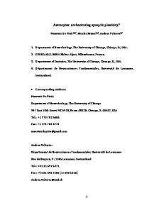
dynamic range of the synapse strength. Thus, for example, insertion or removal of AMPA receptors into or out of ‘weak’ and ‘strong’ synapses would occur via the same exocytic or endocytic mechanisms with similar ease (Figure 1). In the state model, the underlying properties of the synapse change with alterations in synaptic strength (Figure 2). This would result in synapses existing in different and discrete states with regard to plasticity. Recent work suggests that the latter model is most likely: synapses undergoing LTP or LTD do so by moving between different discrete electrophysiologically defined states [23]. Previous studies did not reveal all the mechanisms at work in synaptic plasticity because they recorded activity in large populations of synapses, which can average out the diverse behavior and properties of individual synapses. Hence, the diversity of synaptic states was revealed only when the plastic properties of single or small populations of synapses, for example individual pairs of connected neurons [23], were examined. Five synaptic states have been defined: active, potentiated, depressed, silent, and recently silent. The five states are defined as follows:
Active In the active state, synapses display both AMPA-receptormediated and NMDA-receptor-mediated responses and are pluripotent with regard to plasticity: they can be either potentiated or depressed by the appropriate synaptic activity protocol. Both LTP and LTD arising from the active state are NMDA-receptor-dependent.
745
Potentiated When active synapses undergo LTP, they enter the potentiated state. In the CA3 region, plasticity in the potentiated state differs from that in the active state, because depression (depotentiation) depends not on NMDA receptors but on type-1 mGlu receptors, which are prevalent in this brain region. Mechanisms of plasticity in the potentiated state are thought to differ in the CA1 region [24] and in other areas [25,26], where different mGlu receptor subtypes are prevalent [27].
Depressed When active synapses undergo LTD, they enter the depressed state. According to current published data, this state remains ill-defined, and it might differ little from the active state.
Silent Silent synapses are defined as having normal NMDAreceptor-mediated EPSCs but lacking AMPA-receptormediated EPSCs [18,19,28–31]. In the literature, this lack of AMPA receptor response is generally attributed to a deficiency of AMPA receptors in the postsynaptic membrane [18,19,30–32] (but see also Ref. [29]). Such synapses are termed silent because they have no synaptic responses at normal postsynaptic membrane potentials, as the NMDA receptor is subject to voltage-dependent Mg2C block [33,34]. Because these synapses have no AMPA responses to begin with, their responses cannot be depressed; however, they can be potentiated by the same types of synaptic activity that potentiate active synapses.
AMPA receptor PSD Exocytosis Endocytosis
Potentiated
Depressed
ngth c stre ynapti s f o nuum Conti
TRENDS in Neurosciences
Figure 1. A continuum model to describe synaptic plasticity. In this model, AMPA receptors are continuously cycling into and out of the synaptic membrane: synaptic potentiation [e.g. long-term potentiation (LTP); right] is caused by a relative increase in the rate of AMPA receptor movement into the membrane (thicker green arrow; the relative thicknesses of the red and green arrows indicate the relative rates of exocytosis or endocytosis) compared with the rate of movement out of the membrane (thinner red arrow). Conversely, synaptic depression (left) is caused by a greater rate of AMPA receptor removal (thicker red arrow) than insertion (thinner green arrow). These rate changes will tend towards an equilibrium with more or fewer AMPA receptors in the membrane respectively. Synaptic strengths of intermediate values (middle) reflect more balanced rates for exocytosis and endocytosis of AMPA receptors at the postsynaptic density (PSD). The essential feature of this model is that neither the mechanism nor the ability to undergo synapse strengthening and/or weakening varies with the strength of the synapse. That is, potentiated and depressed synapses differ only in the number of AMPA receptors in the postsynaptic membrane. Of course, this represents an example of only one potential mechanism among many that could underlie the properties of continuous synaptic plasticity. www.sciencedirect.com
Review
746
NMDA receptor
TRENDS in Neurosciences Vol.27 No.12 December 2004
mGlu receptor
Potentiated
30 min
X
Recently silent
NMDA receptor
Active NMDA receptor
Silent LTP
LTD
Depressed Depotentiation
AMPA receptor TRENDS in Neurosciences
Figure 2. The five synaptic plasticity states. The active state is the most plastic of the five states; active synapses have both AMPA receptors and NMDA receptors (not shown in this figure) in the postsynaptic membrane. These synapses can be potentiated, depressed or silenced in an activity-dependent and NMDA-dependent manner. Potentiated synapses transmit a stronger signal because more AMPA receptors are inserted into the postsynaptic membrane. However, the potentiated state is not defined by the strength of synaptic transmission. Indeed, some potentiated synaptic connections transmit more weakly than some active synaptic connections. The potentiated state is defined as a distinct plasticity state in that synaptic depression (depotentiation) from this state occurs via activation of metabotropic glutamate (mGlu) receptors rather than via NMDA receptors. It is not clear whether the depressed state represents a truly distinct state; synapses might vary between active and depressed states in more of a continuous manner. Silent synapses are characterized by the lack of AMPA receptors in the postsynaptic membrane. This state does not, however, represent a part of a continuum because silent synapses cannot be potentiated directly to an active or potentiated state. When awakened, silent synapses must first pass through the recently silent state. This state is characterized by the fact that synaptic depression cannot be induced until 30 min after synapse unsilencing (as depicted by the crossed arrow).
Recently silent Indeed the potentiation of silent synapses leads to the recently silent state. Like active synapses, recently silent synapses have both AMPA-receptor-mediated and NMDAreceptor-mediated responses, but they differ from active synapses in that they cannot undergo synaptic depression (neither LTD nor depotentiation). Recently silent synapses differ from silent synapses because they have AMPAreceptor-mediated responses, and because they undergo a spontaneous transition into the active state w30 min after unsilencing. After this delay, they recover the ability to undergo synaptic depression. This depression has the properties of active rather than potentiated synapses, in that it is NMDA-receptor-dependent. www.sciencedirect.com
Experimentally, synapses in cultured hippocampal slices are almost always in either the active or the silent state; these synapses can be driven to the other three states by synaptic activity protocols that induce LTP or LTD. Because synapses are normally found in either the active or the silent state, these two states can be considered the basal or ground states of glutamatergic excitatory synapses. Why have discrete plastic states? Synaptic plasticity that occurs in a state-dependent manner increases the information-carrying capacity of a synapse, in that the potentiation or depression of a synapse has an historical aspect that is absent from a simple continuum model. In a continuum model, information is coded solely in the current strength of the synapse, whereas a state model adds to the coded information the history of the synapse, because the ability of a synapse to undergo, and mechanisms for undergoing, further plasticity are dictated by previous plastic changes at the synapse. For example, synapses potentiated from the active state (to the potentiated state) differ from synapses potentiated from the silent state (to the recently silent state) in that the first can be depressed and the second cannot. Even unsilenced synapses that have transitioned to the active state retain this memory, because their synaptic depression depends on different receptors from that of potentiated synapses. Discrete states preserve synaptic heterogeneity because no single activity protocol can alter all synapses in the same way, thus maintaining the dynamic range of the synaptic population. Synapses in the silent state were first described several years ago [28] and have been further characterized at CA1 and CA3 synapses in the hippocampus [18,19,29–31] and at thalamocortical synapses [35]. Synapses in this state are crucial to providing circuits with the largest possible dynamic range for increasing synaptic strength. In the recently silent state, synapses are temporarily protected from depression of the AMPA-receptor-mediated response [23]. Because this is the state that silent synapses transition to, synapses in the recently silent state have great potential importance for neural circuitry: they could prevent widespread AMPA receptor downregulation or synapse elimination, thereby protecting any information stored by recently silent synapses. The 30-minute time window of the recently silent state is probably reflected by short-term cellular changes. Transitioning to the active state brings synapses into the most plastic of the states, in that they are the most readily potentiated or depressed in response to incoming neural information. Depression from the potentiated state back to the active state appears to be mGlu-receptor-dependent, whereas depression from the active state to the depressed state is instead NMDAreceptor-dependent [23] (Figure 2). Previous studies have shown that mGlu receptors and NMDA receptors respond preferentially to different synaptic stimulation protocols [36]. This raises the possibility that state transitions could result in state-dependent ‘tuning’ of synapses to listen to different input characteristics. However, because the same synaptic stimulation protocols were employed for
Review
TRENDS in Neurosciences Vol.27 No.12 December 2004
depression at active and at potentiated synapses, statedependent plasticity must invoke additional mGlu receptor regulatory mechanisms. Synapses can be depressed and/or silenced from the active state using identical stimulation protocols [23]. Whether these ‘silenced’ synapses possess the same properties as silent synapses remains to be determined, but such silenced or depressed synapses represent a potential prelude to synapse elimination. Future questions: what molecular changes could define synaptic states? Clearly, synapses can undergo plastic changes by switching between different states, but what cellular mechanisms underlie these states? Currently, each state is defined physiologically by AMPA receptor retrieval from the membrane, and/or the triggering of this retrieval. It is important to appreciate that AMPA receptor regulation is probably not the sole property defining a given state. Many other known presynaptic or postsynaptic processes could play a role in the definition of plastic states but, for the context of this review, discussion will be restricted to recent developments that could provide a molecular framework for regulating postsynaptic AMPA receptor recycling in a state-dependent fashion. Hypothesis 1: AMPA receptor subunit composition as a mechanism of state-dependence AMPA receptor insertion into the synapse is differentially regulated by activity, depending on the subunit composition of the receptor. AMPA receptors are generally thought to be heteromeric channels made up of mixtures of GluR1, GluR2, GluR2 and/or GluR4 subunits; the majority of AMPA receptors in the brain are formed by GluR2 subunits in combination with either GluR1 or GluR3 subunits [37]. GluR1-containing and GluR4-containing receptors are thought to constitute a ‘regulated’ pool of receptors that are driven by synaptic activity to the surface of dendritic shafts and into spines [16,17,38]. By contrast, the delivery of GluR2-containing and GluR3containing receptors to the synapse does not require synaptic stimulation [16]; these receptor subtypes seem to constitute a pool of AMPA receptors that constantly replace existing synaptic AMPA receptors [22]. It will be important to determine which AMPA receptor subunit combinations prevail in different synaptic states, and whether this can define the ability of a synapse to be potentiated or depressed. Because synaptic depression is still evident in GluR2/GluR3 double knockout mice [39], it is likely that mechanisms independent of GluR2 and GluR3 contribute to LTD and the state-dependence of synaptic depression. GluR1 is thought to be sufficient for the expression of synaptic plasticity [39], so this subunit is likely to play an important role in determining the differential state-dependent abilities of AMPA receptors to be removed from postsynaptic membrane (Figure 3). Hypothesis 2: AMPA receptor phosphorylation as a mechanism of state-dependence Synaptic strength can be increased or decreased by changes in glutamate receptor function and/or localization www.sciencedirect.com
747
through post-translational modifications [6,40]. Western blot analysis of large populations of synapses shows that phosphorylation of the Ca2C/calmodulin kinase II (CaMKII) site (Ser831) on GluR1 increases following LTP, whereas dephosphorylation at the protein kinase A (PKA) site (Ser845) occurs following LTD induction to the depressed state, and dephosphorylation of Ser831 occurs following depotentiation [41,42]. In mice expressing mutations at these PKA and CaMKII sites, synaptic depression is lacking [43]. Stabilization of GluR1 during synaptic potentiation by phosphorylation could inhibit internalization of newly inserted AMPA receptors, similar to the recently silent state, whereas their dephosphorylation could mediate the transition to the active state where unfettered internalization of AMPA receptors is allowed. Concerning other AMPA receptor subunits, biochemical analyses show that PSD-95/discs large/zonula occludens (PDZ)-domain-containing proteins show differential binding to GluR2, depending on the phosphorylation state of the receptor [44–46]. Specifically, phosphorylation at the protein kinase C (PKC) site Ser880 in the GluR2 C-terminal sequence IESVKI disrupts GluR2 binding to a PDZ domain in glutamate receptor interacting protein (GRIP1) but not binding to protein interacting with C kinase (PICK1). In neurons, activation of PKC by phorbol esters increases Ser880 phosphorylation, causing recruitment of PICK1 to synapses and increasing internalization of surface GluR2. These data show that the phosphorylation state of the GluR2 subunit can regulate AMPA receptor internalization and play a stabilizing or destabilizing role in statedependent synaptic AMPA receptor expression. Hypothesis 3: changes in the molecular composition of the PSD as a mechanism of state-dependence AMPA receptor insertion, retention and retrieval are intricately controlled by many PDZ domain-containing proteins. PDZ domains are protein–protein interaction sites that bind ligands such as AMPA receptor subunits by their C termini [47,48]. Examples of PSD proteins crucial for AMPA receptor regulation include stargazin [49,50], PICK [51], GRIP [52], and membrane associated guanylate kinases such as PSD-95 [also known as synapseassociated protein (SAP)90] and SAP97 [53]. What role could PSD proteins play in defining synaptic plasticity states? PSD-95 is a key regulator of synaptic AMPA receptor expression, the current defining property of each state. PSD-95 ubiquitination following glutamate receptor activation leads to AMPA receptor internalization [54]; this is consistent with PSD-95 ubiquitination regulating AMPA receptor internalization in discrete synaptic states. PSD proteins are regulated by synaptic activity and plasticity: for example, global changes in synaptic activity lead to changes in PSD protein turnover and ubiquitination [55]. Synaptic protein profiling shows widespread transcriptional changes occurring at the synapse following increased or decreased synaptic activity by chronic bicuculline or tetrodotoxin treatment. Such coordinated regulation of PSD proteins could define distinct synaptic states at a molecular level, resulting in state-dependent differences in synaptic PSD composition.
748
Review
TRENDS in Neurosciences Vol.27 No.12 December 2004
Exocytosis
PSD Endocytosis
Potentiated Time Recently silent
X
Active
LTP
Exocytosis
LTD
Endocytosis
Depotentiation Mobile AMPA receptor 'Reluctant' AMPA receptor Immobile AMPA receptor Silent
Depressed TRENDS in Neurosciences
Figure 3. A model for state-dependent synaptic plasticity. This model illustrates the hypothesis that synaptic plasticity states are specified by the properties of the AMPA receptors that are trafficked into and out of the synaptic membrane. To explain the behavior of synaptic states in a general way, AMPA receptors are specified here as fully mobile (fully able to move either into or out of the membrane), reluctant (able to move but under a restricted set of circumstances or mechanisms) or immobile (unable to leave their current location in the membrane). These properties could be specified by the subunit composition of AMPA receptor heteromers (e.g. GluR1–GluR2 versus GluR2–GluR3 composition), by post-translational modification of AMPA receptors (e.g. phosphorylation), by interactions with binding partners [e.g. proteins containing postsynaptic density (PSD)-95/discs large/zonula occludens (PDZ) domains, such as glutamate receptor interacting protein (GRIP1)], or by other factors. Silent synapses have no detectable AMPA receptors in their postsynaptic membrane; awakening them into the recently silent state results in the appearance of immobile AMPA receptors in the membrane. Over 30 min, these immobile AMPA receptors are either modified to become mobile receptors or exchanged out of the membrane for mobile receptors, because these synapses then become fully plastic (as active state synapses) over that time period [23]. In the active state, AMPA receptors cycling into and out of the membrane are all fully mobile and are readily inserted [the mechanism underlying long-term potentiation (LTP)] or removed [the mechanism underlying long-term depression (LTD)]. Such a pluripotent state could be accounted for by more complicated receptor specifications, but this model is the simplest. It is uncertain whether the mobility of the AMPA receptors changes when synapses become depressed, highlighting the ambiguity of this state as a truly distinct plasticity state. According to the hypothesis, potentiated synapses have both reluctant and immobile receptors in their membranes [synapses can be depotentiated only through an metabotropic glutamate (mGlu)-receptordependent mechanism, and only to, but not beyond, the pre-potentiation transmission level]. Reluctant AMPA receptors can be removed only via the action of mGlu receptors. Immobile receptors represent the population of AMPA receptors that cannot be removed by synaptic activity (i.e. those that are removable in the active state via NMDA receptor activity but not via mGlu receptor activity) [23]. AMPA receptors could be immobile or reluctant either because they have a subunit composition that differs from mobile receptors, or because mobile receptors are modified by LTP induction (e.g. phosphorylated). There are no immobile receptors in the active state as these synapses can be driven to complete silence [23]. Dashed arrows indicate transitions between plasticity states (potentiation in green and depression in red); solid arrows within each synaptic profile indicate receptor exocytosis (green) or endocytosis (red). The relative thickness of the red and green arrows inside each individual profile indicates the rates of endocytosis and exocytosis relative to each other. Bent arrows indicate the exocytic or endocytic process indicated is proposed not to occur in that particular state. The crossed arrow between the recently silent and silent states indicates that this state-transition is not observed.
Many PSD proteins appear to be as dynamic as AMPA receptors in terms of activity-dependent regulation [56,57]. In response to changes in synaptic activity (caused by bath application of tetrodotoxin or glutamate receptor antagonists) synaptic turnover of PSD95 tagged with green fluorescent protein (GFP) is significantly reduced [56]. Synaptic activity can also differentially redistribute PSD proteins: increased synaptic activity (caused by application of high concentrations of extracellular KC) induces a reversible, NMDA-receptor-independent synaptic cluster assembly of GFP-Homer 1c (an mGluR1 interacting protein), whereas glutamate stimulation induces cluster disassembly in a manner that depends on NMDA receptors and voltage-dependent Ca2C channels [57]. Such activity-dependent regulation of these proteins could control the synaptic localization of their glutamate-receptor binding partners in a state-dependent manner. However, because many of these changes were in response to long-term [55,56] or widespread [55–57] changes in activity, it is difficult to relate these changes directly to the state-dependent changes in synaptic strength that occur over the time course of minutes at only a few synapses. www.sciencedirect.com
Concluding remarks That synapses exist in several discrete plasticity states represents a new paradigm for understanding the mechanistic underpinnings of synaptic plasticity, and perhaps also the roles of such plasticity in higher brain functions. Much work remains to be done to define and understand the mechanisms and roles these states play. Emerging data are beginning to elucidate how synaptic plasticity states could arise. Although these states will probably not be specified by a single simple mechanism, it is likely that their underlying mechanisms will arise from differences in the trafficking of different subtypes of AMPA receptors, from regulation of their protein binding partners, and from post-translational modifications of these receptors and partners (e.g. phosphorylation). It will be of primary mechanistic importance to investigate the role of NMDA receptors in the regulation of plasticity states. Recent data show that this receptor, like the AMPA receptor, is subject to activity-dependent regulation [23,36,58]. If NMDA receptor regulation proves to be state-dependent, this would provide a metaplastic aspect [59] to statedependent synaptic plasticity. Despite all that remains to be determined about the underlying mechanisms, the
Review
TRENDS in Neurosciences Vol.27 No.12 December 2004
finding that synapses exist in different plastic states demonstrates that the information-carrying capacity of a single synapse is greater than previously recognized and fundamentally changes our understanding of the way information is processed in neural circuits. Acknowledgements We would like to acknowledge Craig Garner for his insightful comments, and also members of the Madison Laboratory for reading the manuscript and providing their input.
References 1 Bliss, T.V. and Collingridge, G.L. (1993) A synaptic model of memory: long-term potentiation in the hippocampus. Nature 361, 31–39 2 Malinow, R. and Malenka, R.C. (2002) AMPA receptor trafficking and synaptic plasticity. Annu. Rev. Neurosci. 25, 103–126 3 Bliss, T.V. and Lomo, T. (1973) Long-lasting potentiation of synaptic transmission in the dentate area of the anaesthetized rabbit following stimulation of the perforant path. J. Physiol. 232, 331–356 4 Dudek, S.M. and Bear, M.F. (1992) Homosynaptic long-term depression in area CA1 of hippocampus and effects of N-methylD-aspartate receptor blockade. Proc. Natl. Acad. Sci. U. S. A. 89, 4363–4367 5 Mulkey, R.M. and Malenka, R.C. (1992) Mechanisms underlying induction of homosynaptic long-term depression in area CA1 of the hippocampus. Neuron 9, 967–975 6 Benke, T.A. et al. (1998) Collingridge. Modulation of AMPA receptor unitary conductance by synaptic activity. Nature 393, 793–797 7 Weisskopf, M.G. and Nicoll, R.A. (1995) Presynaptic changes during mossy fibre LTP revealed by NMDA receptor-mediated synaptic responses. Nature 376, 256–259 8 Rothman, J.E. (1994) Intracellular membrane fusion. Adv. Second Messenger Phosphoprotein Res. 29, 81–96 9 Nishimune, A. et al. (1998) NSF binding to GluR2 regulates synaptic transmission. Neuron 21, 87–97 10 Noel, J. et al. (1999) Surface expression of AMPA receptors in hippocampal neurons is regulated by an NSF-dependent mechanism. Neuron 23, 365–376 11 Lledo, P.M. et al. (1998) Postsynaptic membrane fusion and long-term potentiation. Science 279, 399–403 12 Man, H.Y. et al. (2000) Regulation of AMPA receptor-mediated synaptic transmission by clathrin-dependent receptor internalization. Neuron 25, 649–662 13 Carroll, R.C. et al. (1999a) Dynamin-dependent endocytosis of ionotropic glutamate receptors. Proc. Natl. Acad. Sci. U. S. A. 96, 14112–14117 14 Carroll, R.C. et al. (1999b) Rapid distribution of glutamate receptors contributes to long-term depression in hippocampal cultures. Nat. Neurosci. 2, 454–460 15 Shi, S.H. et al. (1999) Rapid spine delivery and redistribution of AMPA receptors after synaptic NMDA receptor activation. Science 284, 1811–1816 16 Shi, S. et al. (2001) Subunit-specific rules governing AMPA receptor trafficking to synapses in hippocampal pyramidal neurons. Cell 105, 331–343 17 Kauer, J.A. et al. (1988) A persistent postsynaptic modification mediates long-term potentiation in the hippocampus. Neuron 1, 911–917 18 Isaac, J.T.R. et al. (1995) Evidence for silent synapses: implications for the expression of LTP. Neuron 15, 427–434 19 Montgomery, J.M. et al. (2001) Paired recordings reveal all-silent synaptic connections and the postsynaptic expression of LTP. Neuron 29, 691–701 20 Clarke, K.A. and Collingridge, G.L. (1995) Synaptic potentiation of dual-component excitatory postsynaptic currents in the rat hippocampus. J. Physiol. 482, 39–52 21 Kullmann, D.M. et al. (1996) LTP of AMPA and NMDA receptormediated signals: Evidence for presynaptic expression and extrasynaptic glutamate spillover. Neuron 17, 461–474 22 Daw, M.I. et al. (2000) PDZ proteins interacting with C-terminal GluR2/3 are involved in PKC-dependent regulation of AMPA receptors at hippocampal synapses. Neuron 28, 873–886 www.sciencedirect.com
749
23 Montgomery, J.M. and Madison, D.V. (2002) State-dependent heterogeneity in synaptic depression between pyramidal cell pairs. Neuron 33, 765–777 24 Selig, D.K. et al. (1995) Reexamination of the effects of MCPG on hippocampal LTP, LTD, and depotentiation. J. Neurophysiol. 74, 1075–1082 25 Bashir, Z.I. and Collingridge, G.L. (1994) An investigation of depotentiation of long-term potentiation in the CA1 region of the hippocampus. Exp. Brain Res. 100, 437–443 26 Fitzjohn, S.M. et al. (1998) The potent mGlu receptor antagonist LY341495 identifies roles for both cloned and novel mGlu receptors in hippocampal synaptic plasticity. Neuropharmacology 37, 1445–1458 27 Shigemoto, R. et al. (1997) Differential presynaptic localization of metabotropic glutamate receptor subtypes in the rat hippocampus. J. Neurosci. 17, 7503–7522 28 Faber, D.S. et al. (1991) Silent synaptic connections and their modifiability. Ann. New. York. Acad. Sci. 627, 151–164 29 Kullmann, D.M. (1994) Amplitude fluctuations of dual-component EPSCs in hippocampal pyramidal cells: implications for long-term potentiation. Neuron 12, 1111–1120 30 Liao, D. et al. (1995) Activation of postsynaptically silent synapses during pairing-induced LTP in CA1 region of hippocampal slice. Nature 375, 400–404 31 Durand, G.M. et al. (1996) Long-term potentiation and functional synapse induction in developing hippocampus. Nature 381, 71–75 32 Gomperts, S.N. et al. (1998) Postsynaptically silent synapses in single neuron cultures. Neuron 21, 1443–1451 33 Mayer, M.L. et al. (1984) Voltage-dependent block by Mg2C of NMDA responses in spinal cord neurons. Nature 309, 261–263 34 Nowak, L. et al. (1984) Magnesium gates glutamate-activated channels in mouse central neurons. Nature 307, 462–465 35 Isaac, J.T. et al. (1997) Silent synapses during development of thalamocortical inputs. Neuron 18, 269–280 36 Snyder, E.M. et al. (2001) Internalization of ionotropic glutamate receptors in response to mGluR activation. Nat. Neurosci. 4, 1079–1085 37 Boulter, J. et al. (1990) Molecular cloning and functional expression of glutamate receptor subunit genes. Science 249, 1033–1037 38 Passafaro, M. et al. (2001) Subunit-specific temporal and spatial patterns of AMPA receptor exocytosis in hippocampal neurons. Nat. Neurosci. 4, 917–926 39 Meng, Y. et al. (2003) Synaptic transmission and plasticity in the absence of AMPA glutamate receptor GluR2 and GluR3. Neuron 39, 163–176 40 Derkach, V. et al. (1999) Ca2C/calmodulin-kinase II enhances channel conductance of alpha-amino-3-hydroxy-5-methyl-4-isoxazolepropionate type glutamate receptors. Proc. Natl. Acad. Sci. U. S. A. 96, 3269–3274 41 Lee, H. et al. (2000) Regulation of distinct AMPA receptor phosphorylation sites during bidirectional synaptic plasticity. Nature 405, 955–959 42 Ehlers, M.D. (2000) Reinsertion or degradation of AMPA receptors determined by activity-dependent endocytic sorting. Neuron 28, 511–525 43 Lee, H.K. et al. (2003) Phosphorylation of the AMPA receptor GluR1 subunit is required for synaptic plasticity and retention of spatial memory. Cell 112, 631–643 44 Matsuda, S. et al. (1999) Phosphorylation of serine-880 in GluR2 by protein kinase C prevents its C terminus from binding with glutamate receptor-interacting protein. J. Neurochem. 73, 1765–1768 45 Chung, H.J. et al. (2000) Phosphorylation of the AMPAR subunit GluR2 differentially regulates its interaction with PDZ domaincontaining proteins. J. Neurosci. 20, 7258–7267 46 Perez, J.L. et al. (2001) PICK1 targets activated protein kinase C alpha to AMPA receptor clusters in spines of hippocampal neurons and reduces surface levels of the AMPA-type glutamate receptor subunit 2. J. Neurosci. 21, 5417–5428 47 Kornau, H.C. et al. (1997) Interaction of ion channels and receptors with PDZ domain proteins. Curr. Opin. Neurobiol. 7, 368–373 48 Garner, C.C. et al. (2000) PDZ domains in synapse assembly and signalling. Trends Cell Biol. 10, 274–280
750
Review
TRENDS in Neurosciences Vol.27 No.12 December 2004
49 Hashimoto, K. et al. (1999) Impairment of AMPA receptor function in cerebellar granule cells of ataxic mutant mouse stargazer. J. Neurosci. 19, 6027–6036 50 Chen, L. et al. (2000) Stargazin regulates synaptic targeting of AMPA receptors by two distinct mechanisms. Nature 408, 936–943 51 Xia, J. et al. (1999) Clustering of AMPA receptors by the synaptic PDZ domain-containing protein PICK1. Neuron 22, 179–187 52 Dong, H. et al. (1997) GRIP: a synaptic PDZ domain-containing protein that interacts with AMPA receptors. Nature 386, 279–284 53 Montgomery, J.M. et al. (2004) MAGUKs in synapse assembly and function: an emerging view. Cell. Mol. Life Sci. 61, 911–929 54 Colledge, M. et al. (2003) Ubiquitination regulates PSD-95 degradation and AMPA receptor surface expression. Neuron 40, 595–607
55 Ehlers, M.D. (2003) Activity level controls postsynaptic composition and signaling via the ubiquitin-proteasome system. Nat. Neurosci. 6, 231–242 56 Okabe, S. et al. (1999) Continual remodeling of postsynaptic density and its regulation by synaptic activity. Nat. Neurosci. 2, 804–811 57 Okabe, S. et al. (2001) Rapid redistribution of the postsynaptic density protein PSD-Zip45 (Homer 1c) and its differential regulation by NMDA receptors and calcium channels. J. Neurosci. 21, 9561–9571 58 Nong, Y. et al. (2003) Glycine binding primes NMDA receptor internalization. Nature 422, 302–307 59 Abraham, W.C. and Bear, M.F. (1996) Metaplasticity: the plasticity of synaptic plasticity. Trends Neurosci. 19, 126–130
Important information for personal subscribers Do you hold a personal subscription to a Trends journal? As you know, your personal print subscription includes free online access, previously accessed via BioMedNet. From now on, access to the full-text of your journal will be powered by Science Direct and will provide you with unparalleled reliability and functionality. Access will continue to be free; the change will not in any way affect the overall cost of your subscription or your entitlements. The new online access site offers the convenience and flexibility of managing your journal subscription directly from one place. You will be able to access full-text articles, search, browse, set up an alert or renew your subscription all from one page. In order to protect your privacy, we will not be automating the transfer of your personal data to the new site. Instead, we will be asking you to visit the site and register directly to claim your online access. This is one-time only and will only take you a few minutes. Your new free online access offers you: † Quick search † Basic and advanced search form † Search within search results † Save search † Articles in press † Export citations † E-mail article to a friend † Flexible citation display † Multimedia components † Help files † Issue alerts & search alerts for your journal
http://www.trends.com/claim_online_access.htm www.sciencedirect.com