EMBRYONIC STEM CELLS A cDNA-Based Random RNA Interference Library for Functional Genetic Screens in Embryonic Stem Cells RUI JIAN,a XIAOXING CHENG,a,b JING JIANG,a SHAOLI DENG,a FUQUAN HU,a JUNLEI ZHANGa a
Laboratory of Infection Immunity, Department of Microbiology, Third Military Medical University, Chongqing, People’s Republic of China; bPLA General Hospital, 309th Clinical Division, Beijing, People’s Republic of China
Key Words. RNA interference library • Functional genetic screens • Embryonic stem cells • Self-renewal • Cell differentiation Ubiquitin
ABSTRACT To facilitate high-throughput functional genetic screens in embryonic stem cells, a simple and efficient system to construct cDNA-based random RNA interference (RNAi) library was developed in the study. Previous studies have demonstrated that sequence-specific gene silencing could be induced by long double-stranded RNA (dsRNA) in mouse embryos, mouse oocytes, embryonic stem cells, and other mammalian cells. Based on these findings, a dsRNA-expressing RNAi vector system was designed. This study provided evidence that the vector design could induce efficient knockdown of expression of both exogenous egfp gene and endogenous MTM1 gene in mouse embryonic stem cells. A random RNAi library was established by cloning enzyme-digested cDNA of mouse embryonic stem (ES) cells into the BamHI
site of the convergent dual promoter RNAi vector. Sequencing of 20 randomly selected clones from the library showed that 17 contained inserts and that all of them were unique sequences. A functional genetic screen of genes involving in self-renewal and differentiation with the random RNAi library identified ubiquitin. The ubiquitin knockdown ES cell line generated 20%–30% of undifferentiated colonies in the absence of leukemia inhibitor factor, whereas parental ES cells and control vector pDCont transfectants produced less than 5% of colonies of undifferentiated cells, suggesting that ubiquitin plays a role in ES cell differentiation. The random RNAi library provides a useful tool for investigation of molecular mechanisms of cellular development and differentiation. STEM CELLS 2007;25:1904 –1912
Disclosure of potential conflicts of interest is found at the end of this article.
INTRODUCTION Embryonic stem (ES) cells are excellent model for studying molecular mechanisms of cellular differentiation and development [1], and they have great potential in regenerative medicine [2]. Gene knockout is one of the most important techniques for the study of gene function. However, this technique is technically demanding, labor-intensive, and time-consuming, which limits its routine use in the laboratory, and it cannot be used for large-scale loss-of-function screening in embryonic stem cells [3]. Furthermore, because of low frequency of homologous recombination in mammalian cells, it is difficult to achieve gene knockout in both alleles in embryonic stem cells. Although techniques such as microarray and differential display can be used for large scale identification of genes that are modulated at certain condition, the function of the genes still has to be determined by additional experiments in most of the cases. Direct functional screening at a high-throughput scale represents a new direction in genetic screening. RNA interference (RNAi) libraries have been proved to be a powerful tool for large-scale functional genetic screens in model organisms such as Caenorhabditis elegans [4]. However, their use in mammalian cells was not reported until recently. Since 2003, several RNAi libraries that can be used for loss-offunction screening in mammalian cells have been established. The first reported RNAi library consisted of synthesized small
interfering RNA (siRNA) duplexes that targeted 510 individual genes, most of them encoding kinases [5]. A systematic screening of modulators of tumor necrosis factor-related apoptosisinducing ligand-induced apoptosis with this library identified both known and unknown genes, which proved the worthiness of the RNAi library in functional genetic screens in mammalian cells [5]. Subsequently, screening with several different RNAi libraries has identified new molecules involving many different functions, including tumor suppressors and drug targets [6 – 8], modulators of tumor cell motility [9], and molecules involving signal transduction pathways [10, 11]. Most RNAi libraries reported so far consist of synthesized siRNAs or siRNA vectors that target genes with known sequences or noncoding RNAs [6 – 8, 11–19]. Although synthesized siRNAs transfected into mammalian cell have excellent knockdown efficiency, they are more costly and cannot be used for functional screens that need long-term inhibition of gene expression. RNAi libraries constructed in plasmids or viral vectors can be expanded easily and may provide long lasting knockdown of gene expression. The disadvantage of this kind of RNAi library is that tens of thousands of individual siRNAs or siRNA vectors have to be produced, stored, and used, making the library costly and labor-intensive [19]. A different approach is the construction of random RNAi libraries, either by insertion of enzyme-digested cDNA into RNAi vectors [20 –24] or by cloning randomly synthesized 21-base pair (bp) DNA fragments [19, 25]. In theory, these
Correspondence: Xiaoxing Cheng, Ph.D., Room 6-4-5, PLA General Hospital, 309th Clinical Division, 17 Hei Shan Hu, Beijing 100091, People’s Republic of China. Telephone: 86-23-65226659; e-mail:
[email protected] Received July 19, 2006; accepted for publication March 13, 2007; first published online in STEM CELLS EXPRESS March 22, 2007. ©AlphaMed Press 1066-5099/2007/$30.00/0 doi: 10.1634/stemcells.2006-0448
STEM CELLS 2007;25:1904 –1912 www.StemCells.com
Jian, Cheng, Jiang et al.
1905
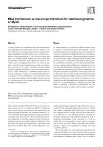
Figure 1. Structure of the double-stranded RNA-expressing RNA interference vector pDoub-neo. Targeting DNA inserts were cloned into the BamHI restriction site between the H1 and U6 promoters.
libraries could target any gene in the mammalian cell. Considering the fact that the targeting sequence is only approximately 21 nucleotides long, it is very difficult to establish a high-quality library that could cover most of the genes within mammalian cells. Previous studies have found that sequence-specific gene silencing could be induced in the mouse embryos [26, 27] and mouse oocytes [28 –31] by long double-stranded RNA (dsRNA). Transgenic expression of long dsRNA targeting the Ski gene in mouse embryos by using a vector with modified RNA polymerase II promoter showed phenotypes similar to Ski-knockout embryos, suggesting that dsRNA expression did not interfere with normal proliferation and differentiation of early embryo cells, since the normal growth of embryo was not affected [27]. Similar specific gene knockdown was also observed in ES cells [32–34], embryonic carcinoma cells [32], and some other mammalian cells [35–39], and dsRNA could be processed into siRNA inside the ES cells [32, 33]. Based on these findings, a novel approach for establishment of cDNAbased random RNAi library was developed in the study. The system used a convergent dual promoter vector with a single BamHI site between the two opposing promoters for cloning of cDNA fragments with compatible stick ends.
MATERIALS
AND
METHODS
ES Cell Culture Mouse ES cell lines CCE [40, 41] and R1 were used in the study and were cultivated on mouse embryonic fibroblasts treated with mitomycin C (Sigma-Aldrich, St. Louis, http://www.sigmaaldrich.com) or gelatin-coated plates in Dulbecco’s modified Eagle’s medium (DMEM) supplemented with 15% knockout serum replacement (KSR), 2 mM L-glutamine, 1 mM sodium pyruvate, 0.1 mM nonessential amino acids, 0.1 mM -mercaptoethanol (all from Invitrogen, Carlsbad, CA, http://www.invitrogen.com), and 10 ng/ml murine leukemia inhibitor factor (LIF) (Chemicon, Temecula, CA, http://www.chemicon.com). For experiments of screening and differentiation, KSR was replaced by fetal bovine serum (Biochrom AG, Berlin, http://www.biochrom.de), and 400 g/ml G418 (Calbiochem, San Diego, http://www.emdbiosciences.com) was added.
Construction of Plasmids with Convergent H1 and U6 Promoters Human H1 and U6 promoters were amplified from genomic DNA of HeLa cells. The primers used for amplification of the H1 promoter were 5⬘-AAGCTTTTCCAAAAACGCTGACGTCATCAACC-3⬘ and 5⬘-GGATCCTTTTTAGAGTGGTCTCATACAGA-3⬘. The polymerase chain reaction (PCR) fragment was cloned into BamHI/HindIII sites of the pBlueScriptII KS(⫺) vector (Stratagene, La Jolla, CA, http://www.stratagene.com), and the resulting plasmid was designated pH1. The U6 promoter sequence was amplified by using following primers: 5⬘-TCTAGAAAAAAAAGGTCGGGCAGGAAGA-3⬘ and 5⬘-GGATCCTTTTTTTCGTCCTTT CCACAA-3⬘. The PCR product was cloned into the XbaI/BamHI sites of the pH1 plasmid to generate plasmid pH1U6. For the purpose of stable transfection, a neomycin gene cassette was amplified from pcDNA3.1 plasmid (Invitrogen), and the primer pair used was 5⬘-CTCGAGCTGTGGAATGTGTG-3⬘ and 5⬘-GGTACCCAGACATGATAAGA-3⬘. The amplified neomycin gene cassette was cloned into XhoI/KpnI sites of the plasmid pH1U6, and the resulting plasmid was designated pDoub-neo (Fig. 1).
www.StemCells.com
Enhanced green fluorescent protein (EGFP) and myotubularin (MTM1) were selected to test efficiency of our RNAi vector design. By using pEGFP-N2 plasmid (Clontech, Palo Alto, CA, http://www. clontech.com) as template, 215-, 326-, and 720-bp fragments within egfp coding sequence were amplified by a set of primers (Table 1). After digestion with BamHI, the DNA fragments were cloned into pDoub-neo vectors, and the resulting plasmids were designated pDGFP215, pDGFP326, and pDGFP720, respectively. A 306-bp fragment of the MTM1 gene was amplified and digested with Sau3AI. The resulting 134- and 170-bp fragments were cloned into the BamHI site of pDoub-neo vector to construct plasmids pDM134 and pDM170, respectively. The ubiquitin-targeting RNAi vector pDUbi was constructed by cloning the Sau3AI-digested 85-bp fragment (coding sequence ⫹6 to ⫹90) of the mouse ubiquitin gene into the BamHI site of pDoub-neo. A control plasmid containing a 209-bp mouse genomic sequence was constructed and designated pDCont. Comparison of the 209-bp insert with sequences in the GenBank found no homology with any known gene.
Transfection of Embryonic Stem Cells The ES cells were transfected with plasmids by using Lipofectamine 2000 (Invitrogen) as directed by the manufacturer’s instructions. In the case of the EGFP knockdown experiment, EGFP expression plasmid pEGFP-N2 and individual RNAi plasmids were cotransfected at a ratio of 1:5. For selection of stable MTM1 gene knockdown cells, the plasmid-Lipofectamine mixture was added to 2 ⫻ 105 ES cells in suspension; the cells were then seeded in 1 well of a gelatin-coated 24-well plate. One day post-transfection, cells were trypsinized and plated onto a 10-cm tissue culture dish, and 400 g/ml G418 was added to the culture medium. Twelve days after selection, cell colonies were isolated and expanded for testing. Ubiquitin-targeting construct pDUbi was transfected into CCE or R1 ES cells, and stable transfectants were selected with 400 g/ml G418. The cells were cultured in ES cell culture medium with 1 ng/ml LIF (1/10 of normal concentration) or without LIF. Control construct pDCont was used as the negative control in all experiments.
Fluorescence-Activated Cell Sorting Analysis and Fluorescence Microscopy The number of EGFP-positive cells and the mean fluorescence intensity were quantified by fluorescence-activated cell sorting (Becton Dickinson Immunocytometry Systems, San Jose, CA, http://www.bd.com) 48 hours after transient transfection. Images of cells were collected and analyzed using a fluorescence microscope (Olympus, Tokyo, http://www.olympus-global.com).
Construction of a cDNA-Based Random RNAi Library Total RNA was extracted from mouse ES cells by TRIzol reagent (Invitrogen), and mRNA was isolated by a PolyATtract system (Promega, Madison, WI, http://www.promega.com). A cDNA synthesis kit (Takara, Otsu, Japan, http://www.takara.co.jp) was used to generate double-stranded cDNA. After digestion with Sau3AI, cDNA fragments were ligated into pDoub-neo vector linearized with BamHI and were transformed into Escherichia coli strain DH5␣. The library contained estimated 2.4 ⫻ 105 primary recombinants. Plasmids were extracted from 20 randomly selected colonies from the original library, and their inserts were sequenced.
Functional Genetic Screening with the Random RNAi Library The random RNAi library (4 g) was introduced into CCE ES cells (2 ⫻ 106) by using Lipofectamine 2000. Considering the fact that
cDNA-Based Random RNAi Library
1906
Table 1. Primers used in this study Gene
Primer sequence
EGFP
a
MTM1
b
MTM1 Oct-4 Nanog
ubiquitin GAPDH
Forward: 5⬘-GGATCCATGGTGAGCAAG-3⬘ Reverse: 5⬘-GGATCCAAGCACTGCACG-3⬘ 5⬘-GGATCCGTCTTGTAGTTG-3⬘ 5⬘-GGATCCTTACTTGTACAG-3⬘ Forward: 5⬘-GATCCCGAAATCGGCTTCCTGTACTGTCGT-3⬘ Reverse: 5⬘-TTAAAGATCTCGCATAAC-3⬘ Forward: 5⬘-ACAAGGCAACAGGAGGAGGATATG-3⬘ Reverse: 5⬘-AGCCGAGTCACAGTTGAATAAGA-3⬘ Forward: 5⬘-GGAAGCCGACAACAATGAGA-3⬘ Reverse: 5⬘-AGAGCAGTGACGGGAACAGA-3⬘ Forward: 5⬘-CAGCCCTGATTCTTCTACCA-3⬘ Reverse: 5⬘-CAGATGCGTTCACCAGATAG-3⬘ Forward: 5⬘-ATGCAGATCTTCGTGAAGAC-3⬘ Reverse: 5⬘-CCTTCTGGATGTTGTAATCA-3⬘ Forward: 5⬘-TTAGCCCCCCTGGCCAAGG-3⬘ Reverse: 5⬘-TCCTGCACCACCAACTGCT-3⬘
Product size (bp)
215 326 720 306 593 429 386 190 550
a
MTM1 primers were used for polymerase chain reaction (PCR) amplification for the construction of plasmids pDM134 and pDM170. MTM1 primers were used for semiquantitative reverse transcription-PCR. Abbreviation: bp, base pairs.
b
the transfection efficiency of ES cells was no more than 20%–30%, as evaluated by observation under a fluorescence microscope, we estimated that 1 ⫻ 105 clones were screened. One day after transfection, cells were digested with trypsin-EDTA solution. Five ⫻ 105 cells were seeded onto a 10-cm sterile bacteriological dish and cultured in DMEM supplemented with 400 g/ml G418 and in the absence of LIF and -mercaptoethanol to form embryoid bodies. The medium was changed at 2-day intervals, and 5 M all-trans retinoic acid (RA) (Sigma-Aldrich) was added from day 5 to day 8. After an 8-day induction, the floating aggregates were transferred to a new sterile bacteriological dish and cultured in suspension for an additional 4 days without RA. At day 12, embryoid bodies were collected, dissociated with trypsin-EDTA, and plated onto gelatincoated tissue culture dishes. In the following days, cells were cultured with standard ES medium lacking LIF until the cell clones formed. Undifferentiated self-renewal colonies were picked and expanded. Total DNA was extracted from the cells, and the inserts were amplified by using primers 5⬘-GCGCGTAATACGACTCACTATAG-3⬘ and 5⬘-GCTCGAGGTCGACGGTATCGATAAG-3⬘, which are located at the T7 promoter and KS primer-binding sites of pBlueScriptII KS(⫺) vector. The PCR products were sequenced and analyzed.
RNA Isolation and Semiquantitative Reverse Transcription-PCR Total RNA was extracted by using TRIzol reagent according to the manufacturer’s protocols. One g of total RNA was converted to cDNA by using oligo(dT)18 primers and the avian myeloblastosis virus reverse transcriptase (BioFlux, Hangzhou, China, http:// www.bioer.com.cn) in a 20-l reaction volume. The reverse transcription (RT) product was diluted to 100 l with sterile, distilled water, and 1 l of cDNA was used in subsequent PCRs, which were performed using Taq DNA polymerase (Tiangen, Beijing, http:// www.tiangen.com) and sets of primers (listed in Table 1). Amplification of GAPDH was used to normalize reactions internally. The RNA template not treated with reverse transcriptase from each sample was also added to the PCRs and run in parallel as negative control to show no contamination of genomic DNA. The PCR conditions were 5 minutes initial denaturation at 95°C; 50 seconds at 94°C, 50 seconds at 60°C, and 50 seconds at 72°C for the cycles (number of cycles used depended on individual genes [Table 1]); followed by 10 minutes of incubation at 72°C. PCR bands were visualized on 1.5% agarose gel stained with ethidium bromide.
Colony-Forming Assay and Immunofluorescence Staining ES cells were dissociated with trypsin to produce a single-cell suspension, and approximately 2 ⫻ 103 cells were seeded into
gelatin-coated 60-mm tissue culture dish. Cells were cultured for 4 – 6 days in the presence of a low concentration of LIF (1 ng/ml) or without LIF. Colonies were stained for the presence of Oct-4 and alkaline phosphatase to determine undifferentiated state of ES cells. For immunofluorescence staining, ES cells were fixed with 4% polyformaldehyde for 20 minutes at room temperature and permeabilized with 0.2% Triton X-100 in phosphate-buffered saline for 5 minutes. After blocking with 1% bovine serum albumin, the cells were incubated with anti-Oct-4 monoclonal antibody (clone 9E3.2; Chemicon) and fluorescein isothiocyanate-conjugated goat antimouse IgG antibodies (Sigma-Aldrich). Images were collected and analyzed by fluorescence microscopy (Olympus). The alkaline phosphatase was examined using a alkaline phosphatase detection kit (Chemicon) according to the manufacturer’s instructions. Colonies consisting entirely of compact, Oct-4-positive and alkaline phosphatase-positive ES cells were considered undifferentiated, whereas colonies consisting entirely of unstained cells with flattened, irregular morphology alone or mixed with stained cells were considered differentiated.
Western Blots Cells were lysed with 1⫻ Laemmli SDS sample buffer and heated in a boiling water bath for 5 minutes. The cell lysates were centrifuged at 17,900g for 5 minutes, and the supernatant was transferred to a new tube and stored at ⫺20°C. Proteins (10 –20 g) were separated by 10% polyacrylamide gel (for detection of ubiquitin and ubiquitinated proteins, 15% separating gel was used) and transferred to polyvinylidene fluoride membrane (Millipore, Billerica, MA, http://www.millipore.com) at 3.0 mA/cm2 for 30 minutes. The membrane was blocked with Tris-buffered saline containing 5% nonfat dry milk and 0.1% Tween-20 for 2 hours, and it was incubated with appropriate primary antibodies at 4°C overnight. The anti-MTM1 rabbit polyclonal antibodies (Abgent, San Diego, http://www.abgent.com), anti--catenin monoclonal antibody (BD Biosciences, San Diego, http://www.bdbiosciences.com), anti-Oct-4 monoclonal antibody (Chemicon), anti-ubiquitin monoclonal antibody (clone 6C1; Sigma-Aldrich), and anti--actin monoclonal antibody (Sigma-Aldrich) were used at dilutions of 1:600, 1:2,000, 1:1,500, 1:1,500, and 1:5,000, respectively. After incubation with peroxidase-conjugated goat anti-mouse IgG or goat anti-rabbit IgG antibodies, signals were detected with an enhanced chemiluminescence system (Santa Cruz Biotechnology Inc., Santa Cruz, CA, http://www.scbt.com) and exposed to x-ray films. The bands on the film were quantified by densitometry analysis.
Statistics Statistical analysis was performed using the SPSS for Windows package, release 10.0 (SPSS, Inc., Chicago, http://www.spss.com).
Jian, Cheng, Jiang et al.
1907
Figure 2. Knockdown of exogenous enhanced green fluorescent protein (EGFP) expression in embryonic stem (ES) cells. (A): Fluorescence microscopy observation. ES cells were cotransfected with following plasmids: (AA) and (AE), pDCont and pEGFP-N2; (AB) and (AF), pDGFP215 and pEGFP-N2; (AC) and (AG), pDGFP326 and pEGFP-N2; (AD) and (AH), pDGFP720 and pEGFP-N2. ES cells cotransfected with egfp-targeting constructs pDGFP215 and pDGFP326 had significantly reduced fluorescence when compared with control. Paired photographs were taken under fluorescence (top row) and normal light (bottom row) of the same field 48 hours posttransfection. Magnification, ⫻100. (B): FACS analysis was performed 48 hours postcotransfection. This is representative of three independent experiments. Both the ratio of EGFPpositive cells and the fluorescence intensity were decreased in ES cells cotransfected with egfp-targeting double-stranded RNA expression vectors. The ratios of EGFP-positive ES cells were as follows: pDCont and pEGFP-N2 cotransfected cells, 70.69%; pDGFP215 and pEGFP-N2 cotransfected cells, 50.23%; pDGFP326 and pEGFP-N2 cotransfected cells, 55.48%; pDGFP720 and pEGFP-N2 cotransfected cells, 61.25%. The mean fluorescence intensities of EGFP-positive ES cells (M1) were as follows: pDCont and pEGFP-N2 cotransfected cells, 898.84; pDGFP215 and pEGFP-N2 cotransfected cells, 146.36; pDGFP326 and pEGFP-N2 cotransfected cells, 225.34; pDGFP720 and pEGFP-N2 cotransfected cells, 522.41. (C): Significantly lower fluorescence intensity in ES cells cotransfected with egfp-targeting vectors as determined by FACS analysis. ES cells were cotransfected with following plasmids (columns from left to right): pDCont and pEGFP-N2, pDGFP215 and pEGFP-N2, pDGFP326 and pEGFP-N2, and pDGFP720 and pEGFP-N2. Data are expressed as mean ⫾ SD of three experiments. ⴱ, p ⬍ .01 compared with control.
Student’s t test was used to analyze the statistical differences. All results were expressed as mean ⫾ SD, and p ⬍ .05 was considered to be statistically significant.
RESULTS Strategy for Construction of cDNA-Based Random RNAi Library Previous studies have proved that long dsRNA could achieve specific gene knockdown in embryonic stem cells [32–34] and some mammalian cells [32, 35–39]. Based on these findings, a simple and efficient system to generate random RNAi library was designed. The system used a convergent H1 and U6 dual promoter vector, pDoub-neo, for the expression of dsRNA (Fig. 1). In this vector design, the last five nucleotides of each promoter were replaced by 5 adenines, which served as a termination signal for the opposing promoter [42]. A BamHI restriction enzyme site was added between these two convergent promoters to facilitate cloning of any DNA fragment with GATC stick ends (Fig. 1). www.StemCells.com
RNAs are expected to be transcribed from both strands by two convergent promoters to form dsRNA, which induces knockdown of target genes. The random RNAi library could be established by following a few simple steps. The RNAi vector pDoub-neo was linearized with BamHI and was treated with alkaline phosphatase to prevent self-ligation. cDNAs were digested by Sau3AI, and DNA fragments ranging from 50 to 500 bp were purified from the gel. Following ligation of the vectors with cDNA fragments, the recombinants were transformed into E. coli. The plasmids containing the random RNAi library were extracted from the bacteria and kept at ⫺20°C until needed. To determine whether the system works in embryonic stem cells, dsRNA expression vectors targeting egfp and MTM1 were constructed by the system described above, and their knockdown efficacy was studied.
Knockdown of EGFP Expression by dsRNA Expression Vector in Embryonic Stem Cells To test whether dsRNA transcribed from the convergent U6 and H1 promoter vector could induce knockdown of exogenous gene expression, DNA fragments of 215, 326, and 720 bp that
cDNA-Based Random RNAi Library
1908
pression compared with those cotransfected with control plasmid pDCont and pEGFP-N2 (Fig. 2A). A similar reduction rate of EGFP expression was also observed in ES cells cotransfected with pDGFP326 and pEGFP-N2. pDGFP720 had less effect on the expression of EGFP (Fig. 2A). Despite transfection of dsRNA expression vectors, the ES cells grew normally, without apparent toxic effect. The transfected ES cells were subjected to flow cytometry analysis as well. Compared with ES cells cotransfected with pDCont and pEGFP-N2, both pDGFP215 and pDGFP326 led to a dramatic decrease in both the ratio of EGFP-positive cells and the fluorescence intensity of all cells (Fig. 2B, 2C). The pDGFP720 caused less reduction on EGFP expression (Fig. 2B, 2C). These results demonstrated that dsRNA expressed from the convergent dual promoter vectors could induce efficient inhibition of exogenous EGFP expression in embryonic stem cells. This observation is consistent with previous reports using an siRNA-based approach [43, 44]. Figure 3. Inhibition of MTM1 expression by MTM1-targeting doublestranded RNA expression vectors as analyzed by Western blot (A) and semiquantitative reverse transcription-polymerase chain reaction (RTPCR) (B). (A): Western blot showing knockdown of endogenous MTM1 expression by MTM1-targeting vectors pDM134 and pDM170. Embryonic stem (ES) cells were untreated (lane 1) or transfected with control plasmid pDCont (lane 2), MTM1-targeting plasmid pDM134 (lane 3), or pDM170 (lane 4). The blot was probed with anti-MTM1 rabbit polyclonal antibodies. -Actin was used as a loading control. (B): Semiquantitative RT-PCR showing knockdown of endogenous MTM1 expression by MTM1-targeting vectors pDM134 and pDM170. RNA was extracted from normal CCE ES cells (lane 1) or from ES cells transfected with control plasmid pDCont (lane 2), MTM1-targeting plasmid pDM134 (lane 3), or pDM170 (lane 4). Semiquantitative RTPCR was performed using GAPDH as internal control. Abbreviations: GAPDH, glyceraldehyde-3-phosphate dehydrogenase; MTM1, myotubularin.
were located within the coding region of egfp gene were amplified by PCR and cloned into the BamHI site of pDoub-neo vectors by the strategy described above. The resulting constructs were designated pDGFP215, pDGFP326, and pDGFP720, respectively. Fluorescence microscopy observation showed that CCE ES cells cotransfected with pDGFP215 and EGFP expression plasmid pEGFP-N2 had significantly reduced EGFP ex-
Knockdown of Endogenous MTM1 Expression by dsRNA Expression Vector in Embryonic Stem Cells We then asked whether this dsRNA expression vector could inhibit expression of endogenous gene. The target we chose was the MTM1 gene. MTM1 encodes myotubularin, a ubiquitously expressed protein that may play an important role in muscle cell differentiation [45, 46]. In our previous study, we found that MTM1 was expressed in undifferentiated embryonic ES cells. MTM1-targeting vectors pDM134 and pDM170, which were expected to transcribe 134- and 170-bp dsRNA that targets different regions of the MTM1 coding sequence, were transfected into CCE ES cells and selected in the presence of G418. Western blot analysis showed that both pDM134- and pDM170transfected ES cells had more than 70% reduction of MTM1 expression compared with ES cells transfected with control plasmid pDCont alone, as determined by densitometry analysis (Fig. 3A). The inhibition was maintained for at least 3 months in ES cells. Results obtained with semiquantitative RT-PCR were consistent with the result of Western blot (Fig. 3B). These results indicated that our vector design could induce potent knockdown of endogenous gene in ES cells as well, and the inhibition was not due to toxic effect to the ES cells because
Table 2. Sequences of randomly selected clones from the cDNA-based RNA interference library Sequence location
Length (bp)
Gene
Accession number
596–701 227–317 8,491–8,598 8,671–8,736 360–211 588–657 1,236–1,134 2,964–2,907 4,117–4,079 1,175–1,270
106 91 108 66 150 70 103 58 39 96
BC032050 AY248756 AB114630 AB114630 NM_026684 NM_027422 NM_021422 NM_144921 NM_008057 NM_008871
975–1,217
243
959–1,170 1,367–1,266 2,373–2,524 22,434–22,516 742–440 946–767
212 101 152 83 303 180
IMAGE:3486529 18S ribosomal RNA-like mRNA Cadherin-related neuronal receptor Cadherin-related neuronal receptor NADH dehydrogenase (ubiquinone) 1 subcomplex RIKEN cDNA 2700087H15 DnaJ (Hsp40) homolog, subfamily A, member 4 (Dnaja4) ATPase, Na⫹/K⫹ transporting, ␣ 3 polypeptide Frizzled homolog 7 (Drosophila) (Fzd7) Serine (or cysteine) peptidase inhibitor, clade E, member 1 (Serpine1) ATP synthase, H⫹ transporting, mitochondrial F1 complex, ␣ subunit, isoform 1 (Atp5a1) Tubulin, ␣ 2 (Tuba2) Pituitary tumor-transforming 1 interacting protein Neuronal development-associated protein 7 (Ndap7) Chromosome 1, clone RP23–469D21 Dynactin 3 RNA cyclase homolog
Abbreviation: bp, base pairs.
NM_007505 NM_011654 BC029144 AF361435 AC102776 BC061120 BC053404
Jian, Cheng, Jiang et al.
Figure 4. Western blot showing knockdown of ubiquitin expression by ubiquitin-targeting construct pDUbi in embryonic stem (ES) cells. CCE ES cells were transfected with C or U and were cultured in the presence of 1 ng/ml LIF or in the absence of LIF (0). The blot was probed with anti-ubiquitin monoclonal antibody. -Actin was used as a loading control. Abbreviations: C, control plasmid pDCont; HMW, high molecular weight; LIF, leukemia inhibitor factor; U, reconstructed ubiquitintargeting plasmid pDUbi; Ub, ubiquitin; ub-conj, ubiquitin-conjugates.
neither -actin nor glyceraldehyde-3-phosphate dehydrogenase (GAPDH) expression was affected, as judged by both Western blot and semiquantitative RT-PCR.
Generation and Characterization of a cDNA-Based Random RNAi Library Having verified that dsRNA could induce potent RNAi in ES cells, we constructed a cDNA-based random RNAi library by using this convergent promoter vector system. The library we constructed was estimated to have 2.4 ⫻ 105 recombinants. To validate the library, 20 recombinant bacterial colonies randomly selected from the library were subjected to sequencing. Sequence analysis indicated that 17 had sequences identical to GenBank entries, with size of inserts ranging from 39 to 303 bp (Table 2). Of these 17 clones, two had inserts targeting the same gene but located at different regions, and one was a DNA fragment from genomic DNA. The result indicated that the library contained clones with good diversity and size range.
Screening for the Suspected Modulator Involved in Mouse ES Cell Differentiation Next, we sought to apply this random RNAi library to functional genetic screening in mouse ES cells. To reduce background, a 12-day culture process of CCE ES cells in the absence LIF and with addition of all-trans RA was used. By using this induced differentiation condition, the mouse ES cells transfected with control plasmid pDCont underwent nearly complete differentiation, and the isolated cell colonies were unpassageable. In contrast, six morphologically undifferentiated colonies were obtained from ES cells transfected with the random RNAi library. Two unique genes were identified by sequencing of the inserts and BLAST analysis. One clone contained a sequence that matches the mouse ubiquitin gene (GenBank accession number X51703), and the other one contained a sequence that matches the retinoic acid-induced 17 gene (Rai17) (GenBank accession number BC058646). www.StemCells.com
1909
We reconstructed the ubiquitin-targeting RNAi vector, which was designated pDUbi. ES cells were transfected with pDUbi or the control vector pDCont, and free ubiquitin and ubiquitinated proteins were detected by Western blot with antiubiquitin monoclonal antibody. As shown in Figure 4, most of the ubiquitinated proteins were of high molecular mass. The size of free ubiquitin monomer is approximately 8 kDa, which can be seen in the blot. Compared with controls, the expression of free ubiquitin and the amount of ubiquitinated proteins were significantly reduced in the ES cells transfected with pDUbi, in either the presence or the absence of LIF (Fig. 4). A similar reduction of ubiquitin expression was also observed by semiquantitative RT-PCR analysis (data not shown). This result demonstrated that ubiquitin-targeting vector pDUbi could inhibit the expression of ubiquitin in ES cells. The self-renewal capacity of CCE ES cells transfected with pDUbi was evaluated by a colony-forming assay. Colonies stained positive for Oct-4 and alkaline phosphatase were classified as undifferentiated. In the presence of 1 ng/ml LIF (1/10 of normal concentration), parental ES cells and control vector pDCont transfectants produced an estimated 20% of ES cell colonies of undifferentiated cells. In the absence of LIF, this ratio reduced to no more than 5% at the end of the 6-day assay (Fig. 5). In contrast, the ubiquitin knockdown cell line generated an 80%–90% of undifferentiated colonies in the presence 1 ng/ml LIF and formed typical compact morphology. Without LIF, 20%–30% of colonies were undifferentiated (Fig. 5). To determine whether knockdown of ubiquitin affects proliferation of ES cells, the cell number was counted over a 6-day period. Compared with controls, ubiquitin knockdown ES cells had a significantly increased growth rate at days 4 and 6 (p ⬍ .01; n ⫽ 6) in either the presence of 1 ng/ml LIF or the absence of LIF (Fig. 5E). The expression of Oct-4 and -catenin was examined by Western blot (Fig. 6) and RT-PCR (data not shown) in pDUbitransfected ES cells. The expression of both proteins was increased in pDUbi transfectants when compared with parental ES cells and control vector transfectants (Fig. 6). Similar results were also observed when R1 ES cells were used. These results confirmed that knockdown of ubiquitin promoted self-renewal of ES cells.
DISCUSSION Most RNAi libraries reported so far consist of a great array of individual siRNAs or siRNA expression vectors, and their construction and application were costly and labor-intensive [5–14, 19, 20]. Random siRNA libraries based on enzyme digestion of cDNA fragments might target any genes and were cost-effective, but their construction requires complicated steps to form siRNA expression vectors [22–24], making it a demanding task. An RNAi library established by cloning randomly synthesized 21–23-bp DNA fragments [19, 25] may contain great amounts of siRNAs that may not match any sequence in the genome of the cell. In this study, a simple and efficient new system to construct a cDNA-based random RNAi library was established. In our system, an RNAi library could be established simply by cloning enzyme-digested cDNAs into the BamHI site of the dual promoter RNAi vector. Our study demonstrates that the library can be used for functional genetic screens in ES cells. In mouse oocytes and embryos, long dsRNA could induce gene-specific RNAi, and the phenotypes exhibited were highly similar to those of gene-deficient mutants [26, 27]. This dsRNAinduced gene-specific knockdown was also observed in ES cells [32–34] and some other mammalian cells [32, 35–39]. We
1910
cDNA-Based Random RNAi Library
Figure 5. Knockdown of mouse ubiquitin promoted self-renewal of embryonic stem (ES) cells. (A): Morphology of representative colonies of CCE ES cells transfected with control vector pDCont or ubiquitin-targeting vector pDUbi 6 days after plating. The ES cells were cultured in the presence of 1 ng/ml LIF (pDCont(LIF) and pDUbi(LIF)) or in the absence of LIF (pDCont(0) and pDUbi(0)). Magnification, ⫻400. (B): Immunofluorescence staining of ES cell colonies with anti-Oct-4 monoclonal antibody (clone 9E3.2; Chemicon) as analyzed by fluorescence microscopy. ES cells were transfected with control vector pDCont or ubiquitin-targeting vector pDUbi and cultured in the presence of 1 ng/ml LIF (pDCont(LIF) and pDUbi(LIF)) or in the absence of LIF (pDCont(0) and pDUbi(0)). Strong fluorescence was observed in undifferentiated ES cell colonies. Magnification, ⫻400. (C): Alkaline phosphatase staining of ES cell colonies 6 days after plating. ES cells were transfected with control vector pDCont or reconstructed ubiquitin-targeting vector pDUbi. The cells were cultured in the presence of 1 ng/ml LIF (pDCont(LIF) and pDUbi(LIF)) or in the absence of LIF (pDCont(0) and pDUbi(0)). Undifferentiated colonies were stained red by alkaline phosphatase substrate. Magnification, ⫻100. (D): There was a significantly increased number of undifferentiated ES cell colonies in ubiquitin knockdown CCE ES cells 6 days after plating. Parental ES cells, control vector transfectants (pDCont), and ubiquitin-targeting construct transfectants (pDUbi) were cultured in the presence of 1 ng/ml LIF (LIF) or in the absence of LIF (0). The number of undifferentiated colonies was scored as a percentage of the total number of colonies. Data are expressed as mean ⫾ SD of four independent experiments. ⴱ, p ⬍ .01 compared with parental ES cells and control. (E): Increased growth rate of ubiquitin knockdown ES cells. ES cells were transfected with control vector pDCont or ubiquitin-targeting construct pDUbi and were cultured in the presence of 1 ng/ml LIF (LIF) or in the absence of LIF (0). Data are expressed as mean ⫾ SD of six independent experiments. At days 4 and 6, the difference was significant either in the presence of 1 ng/ml LIF (LIF) or in the absence of LIF (0) (p ⬍ .01). Abbreviation: LIF, leukemia inhibitor factor.
Jian, Cheng, Jiang et al.
Figure 6. Western blot showing increased expression of Oct-4 and -catenin following knockdown of ubiquitin expression. CCE embryonic stem cells were transfected with C or U and cultured in the presence of 1 ng/ml LIF (1/10 of normal concentration) (LIF) or in the absence of LIF (0). The blot was probed with anti-Oct-4 and anti--catenin monoclonal antibodies. -Actin was used as a loading control. Abbreviations: C, control plasmid pDCont; LIF, leukemia inhibitor factor; U, reconstructed ubiquitin-targeting plasmid pDUbi.
observed potent gene knockdown of both exogenous and endogenous genes in mouse ES cells. This knockdown of gene expression appeared to be specific, because the transfected ES cells grew normally, with healthy morphological appearance, and the expression of -actin and GAPDH was not affected. The observation demonstrated that this dsRNA expression system was functional in ES cells. Although it has been proved that dsRNA can induce efficient gene-specific RNAi in undifferentiated ES cells, the RNAi library may not be applicable to some differentiated cells due to nonspecific responses [34]. Three egfp-targeting dsRNA expression vectors with insert sizes ranging from 215 to 720 bp were tested for their efficacy in the knockdown of exogenous EGFP expression. The two vectors with insert sizes of 215 and 326 bp could efficiently inhibit the expression of EGFP in ES cells, whereas the vector with the 720-bp insert had less effect on EGFP expression. The explanation could be that the 720-bp insert may be too long to be transcribed efficiently by RNA polymerase III promoters, such as H1 and U6, which affects the formation of dsRNA in cells [18, 47]. A functional genetic screen with the random RNAi library was carried out by using a feeder-independent ES cell line, CCE cells [40, 41]. In monolayer culture, CCE cells differentiate spontaneously under conditions depleted of LIF and feeder cells. However, mixed colonies containing undifferentiated ES cells were always yielded, even after formation of three-dimensional embryoid bodies. To eliminate the background, an embryoid body formation combined with RA-induced differentiation was used in this study, and nearly all of the ES cells were differentiated under this culture condition. The transfection efficiency of ES cells estimated in the study varies, from approximately 20% to approximately 70%, depending upon the methods used for the evaluation. If transfection efficiency is evaluated by observation under a fluorescence microscope, only cells with clear visible fluorescence are con-
REFERENCES
1911
sidered positive. Flow cytometry is a highly sensitive technique, and cells with minimal expression of EGFP were still classified as positive. Many of the cells with fluorescence density close to the cut-off value may not have functional significance, and they may not show fluorescence when observed under fluorescence microscope. Therefore, we believe that functional transfection efficiency should be much lower than those obtained by flow cytometry. Ubiquitin is a conserved protein that is a component of the ubiquitin-dependent proteolysis pathway and functions by attaching to proteins to mark them for destruction. It also plays an important role in the controlling of cellular mRNA synthesis through both proteasome-dependent and proteasome-independent mechanisms [48]. Many studies have demonstrated that the level of cytoplasmic ubiquitin was elevated during cell differentiation [49 –54], and the ubiquitin-dependent proteolysis pathway plays an important role in the removal of proteins responsible for proliferation. Recent studies have reported that Oct-4, the master regulator affecting the fate of pluripotent embryonic stem cells, could be post-translationally modified by ubiquitination, which dramatically suppresses its transcriptional activity [55]. Ubiquitination is also involved in negative regulation of Wnt/-catenin signaling pathway [56]. Therefore, knockdown of ubiquitin could lead to increased number of ES cells that are maintained in an undifferentiated state, as reflected by elevated expression of both Oct-4 and -catenin, which could explain our finding that knockdown of ubiquitin promoted self-renewal of ES cells. The undifferentiated state of ES cells was confirmed by both immunofluorescence staining for Oct-4 and alkaline phosphatase staining. The antibody we used for the immunofluorescence staining is an anti-Oct-4 monoclonal antibody from Chemicon (product MAB4305, clone 9E3.2). Our experiments indicated that although the staining seems to be mainly cytosolic, it is specific for Oct-4. One possible explanation for cytosolic staining is that this antibody might react mainly with the newly synthesized cytoplasmic form of Oct-4. In conclusion, a novel approach to constructing a cDNAbased random RNAi library was developed in this study, and it provides a useful tool for investigation of molecular mechanisms of cellular differentiation and development.
ACKNOWLEDGMENTS This work was supported by Grants 30370603 and 30470094 from the National Natural Science Foundation of China; a grant from the Scientific Research Foundation for the Returned Overseas Scholars, Ministry of Education, China; and a grant from the Third Military Medical University, Chongqing, China.
DISCLOSURE
2 3
Keller G. Embryonic stem cell differentiation: Emergence of a new era in biology and medicine. Genes Dev 2005;19:1129 –1155. Mayhall EA, Paffett-Lugassy N, Zon LI. The clinical potential of stem cells. Curr Opin Cell Biol 2004;16:713–720. Torres RM, Kuhn R. Laboratory Protocols for Conditional Gene Targeting. New York: Oxford University Press, 1997.
www.StemCells.com
CONFLICTS
The authors indicate no potential conflicts of interest.
4 5
1
OF POTENTIAL OF INTEREST
6 7
Kamath RS, Fraser AG, Dong Y et al. Systematic functional analysis of the Caenorhabditis elegans genome using RNAi. Nature 2003;421:231–237. Aza-Blanc P, Cooper CL, Wagner K et al. Identification of modulators of TRAIL-induced apoptosis via RNAi-based phenotypic screening. Mol Cell 2003;12:627– 637. Kolfschoten IG, van Leeuwen B, Berns K et al. A genetic screen identifies PITX1 as a suppressor of RAS activity and tumorigenicity. Cell 2005;121:849 – 858. Westbrook TF, Martin ES, Schlabach MR et al. A genetic screen for candidate tumor suppressors identifies REST. Cell 2005;121:837– 848.
1912
8 9 10 11 12 13 14 15 16 17 18 19 20 21 22 23 24 25 26 27 28 29 30 31 32
Ngo VN, Davis RE, Lamy L et al. A loss-of-function RNA interference screen for molecular targets in cancer. Nature 2006;441:106 –110. Collins CS, Hong J, Sapinoso L et al. A small interfering RNA screen for modulators of tumor cell motility identifies MAP4K4 as a promigratory kinase. Proc Natl Acad Sci U S A 2006;103:3775–3780. Bjo¨rklund M, Taipale M, Varjosalo M et al. Identification of pathways regulating cell size and cell-cycle progression by RNAi. Nature 2006; 439:1009 –1013. Berns K, Hijmans EM, Mullenders J et al. A large-scale RNAi screen in human cells identifies new components of the p53 pathway. Nature 2004;428:431– 437. Moffat J, Grueneberg DA, Yang X et al. A lentiviral RNAi library for human and mouse genes applied to an arrayed viral high-content screen. Cell 2006;124:1283–1298. Silva JM, Li MZ, Chang K et al. Second-generation shRNA libraries covering the mouse and human genomes. Nat Genet 2005;37:1281–1288. Willingham AT, Orth AP, Batalov S et al. A strategy for probing the function of noncoding RNAs finds a repressor of NFAT. Science 2005; 309:1570 –1573. Hsieh AC, Bo R, Manola J et al. A library of siRNA duplexes targeting the phosphoinositide 3-kinase pathway: Determinants of gene silencing for use in cell-based screens. Nucleic Acids Res 2004;32:893–901. Paddison PJ, Silva JM, Conklin DS et al. A resource for large-scale RNAinterference-based screens in mammals. Nature 2004;428:427– 431. Silva JM, Mizuno H, Brady A et al. RNA interference microarrays: High-throughput loss-of-function genetics in mammalian cells. Proc Natl Acad Sci U S A 2004;101:6548 – 6552. Zheng L, Liu J, Batalov S et al. An approach to genomewide screens of expressed small interfering RNAs in mammalian cells. Proc Natl Acad Sci U S A 2004;101:135–140. Cheng X, Jian R, Deng S et al. RNA interference library and its application in functional genomics. Progress Biochem Biophys 2005;32: 195–198. Kittler R, Buchholz F. Functional genomic analysis of cell division by endoribonuclease-prepared siRNAs. Cell Cycle 2005;4:564 –567. Kittler R, Putz G, Pelletier L et al. An endoribonuclease-prepared siRNA screen in human cells identifies genes essential for cell division. Nature 2004;432:1036 –1040. Luo B, Heard AD, Lodish HF. Small interfering RNA production by enzymatic engineering of DNA (SPEED). Proc Natl Acad Sci U S A 2004;101:5494 –5499. Sen G, Wehrman TS, Myers JW et al. Restriction enzyme-generated siRNA (REGS) vectors and libraries. Nat Genet 2004;36:183–189. Shirane D, Sugao K, Namiki S et al. Enzymatic production of RNAi libraries from cDNAs. Nat Genet 2004;36:190 –196. Chen M, Zhang L, Zhang HY et al. A universal plasmid library encoding all permutations of small interfering RNA. Proc Natl Acad Sci U S A 2005;102:2356 –2361. Wianny F, Zernicka-Goetz M. Specific interference with gene function by double-stranded RNA in early mouse development. Nat Cell Biol 2000;2:70 –75. Shinagawa T, Ishii S. Generation of Ski-knockdown mice by expressing a long double-strand RNA from an RNA polymerase II promoter. Genes Dev 2003;17:1340 –1345. Svoboda P, Stein P, Filipowicz W et al. Lack of homologous sequencespecific DNA methylation in response to stable dsRNA expression in mouse oocytes. Nucleic Acids Res 2004;32:3601–3606. Svoboda P, Stein P, Hayashi H et al. Selective reduction of dormant maternal mRNAs in mouse oocytes by RNA interference. Development 2000;127:4147– 4156. Stein P, Zeng F, Pan H et al. Absence of non-specific effects of RNA interference triggered by long double-stranded RNA in mouse oocytes. Dev Biol 2005;286:464 – 471. Stein P, Svoboda P, Schultz RM. Transgenic RNAi in mouse oocytes: A simple and fast approach to study gene function. Dev Biol 2003;256: 187–193. Billy E, Brondani V, Zhang H et al. Specific interference with gene expression induced by long, double-stranded RNA in mouse embryonal teratocarcinoma cell lines. Proc Natl Acad Sci U S A 2001;98:14428 – 14433.
cDNA-Based Random RNAi Library
33 Yang S, Tutton S, Pierce E et al. Specific double-stranded RNA interference in undifferentiated mouse embryonic stem cells. Mol Cell Biol 2001;21:7807–7816. 34 Paddison PJ, Caudy AA, Hannon GJ. Stable suppression of gene expression by RNAi in mammalian cells. Proc Natl Acad Sci U S A 2002;99: 1443–1448. 35 Park WS, Miyano-Kurosaki N, Hayafune M et al. Prevention of HIV-1 infection in human peripheral blood mononuclear cells by specific RNA interference. Nucleic Acids Res 2002;30:4830 – 4835. 36 Gan L, Anton KE, Masterson BA et al. Specific interference with gene expression and gene function mediated by long dsRNA in neural cells. J Neurosci Methods 2002;121:151–157. 37 Yi CE, Bekker JM, Miller G et al. Specific and potent RNA interference in terminally differentiated myotubes. J Biol Chem 2003;278:934 –939. 38 Konstantinova P, de Vries W, Haasnoot J et al. Inhibition of human immunodeficiency virus type 1 by RNA interference using long-hairpin RNA. Gene Ther 2006;13:1403–1413. 39 Strat A, Gao L, Utsuki T et al. Specific and nontoxic silencing in mammalian cells with expressed long dsRNAs. Nucleic Acids Res 2006; 34:3803–3810. 40 Keller G, Kennedy M, Papayannopoulou T et al. Hematopoietic commitment during embryonic stem cell differentiation in culture. Mol Cell Biol 1993;13:473– 486. 41 Robertson E, Bradley A, Kuehn M et al. Germ-line transmission of genes introduced into cultured pluripotential cells by retroviral vector. Nature 1986;323:445– 448. 42 Jian R, Peng T, Deng S et al. A simple strategy for generation of gene knockdown constructs with convergent H1 and U6 promoters. Eur J Cell Biol 2006;85:433– 440. 43 Velkey JM, Slawny NA, Gratsch TE et al. Gene silencing using RNA interference in embryonic stem cells. Methods Mol Biol 2006;329:233–261. 44 Tang FC, Meng GL, Yang HB et al. Stable suppression of gene expression in murine embryonic stem cells by RNAi directed from DNA vector-based short hairpin RNA. STEM CELLS 2004;22:93–99. 45 Taylor GS, Maehama T, Dixon JE. Inaugural article: Myotubularin, a protein tyrosine phosphatase mutated in myotubular myopathy, dephosphorylates the lipid second messenger, phosphatidylinositol 3-phosphate. Proc Natl Acad Sci U S A 2000;97:8910 – 8915. 46 Buj-Bello A, Laugel V, Messaddeq N et al. The lipid phosphatase myotubularin is essential for skeletal muscle maintenance but not for myogenesis in mice. Proc Natl Acad Sci U S A 2002;99:15060 –15065. 47 Tuschl T. Expanding small RNA interference. Nat Biotechnol 2002;20: 446 – 448. 48 Conaway RC, Brower CS, Conaway JW. Emerging roles of ubiquitin in transcription regulation. Science 2002;296:1254 –1258. 49 Zavrski I, Krebbel H, Wildemann B et al. Proteasome inhibitors abrogate osteoclast differentiation and osteoclast function. Biochem Biophys Res Commun 2005;333:200 –205. 50 Kagan WA, Siegal FP, Gupta S et al. Early stages of human marrow lymphocyte differentiation: Induction in vitro by thymopoietin and ubiquitin. J Immunol 1979;122:686 – 691. 51 Chen CY, Pajak L, Tamburlin J et al. The effect of proteasome inhibitors on mammalian erythroid terminal differentiation. Exp Hematol 2002;30: 634 – 639. 52 Nahreini P, Andreatta C, Prasad KN. Proteasome activity is critical for the cAMP-induced differentiation of neuroblastoma cells. Cell Mol Neurobiol 2001;21:509 –521. 53 Shang F, Gong X, McAvoy JW et al. Ubiquitin-dependent pathway is up-regulated in differentiating lens cells. Exp Eye Res 1999;68:179 –192. 54 La Rosa FG, Kumar S, Prasad KN. Increased expression of ubiquitin during adenosine 3⬘,5⬘-cyclic monophosphate-induced differentiation of neuroblastoma cells in culture. J Neurochem 1996;66:1845–1850. 55 Xu HM, Liao B, Zhang QJ et al. Wwp2, an E3 ubiquitin ligase that targets transcription factor Oct-4 for ubiquitination. J Biol Chem 2004; 279:23495–23503. 56 Angers S, Thorpe CJ, Biechele TL et al. The KLHL12-Cullin-3 ubiquitin ligase negatively regulates the Wnt-beta-catenin pathway by targeting Dishevelled for degradation. Nat Cell Biol 2006;8:348 –357.