The Open Orthopaedics Journal, 2008, 2, 33-39
33
A Finite Element Model of Electrode Placement During Stimulus Evoked Electromyographic Monitoring of Iliosacral Screw Insertion M.A. Kopec1, B.R. Moed2 and D.W. Barnett*,1 1
Department of Biomedical Engineering, Saint Louis University, St. Louis, MO, USA
2
Department of Orthopaedic Surgery, Saint Louis University School of Medicine, St. Louis, MO, USA Abstract: Pelvic ring fractures that occur as a result of substantial orthopedic trauma are frequently repaired using iliosacral screws to stabilize the fracture. Stimulus evoked electromyography, using pulsed current stimuli provided through the drill bit cathode, has been advocated to prevent nerve root injury during iliosacral screw insertion. Our objective was to examine the effects of anode location, drill bit position, and anatomical structure on the nerve monitoring technique. A three-dimensional finite element model was constructed from computed tomography data to evaluate the effectiveness of five anode locations at four stations of drill bit insertion. Results indicate that the anode location should be at the midline or on the side contralateral to drill bit insertion. Locating the anode at other positions, such that the nerve root is outside of the primary electromagnetic field, leads to an attenuated electromyographic response that will ultimately lead to the failure of the monitoring technique.
Keywords: Finite element modeling; spinal nerve roots; stimulus-evoked electromyography. INTRODUCTION Pelvic ring fractures resulting from motor vehicle accidents or substantial falls are a relatively common form of orthopaedic trauma. When the posterior portion of the ring is displaced and/or unstable, a common method of restoring mechanical stability is the insertion of iliosacral screws. However, the proximity of the ideal screw trajectory to the fifth lumbar (L5) and first sacral nerve roots (S1), as well as the spinal canal, make neural tissue injury a potential hazard. Screw trajectory deviation of as little as 4° has been reported to direct the screw into the S1 nerve root [1]. To assist in proper screw placement, guide-wires are inserted under fluoroscopic guidance followed by the screws. However, fluoroscopy alone has not been shown to be sufficient for avoiding nerve root injury, which has been reported to range from 1% to as many as 18% of cases [2, 3]. Newer image navigation systems, such as fluoroscopy-based 3-D imaging and fluoroscopically registered computer-assisted surgery techniques, have yet to demonstrate any improvement over standard fluoroscopic techniques [4, 5]. As an alternative to these imaging methods, a simple technique to reduce the risk of surgical complications based on an evoked electromyographic (EMG) response may be used to determine the proximity of the drill bit to the neural structure [3, 6-8]. The drill bit’s depolarizing stimulus, created by a small current (< 50 mA searching current delivered in 0.2 ms duration pulses three times per second) between the tip of the dill-bit and a reference electrode [7, 8], causes action potential propagation from the nervous tissue towards the muscles they innervate. The magnitude of the stimulus required to induce a 20 mV EMG response (the current threshold) can be used to infer the distance between the tip of the drill bit *Address correspondence to this author at the Department of Biomedical Engineering, Saint Louis University, St. Louis, MO, USA; E-mail:
[email protected]
1874-3250/08
and the neural structure intraoperatively. The location of the reference electrode relative to the drill bit determines the path which current flows through the body, and therefore the potential at any point on the nervous structure. The significance of anode location on the effectiveness of the nerve monitoring technique has been identified as an important variable [6, 9, 10]. This study aims to computationally investigate the effect of anode location on the current density observed in the first sacral nerve root (S1) using finite element modeling (FEM). This method has been employed extensively when modeling electrical activity in the torso and cranium, but not in the midsection [11-14]. Examination of different reference electrode locations is straightforward using a FEM. The modeling approach provides a lucid illustration of the current emanating from the cathode that is easily reconfigurable for examining multiple configurations. The insight provided by the FEM provides a clear scientific justification for the selection of an optimal anode location. MATERIALS AND METHODOLOGY Ninety-three axial computed tomography (CT) slices from the level of the fourth lumbar vertebrae to the diaphysis of the femur were obtained from a healthy female subject with 0.08 cm x 0.08 cm x 0.25 cm resolution. The voxel data were segmented by tissue type in a commercially available software package (Amira 3.1, Mercury Computer Systems Inc., Chelmsford, MA). These data were used to create tissue surfaces, which were compiled to construct a solid tetrahedral mesh in the same software environment. The number of elements of each tissue type and their conductivity values, drawn from well-accepted studies, are shown in Table 1. The final mesh was imported into the FEM solver for analysis (Algor V16, Algor Inc., Pittsburgh, PA). The changes in tissue impedance that occur with changes in frequency were ignored for this analysis since the bandwidth of the stimulus is approximately 5 kHz and reported tissue impedances show little change over the low frequency ranges considered here [13]. Anisotropy of the tissue conductance was also ignored. 2008 Bentham Science Publishers Ltd.
34 The Open Orthopaedics Journal, 2008, Volume 2
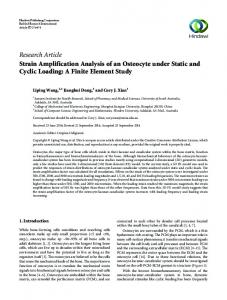
These assumptions allowed the electrostatics module in Algor, which solves the governing equation (Poisson) over the entire computational domain, to be used to calculate the current density throughout the model. The anode was assigned the ground potential and the stimulus current was generated in elements located at the end of the 6.5 mm diameter drill bit (representative of commercially available systems). Analysis time was approximately ten minutes on a 3.4 GHz Dell Dimension 370 (Dell Inc., Round Rock, TX). The number of elements in each tissue and their conductivity values are shown in Table 1. The current density in the S1 nerve root was studied in two basic configurations. First, the five anode locations depicted in Fig. (1) were examined with the drill bit (cathode) in the normal final resting position (station III). The five anode locations were the contralateral anterior superior iliac spine (CASIS), the ipsilateral anterior superior iliac spine (IASIS), the dorsal and ventral midlines (DM, VM) at the level of the umbilicus, and 2 cm anterior to the insertion site (IS). Regression data from animal studies [8] were used to determine the applied current (7.28 mA) based on a 5 mm perpendicular distance from the drill bit to the nerve root, in the transverse plane, with the anode at the VM location. Second, using the ventral midline anode location, models were created to examine four stations of screw insertion: station I: the sacroiliac joint; station II: prior to the tip of the drill bit crossing anteromedial to the nerve root; station III: the normal final screw resting position (drill bit tip located approximately on the midline of the body); and station IV: the drill bit tip approaching the contralateral nerve root. As with the anode location comparison, the minimum perpendicular drill bit to nerve root distance was 5 mm and all models were subjected to the same stimulus level of 7.28 mA. Finally, with the drill bit at station III, the insertion pathway was adjusted such that the drill bit passed at a disTable 1.
Kopec et al.
tance of 2 mm, 5 mm or 10 mm from the nerve root as measured orthogonal to the direction of the screw insertion, see Fig. (1). Once again, the anode was located at the VM location and the stimulus level was either held constant at 8 mA, or selected, based on the distance [8], as 4.71 mA for the 2mm model, 7.28 mA for the 5 mm model, and 10.78 mA for the 10 mm model.
Fig. (1). A CT image showing the five simulated anode locations and four drill bit positions of surgical interest along the path described as insertion motion. Translation of the drill bit from the nerve root towards the anterior cortex of the sacrum, identified as perpendicular motion, is also modeled at three distances (2 mm, 5 mm, and 10 mm).
The nerve root current density magnitude was exported for analysis in Matlab (The Mathworks, Natick, MA). The maximum current density magnitude (|Jmax|) at an axial cross section of the ipsilateral S1 nerve root was examined under each of the test conditions described previously. It is important to note that an increase in peak |Jmax| coincides with a lower current threshold for evoking an EMG re-
Model Element Count and Conductivity Data by Tissue Type Tissue
Conductivity (S/m)
Number of Elements
Fat
0.0400 a
86751
Intestine
0.1000 a
26021
Isotonic Soft Tissue
1.20000
13245
Muscle
0.1050 a
105309
Bone
0.0057 b
74759
Vasculature (Whole blood)
0.6250 c
5672
Nervous Tissue
0.1736 d
50829
Vertebral Disc
0.7355 e
659
Cerebrospinal Fluid
1.4580 d
14284
Bladder (Urine)
3.0000 f
3273
Drill Bit (Ti6Al4V)
571600
2822
Boundary and Current Source Elements
70
Total
383694
a See Schwan and Kay [15]; b Averaged from Saha and Williams [16]; c See Geddes and Sadler [17]; d See Geddes and Baker [18]; e See Gu et al. [19]; f See Bichonski and Pawelek [20]. The number of elements shown is for the model with drill bit to nerve root distance of 5 mm, station III of its insertion path, and the VM anode location.
A Finite Element Model of Electrode Placement
The Open Orthopaedics Journal, 2008, Volume 2
sponse. Although not presented here, the sensitivity of the model was assessed in order to eliminate any modeldependent results due to mesh size and conductivity parameters. Specifically, the number of tetrahedral elements for each model was selected such that further increasing the mesh density did not impact the resultant current density (