RESEARCH ARTICLE
A molecular dynamics-based algorithm for evaluating the glycosaminoglycan mimicking potential of synthetic, homogenous, sulfated small molecules Balaji Nagarajan1, Nehru Viji Sankaranarayanan1, Bhaumik B. Patel2,3, Umesh R. Desai1*
a1111111111 a1111111111 a1111111111 a1111111111 a1111111111
OPEN ACCESS Citation: Nagarajan B, Sankaranarayanan NV, Patel BB, Desai UR (2017) A molecular dynamics-based algorithm for evaluating the glycosaminoglycan mimicking potential of synthetic, homogenous, sulfated small molecules. PLoS ONE 12(2): e0171619. doi:10.1371/journal.pone.0171619 Editor: Nikos K Karamanos, University of Patras, GREECE Received: December 1, 2016 Accepted: January 23, 2017 Published: February 9, 2017 Copyright: © 2017 Nagarajan et al. This is an open access article distributed under the terms of the Creative Commons Attribution License, which permits unrestricted use, distribution, and reproduction in any medium, provided the original author and source are credited. Data availability statement: All relevant data are within the paper and its Supporting Information files.
1 Institute for Structural Biology, Drug Discovery and Development and Department of Medicinal Chemistry, Virginia Commonwealth University, Richmond, Virginia, United States of America, 2 Hunter Holmes Muire VA Medical Center, Richmond, Virginia, United States of America, 3 Division of Hematology, Oncology, and Palliative Care, Department of Internal Medicine and Massey Cancer Center, Virginia Commonwealth University, Richmond, Virginia, United States of America *
[email protected]
Abstract Glycosaminoglycans (GAGs) are key natural biopolymers that exhibit a range of biological functions including growth and differentiation. Despite this multiplicity of function, natural GAG sequences have not yielded drugs because of problems of heterogeneity and synthesis. Recently, several homogenous non-saccharide glycosaminoglycan mimetics (NSGMs) have been reported as agents displaying major therapeutic promise. Yet, it remains unclear whether sulfated NSGMs structurally mimic sulfated GAGs. To address this, we developed a three-step molecular dynamics (MD)-based algorithm to compare sulfated NSGMs with GAGs. In the first step of this algorithm, parameters related to the range of conformations sampled by the two highly sulfated molecules as free entities in water were compared. The second step compared identity of binding site geometries and the final step evaluated comparable dynamics and interactions in the protein-bound state. Using a test case of interactions with fibroblast growth factor-related proteins, we show that this three-step algorithm effectively predicts the GAG structure mimicking property of NSGMs. Specifically, we show that two unique dimeric NSGMs mimic hexameric GAG sequences in the protein-bound state. In contrast, closely related monomeric and trimeric NSGMs do not mimic GAG in either the free or bound states. These results correspond well with the functional properties of NSGMs. The results show for the first time that appropriately designed sulfated NSGMs can be good structural mimetics of GAGs and the incorporation of a MD-based strategy at the NSGM library screening stage can identify promising mimetics of targeted GAG sequences.
Funding: This work was supported by grants from the NIH, including HL107152, HL090586 and HL128639, and internal funds from VCU. Competing interests: The authors have declared that no competing interests exist.
PLOS ONE | DOI:10.1371/journal.pone.0171619 February 9, 2017
1 / 23
GAG mimicking potential of small molecules
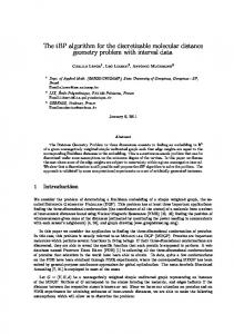
Introduction Glycosaminoglycans (GAGs), major constituents of the extracellular matrix, participate in regulating many different physiological and pathological processes by targeting a broad spectrum of proteins [1,2]. These negatively charged polymers recognize target proteins on the basis of number, density and distribution of sulfate groups. Because the biosynthesis of GAGs is a template-less process, nature tends to produce a large number of sulfation patterns in GAGs. Further, conformational biases of individual saccharide residues, e.g., 1C4 and 2SO of iduronic acid, introduce additional diversity to the GAG scaffold. This implies that a GAG sequence as small as a hexasaccharide can exist in thousands of possible distinct topologies [3] Although not all of these sequences are likely to induce a biological function, the massive heterogeneity present in a typical GAG population appears to be important for ensuring a higher probability of functional success, e.g., cell growth and migration, morphogenesis, inflammation and immunity [2,4]. A key feature of majority of GAG–protein systems is that the affinity and specificity of interaction relies primarily on the surface exposed sulfate groups and minimally on backbone atoms of GAGs. Some time ago we hypothesized that it may be more advantages to develop homogeneous, synthetic molecules containing appropriate number of sulfate groups as functional mimetics of GAGs because of the well-known difficulties with synthesizing or preparing/purifying GAG sequences [5]. This hypothesis then led to the synthesis of several homogeneous non-saccharide glycosaminoglycan mimetics (NSGMs, see Fig 1), which displayed major promise in modulating a number of biological processes [6–9]. The NSGMs studied so far have been found to engage their protein targets by means of hydrophobic, hydrogen bonding and Columbic forces, thereby affording considerable selectivity of recognition [5]. NSGMs prepared to date possess an aromatic backbone, whereas GAGs are based on the saccharide scaffold. NSGMs bear sulfate groups on phenolic groups, whereas GAGs bear sulfates on alcoholic or amine groups. NSGMs are generally achiral, whereas GAGs possess multiple chiral centers. These diametric differences bring forth a fundamental question on whether NSGMs are structurally equivalent or similar to GAGs. In fact, the number of NSGMs being studied in the literature is growing fast [6–12] and there is a strong possibility of developing drugs and/or chemical probes based on the promising NSGM leads identified to date. Further, the future of NSGMs as a technology is also promising because of their easier synthesis [5,7,9,11,13] and ability to apparently mimic GAG sequences at will. Thus, it is important to address the question on whether NSGMs structurally mimic GAGs effectively. Yet, tools for addressing this have not been established. In fact, availability of such tools may catapult the design of better NSGMs. Recently, we studied a small library of NSGMs for inhibition of cancer stem cells (CSCs) [11]. Screening a library of 53 NSGMs from 12 different scaffolds resulted in identification of G2.1 and G2.2 (Fig 1) as inhibitors of CSCs. Interestingly, neither G1.1 nor G4.1 (Fig 1) were found to exhibit this activity suggesting a high level of selectivity. Why do G2.1 and G2.2 function as CSCs inhibitors but not G1.1 and G4.1? Is it possible that G2.1 and G2.2 mimic a GAG sequence of defined chain length, e.g., a heparan sulfate (HS) hexasaccharide (HS06)? This may be the case because HS sequences are known to regulate growth by binding to fibroblast growth factor 2 (FGF2) and/or its receptor (FGFR1) [14,15]? A traditional approach to evaluate structural equivalence of GAGs is to compare similarity of sulfate groups in static structures [16,17]. However, sulfate moieties, especially present on GAGs, exhibit considerable dynamism, which is likely to be important in inducing functional properties. Thus, we utilized molecular dynamics (MD) to study the behavior of NSGMs (i.e.,
PLOS ONE | DOI:10.1371/journal.pone.0171619 February 9, 2017
2 / 23
GAG mimicking potential of small molecules
Fig 1. Structures of heparan sulfate (HS) sequences of varying lengths (HS02 to HS08) and non-saccharide glycosaminoglycan mimetics (NSGMs) G1.1, G2.1, G2.2 and G4.1. G1.1 can be considered as monomeric NSGM; G2.1 and G2.2 are approximately dimeric and G4.1 can be thought of as tetrameric NSGM. doi:10.1371/journal.pone.0171619.g001
G1.4, G2.1, G2.2 and G4.1) and compared it with HS06 in free and protein-bound states This led to a three-step algorithm that helped predict GAG mimicking ability of different NSGMs. The algorithm predicted that although G2.1 and G2.2 have very similar structures, G2.2 mimics HS06 better than G2.1 in solution, while G1.1 and G4.1 exhibit a completely different profile. In the protein-bound form (FGF2 and FGF2–FGFR1 complex), G2.2 is predicted to interact with residues that engage HS06 and these complexes are better stabilized than equivalent ones with G2.1. These results provide a structural foundation for the literature report on the functional activity of NSGMs as anti-CSC agents. This is the first report on the development of a detailed computational algorithm for assessing equivalence of NSGMs with natural GAG sequences. We posit that this algorithm, or variants thereof, can now be implemented on a high-throughput scale for discovering/designing novel NSGMs.
Methods Molecular dynamics (MD) of NSGMs The initial models of NSGMs were built using SYBYLX2.1 (Tripos Associates, St. Louis, MO). Gasteiger-Hu¨ckel charges were assigned to the molecules and then each NSGM was minimized using conjugate gradient method for 10,000 iterations. Explicit MD simulations were then carried out using the AMBER14 package [18,19], using periodic boundary condition and long range interactions were calculated using particle Mesh Ewald method, which utilizes a generalized Amber force field (GAFF) for small organic molecules [20]. This force field has been optimized for a diverse range of organic scaffolds by employing a semi-empirical AM1-BCC charge model [21]. Initial models of NSGMs were loaded to the antechamber module of AMBER14 to assign appropriate charge and torsional angle parameters [22] and then neutralized by Na+ counter ions to give a net charge of zero. The solute molecules were set in
PLOS ONE | DOI:10.1371/journal.pone.0171619 February 9, 2017
3 / 23
GAG mimicking potential of small molecules
center and were solvated in a cuboid periodic box of TIP3P water molecules [23] with a minimum distance of 10 Å between the wall and any atom of the solute. Initial parameters and coordinates files were generated followed by a two-step minimization process. In first step the solute and the Na+ ions were restrained using a harmonic potential of 100 kcal/(mol Å2). The water molecules were relaxed using 500 cycles of steepest descent and 1500 cycles of conjugate gradient method. In the second step the whole system was relaxed to 2500 cycles of conjugate gradient minimization. Following this the system was brought to constant temperature (300 K) using the Berendsen temperature coupling with time constant 2 ps and then brought to constant pressure (1 atm). Finally the system was equilibrated (at NPT) without any restraints. All these phases were performed for a total time period of 1 ns with 2 fs integration time step. The NPT MD simulation was carried out after equilibrating the system, with integration time step of 1 fs for a total time period of 20 ns, during which the ensemble coordinates were collected at every 1 ps. The covalent bonds involving hydrogen atoms were constrained using SHAKE algorithm throughout the simulation.
MD of heparan sulfate hexasaccharide (HS06) The exploration of HS06 dynamics in explicit water was performed using AMBER14 with GLYCAM06h force field parameters [24]. Two independent initial HS06 starting structures for this simulation were taken from the experimental NMR structure (PDBID: 1HPN) [25]. This structure has a repeating sequence of (IdoA2S-GlcNS6S)3 with IdoA2S existing in either chair 1C4 or skew boat 2SO conformations, as shown in (Fig A in S1 File). The initial structures were loaded in Leap and the system was neutralized with appropriate number of Na+ counter ions. The molecules were solvated in a periodic box of TIP3P water molecules [23] with a distance of 10 Å between the box edge and solute surface. A protocol similar to that of NSGM simulations was followed here (see the section above), except for an additional weak torsional restrain to keep the pyranose ring of IdoA2S in either 2SO or 1C4 conformation throughout the simulation [26,27].
Molecular docking of NSGMs binding to proteins GOLD v5.2 (from Cambridge Crystallographic Data Center, Cambridge, UK) was employed to study the interaction of NSGMs with proteins [28]. The three dimensional structures of FGF2 (PDBID: 1BFC) [29] and FGF2–FGFR1 (PDBID: 1FQ9) [30] complex were obtained from Protein Data Bank (http://www.rcsb.org). Protein preparation was carried out using SYBYL X2.1, which included addition of hydrogen and missing atoms, protonation of residues, removal of steric clashes and energy minimization. The centroid of the GAG-binding sites in these proteins (FGF2 and FGF2–FGFR1 complex) was taken as the center and 18 Å radius was defined as the binding site. Docking was performed for 300 genetic algorithm runs with 100,000 iterations and early termination option was disabled. The GOLD fitness score was calculated from the contributions of hydrogen bond and van der Waals interactions between the protein and ligand [28]. From the GOLD based docking, the best sampled pose (highest GOLD score) with