A Molecular Dynamics Simulation Based Prediction of Deleterious Angiogenin Mutations Causing Amyotrophic Lateral Sclerosis Aditya K. Padhi1, B. Jayaram1, 2, 3, James Gomes1* 1
Kusuma School of Biological Sciences, 2 Department of Chemistry, 3 Supercomputing Facility for Bioinformatics and Computational Biology, Indian Institute of Technology Delhi, India. ABSTRACT
Amyotrophic lateral sclerosis (ALS) is a fatal neurodegenerative disorder characterized by the selective death of motor neurons leading to paralysis and death between 3-5 years of diagnosis. Through whole genome association studies, several single nucleotide polymorphisms (SNPs) encoding missense mutations in angiogenin (ANG) protein have been identified as one of the primary factors causing ALS. Structural studies of ANG show that catalytic triad comprising His13, Lys40 and His114 residues imparts ribonucleolytic activity while nuclear localization signal residues 31RRR33 are responsible for nuclear translocation activity. Loss of either ribonucleolytic activity or nuclear translocation activity or both of these functions due to mutations cause ALS. However, the mechanisms of loss-of-functions of ANG mutants are not completely understood. Here we present a cohesive and comprehensive picture of functional loss mechanisms of all known ALS associated ANG mutants by extensive molecular dynamics (MD) simulations. Our studies show that conformational switching of catalytic residue His114 is responsible for the loss of ribonucleolytic activity while reduction in solvent accessible surface area (SASA) of 31RRR33 as a result of local folding is responsible for the loss of nuclear translocation activity. Our prediction of loss-of-functions of 17 ANG mutants correlated positively with the reported experimental results. We have subsequently developed a fast molecular dynamics method based on certain global attributes / dynamic markers that can be used to determine whether a mutation is deleterious or benign. To make our method accessible to researchers and clinicians, we created a web server based tool, ANGDelMut, freely available at http://bioschool.iitd.ernet.in/research.htm, where a user can submit new mutations to ascertain whether they cause ALS. We hope that our method will benefit the community at large and will pave the way for the development of a successful therapy for patients suffering from ALS. ANGDelMut
INTRODUCTION ALS is a fatal progressive neurodegenerative disorder affecting motor neurons in the motor cortex, brain stem and spinal cord, consequently leading to paralysis and death, usually between 3-5 years of diagnosis. Mutations in several genes such as SOD1, FUS/TLS, TARDBP, OPTN, VAPB, ANG, FIG4 and a hexanucleotide repeat expansion (GGGGCC) in the C9ORF72 have been identified as the causative genetic factors of adult onset ALS. ANG has emerged as one of the most frequently mutated genes in ALS cases. ANG, a 14.1 kDa protein, encoded by ANG gene is a member of the pancreatic ribonuclease A superfamily and stimulates angiogenesis. Structural study revealed that ANG has three functional sites that includes the receptor binding site (59NKNGNPHREN68), catalytic triad (His13, Lys40 and His114) and nuclear localization signal (NLS) consists of 29IMRRRGL35, necessary for its angiogenic activity. Catalytic triad imparts the ribonucleolytic activity and NLS is responsible for nuclear translocation activity of ANG. Loss of either RA, NTA or both of these functions of ANG due to missense mutations lead to ALS pathogenesis. Mechanisms of functional loss of ANG mutants and hence disease pathogenesis was not completely understood.
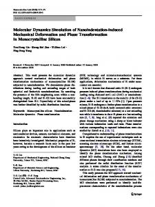
Figure. Structure of human ANG with functional sites. catalytic triad residues are represented as stick model, nuclear localization signal (NLS) is represented as lime green color and receptor binding site (RBS) is represented as violet color.
Figure. Cartoon representation of human angiogenin showing missense mutations found in ALS patients. All the reported ALS associated angiogenin (PDB ID: 1B1I) mutations are labelled and represented as stick model (lime green colour).
DISCUSSION
RESULTS
(A) Investigating the loss of ribonucleolytic activity
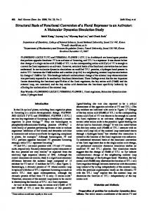
Figure. Conformational switching of His114 in ALS associated ANG mutants. (A) Observed stable and native conformation of catalytic residue His114 during MD simulations of WT-ANG and R121H, K54E, R121C, V103I, F100I-ANG mutants (B) Observed conformational switching of catalytic residue His114 in I46V, K17E, R31K, D22G, T80S and N49S-ANG mutants from MD simulations.
Quantification of His114 dihedral angle
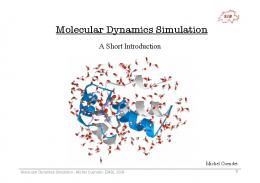
Hydrogen bond interaction path Molecular Docking results ANG WT-ANG* I46V K17E R31K D22G N49S T80S V103I* K54E* F100I* R121C* R121H*
To investigate the mechanisms of loss-offunctions of ANG mutants using MD simulations. To predict deleterious ANG mutations causing ALS rapidly using a faster method ahead of experiments.
ParDOCK binding energy scores Native His114 conformation Altered His114 conformation -4.96 kcal/mol -3.99 kcal/mol -2.94 kcal/mol -3.58 kcal/mol -2.61 kcal/mol -3.10 kcal/mol -1.86 kcal/mol -3.36 kcal/mol -2.47 kcal/mol -3.98 kcal/mol -2.05 kcal/mol -4.15 kcal/mol -1.93 kcal/mol -4.01 kcal/mol -3.91 kcal/mol -4.11 kcal/mol -4.00 kcal/mol -3.89 kcal/mol
(*) represents ANG types that do not exhibit His114 conformational switching. Figure. HA-CA-CB-CG dihedral angle change of His114 over MD simulations of ANG mutants.
Figure. Hydrogen bond interaction path from the site of mutation to His114 for I46V & D22G mutants.
(B) Investigating the loss of nuclear translocation activity
Molecular Dynamics simulation
Loss of ribonucleolytic activity Conformational switching of catalytic residue His114 is responsible for loss of ribonucleolytic activity. Simulation results show only His114 exhibit conformational change, and is supported by experimental report “H114N-ANG mutant had lost 3,300 fold catalytic activity”. Hydrogen bond interaction path from the site of mutation to His114 mediated through Leu115 seems to be the molecular level marker favorable for His114 conformational change (Allosteric regulation ???). Binding energy scores obtained from docking studies ascertains conformational switching of His114 causes loss of ribonucleolytic activity.
Loss of nuclear translocation activity Certain ANG mutants exhibit loss of nuclear translocation activity due to local misfolding of 31RRR33 residues and hence ALS causative.
Structure Preparation and computational methodology: The crystal structure of human angiogenin (PDB: 1B1I) was used as the starting point. Mutants proteins were generated in silico keeping the secondary structure intact. MD simulations were performed using AMBER 10. ff99SB force field in presence of explicit TIP3P water molecules within 10 Å of the protein in an octahedral box was used for 25 ns MD simulations. PTRAJ module was used for analysis of simulation trajectories. Simulations were performed on a 320 processors SUN Microsystems clusters at Supercomputing Facility for Bioinformatics & Computational Biology (SCFBio) at Indian Institute of Technology Delhi. Molecular Docking was carried out using ParDOCK and binding free energies were obtained.
OBJECTIVES
METHODS
A fast method to predict deleterious ANG mutation causing ALS. A newly identified ANG mutation can be submitted to ANGDelMut
Comprehensive analysis of all ANG mutants First attribute: characteristic conformational switching of His114 loss of ribonucleolytic activity (Table 1). Second attribute: local folding of 31RRR33, and hence reduction in SASA loss of nuclear translocation activity. Our study shows how molecular simulations on protein tertiary structures can be used to infer functional implications of mutations. A faster method has been developed to predict deleterious ANG mutations.
CONCLUSION
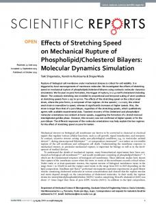
Figure. Structural and dynamic changes of 31RRR33 in ALS associated ANG mutants. (A) & (C) Snapshots of WT-ANG and D22G mutant showing loose packing of nuclear localization signal residues 31RRR33 and (B) Snapshot of V103I mutant showing local folding of 31RRR33 resulting close packing of residues observed from simulations. Comprehensive study of all ANG mutations Quantification of SASA of 31RRR33
Figure. Computed SASA values of R31 (black), R32 (red) and R33 (green) of WT-ANG and mutants during the course of MD simulations.
Table 1. Functional loss results of all ALS associated ANG mutants obtained from MD simulations. These results have been found to be correlated with experimental reports .
Albany 2013: The 18th Conversation June 11th to 15th’ 2013, State University of New York at Albany
For the first time, our study provides mechanisms of lossof-functions of ALS associated ANG mutants. We developed a methodology to predict deleterious ANG mutations, that may cause ALS.
REFERENCES 1. Padhi AK et al. Mechanisms of loss of functions of human angiogenin variants implicated in amyotrophic lateral sclerosis. PLoS One 7, e32479 (2012). 2. Padhi AK et al. Prediction of Functional Loss of Human Angiogenin Mutants Associated with ALS by Molecular Dynamics Simulations. Scientific Reports 3, 1225 (2013). 3. Wu D et al. Angiogenin loss-of-function mutations in amyotrophic lateral sclerosis. Annals of Neurology 62, 609617 (2007). 4. Jayaram B et al. "Sanjeevini: A Freely Accessible WebServer for Target Directed Lead Molecule Discovery". BMC Bioinformatics 13, (Suppl 17):S7 (2012). 5. Thiyagarajan N et al. Structural and molecular insights into the mechanism of action of human angiogenin-ALS mutants in neurons. Nature Communications 3, 1121 (2012). 6. Padhi AK et al. Fast prediction of deleterious angiogenin mutations causing amyotrophic lateral sclerosis. FEBS Letters, doi: 10.1016/j.febslet.2013.04.022. (2013). *E-mail:
[email protected]