Sep 8, 2015 - The biodistribution of biodegradable nanoparticles can be difficult to quantify. We report a method using time resolved fluorescence (TRF) from ...
www.nature.com/scientificreports
OPEN
received: 14 April 2015 accepted: 07 July 2015 Published: 08 September 2015
A Simple and Sensitive Method to Quantify Biodegradable Nanoparticle Biodistribution using Europium Chelates Lindsey Crawford1, Jaclyn Higgins2 & David Putnam1,3 The biodistribution of biodegradable nanoparticles can be difficult to quantify. We report a method using time resolved fluorescence (TRF) from a lanthanide chelate to minimize background autofluorescence and maximize the signal to noise ratio to detect biodegradable nanoparticle distribution in mice. Specifically, antenna chelates containing europium were entrapped within nanoparticles composed of polylactic acid-polyethylene glycol diblock copolymers. Tissue accumulation of nanoparticles following intravenous injection was quantified in mice. The TRF of the nanoparticles was found to diminish as a second order function in the presence of serum and tissue compositions interfered with the europium signal. Both phenomena were corrected by linearization of the signal function and calculation of tissue-specific interference, respectively. Overall, the method is simple and robust with a detection limit five times greater than standard fluorescent probes.
Biodegradable nanoparticles (NPs), such as those derived from diblock copolymers of polylactic acid and polyethylene glycol (PLA-PEG), are valuable in the field of targeted drug delivery. The hydrophobic PLA core can entrap hydrophobic compounds to serve as a carrier for medicines that can be difficult to deliver intravenously1,2. An important step in the development of PLA-PEG NP drug delivery systems is their detection following injection in the blood stream. Common methods to detect the biodistribution of NPs include radiolabels and fluorescent markers. Fluorescence is a safer alternative to radiolabeling; however, the autofluorescence of biological tissues makes it challenging3,4. Specifically, amino acids in biological tissues contribute to autofluorescent signals by absorbing and emitting light at frequencies that overlap with those of common fluorescent markers5. Therefore, the level of detection and the signal to noise ratios of common fluorescent markers is compromised leading to decreased sensitivity6–8. Another approach to the detection of NPs in tissues is to use markers that absorb and emit light in the longer wavelength near-infrared spectrum to minimize autofluorescence; however, these compounds are prone to degradation and photobleaching9. As an alternative strategy to overcome autofluorescence in the detection of structures in biological tissues, time resolved fluorescence (TRF) has been explored. Elements in the lanthanide III series exhibit TRF. Their large Stokes shifts and long decay lifetimes allow them to be detected distinct from competing autofluorescence signals10. They are also less susceptible to photobleaching than traditional organic dyes11–13. Earlier reports have described the utility of lanthanide chelates for the detection of non-degradable NPs both in vivo and ex vivo, but degradable NPs, like those based on PLA-PEG, have unreported characteristics that makes their detection unique and should be considered to accurately quantify their concentration in tissues14–16. 1
School of Chemical and Biomolecular Engineering, Cornell University, Ithaca NY. 2Department of Biological and Environmental Engineering, Cornell University, Ithaca NY. 3Department of Biomedical Engineering, Cornell University, Ithaca NY. Correspondence and requests for materials should be addressed to D.P. (email: dap43@ cornell.edu) Scientific Reports | 5:13177 | DOI: 10.1038/srep13177
1
www.nature.com/scientificreports/
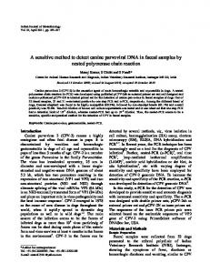
Figure 1. Characteristics of PLA-PEG nanoparticles doped with europium chelate. (a) representative structure of Eu(NTA)3 chelate. (b) excitation and emission spectra for 1 mg/mL nanoparticle suspension in PBS (solid lines) and 0.1 mg/mL chelate solution in acetone (dotted lines) under time resolved conditions.
This communication outlines a simple approach to the detection and quantitation of PLA-PEG NPs in tissues following intravenous injection. A hydrophobic europium chelate was encapsulated within the PLA-PEG core. The TRF of the europium-doped NPs is significantly above tissue autofluorescence, and the particles are detectable in harvested organs ex vivo with minimal post-processing of the tissues. Two important characteristics of the encapsulated europium were identified when quantifying NPs in tissues. First, the TRF of europium diminishes as a second-order function upon exposure to water. Europium chelate located toward the outer surface of the particle is susceptible to water penetration along the same time scale as the biodistribution studies; therefore, the rate of signal decrease upon water exposure is an important parameter in the biodistribution calculations. Second, the TRF of europium is compromised by iron ions; therefore, the signal must be corrected for each tissue using doped tissue blanks. The described method was used to assess the biodistribution of the widely used PLA-PEG-based NP drug delivery system.
Results
Nanoparticle fabrication and characterization. Polylactic acid–mono methoxy polyethylene gly-
col (PLA-PEG) diblock copolymers were synthesized by ring opening polymerization. Mn was consistent between both GPC and 1H NMR (21,000 and 20,600, respectively). Nanoparticle fabrication yielded particles 106 ± 6.5 nm in diameter and − 1.45 ± 0.25 mV surface charge with a polydispersity of 0.079 ± 0.03. Excitation and emission spectra for the particles and chelate (Fig. 1), show maximum excitation at 340 nm and a maximum emission at 610 nm. Both the unencapsulated chelate and the chelate-containing nanoparticles show a large Stokes shift, which is a hallmark characteristic of europium chelates. Release studies of europium chelate from PLA-PEG nanoparticles (Figure S1) show no detectable chelate release over nine hours.
Sensitivity Analysis. The sensitivity limit of the Eu(NTA)3 doped PLA-PEG NPs was determined
and compared to RhoB loaded PLA-PEG NPs (Fig. 2). Encapsulation efficiencies were determined to be 87.0 ± 14% and 7.82 ± 1.7% for europium chelate and RhoB doped nanoparticles, respectively. Using the encapsulation efficiency, the europium doped NPs were detectable at as little as 8.7 × 10⁻⁶ mg/mL of europium chelate above the sensitivity limit compared to 3.9 × 10⁻⁵ mg/mL of RhoB for RhoB doped
Scientific Reports | 5:13177 | DOI: 10.1038/srep13177
2
www.nature.com/scientificreports/
Figure 2. Sensitivity analysis of (a) Eu(NTA)3 doped nanoparticles and (b) rhodamine B doped nanoparticles. Dotted lines correspond to the sensitivity limit. Eu(NTA)3 doped nanoparticles are more sensitive than rhodamine B doped nanoparticles. *Lowest concentration that is statistically different from sensitivity limit at p