We managed a total of 212 tympano- jugular paragangliomas (TJPs) and 22 vagal paragangliomas (VPs) from 1988 to 2009 at the Gruppo Otologico, Piacenza, ...
Advanced techniques in the management of complex head and neck paragangliomas Mario Sanna MD, Gruppo Otologico, Piacenza, Italy.
Sampath Chandra Prasad MS, Gruppo Otologico, Piacenza, Italy. Correspondence E: sampathcp@ yahoo.co.in Declaration of Competing Interests None declared.
Introduction Head and neck paragangliomas (HNPs) are slow growing neuroendocrine tumours arising from the parasympathetic autonomic nervous system with a strong vascularisation. In the process of this slow and insidious growth, they often invade and erode the bone of the skull base, infiltrate the regional cranial nerves (CNs), constrict the major blood vessels to the brain either by encasement or direct infiltration, and transgress the dural barrier [1]. The ideal primary treatment of these tumours is total surgical extirpation [2]. The evolution of the present surgical approach to this region was initiated in 1949 with the proposal to resect the jugular bulb to treat these tumours [3]. It took nearly three decades of innovation by a multitude of workers for the surgery of these tumours to evolve to the approach that is practised today. However, surgery in this delicate region is still fraught with danger, requiring the ability to accurately assess varied interrelated factors, their impact on the ultimate result and their management [4]. We managed a total of 212 tympanojugular paragangliomas (TJPs) and 22 vagal paragangliomas (VPs) from 1988 to 2009 at the Gruppo Otologico, Piacenza, Italy. The object of this article is to present our management protocols in the treatment of HNPs, specifically the more complex tumours involving the skull base including the cranio-cervical neuro vasculature.
General pre-operative and intra-operative protocol At the Gruppo Otologico, all patients with HNPs undergo routine ENT examination with at least one hearing assessment in the form of pure tone audiometry (PTA) and a clinical photograph of the otoscopy findings. We also document the facial nerve (FN) by House Brackmann grading and by taking pictures of four facial postures during each of the patient’s visits. All patients are subjected to high resolution computed tomography (HRCT) with bone windows, multi-resonance imaging with Gadolinium (MRI-Gd) and four-vessel angiography along with the vertebrobasilar system and manual cross-compression test of the internal carotid artery
(ICA). Intra-operatively, we have been deploying the FN monitor in all skull-base cases in our institution since 1994. The primary surgical approach used is infratemporal fossa approach – Type A (ITFA–A) [4,5]. Much of the dissection of the tumour is done using the operating microscope. Post-operatively, patients are usually discharged within seven days and followed up for a period of five to 10 years.
Tympanojugular paragangliomas (TJPs) 212 patients with TJPs were treated at our centre between 1988 and 2009 [6]. Of these, 60 were considered to be complex due to the following conditions: Very large size – As TJPs grow, they extend either into the carotid canal and the petrous apex or into the intradural space through the medial wall of the jugular bulb, thereby involving the lower cranial nerves (LCNs). Large size obviously brings into play the other factors mentioned above that complicate the presentation of TJPs discussed below. Fisch class C3 and C4 tumours are generally considered as large tumours. We managed 35 patients with tumours considered to be of a large size of which 27 were Fisch C3, five were C4 and three were De2. As a rule, ITFA-A is used for C2 tumours and ITFA-A or ITFA-A combined with ITFA-B can be used for C3/C4 tumours. If the tumour involves the clivus, occipital condyle, or foramen magnum, additional procedures such as modified trans-condylar approach (MTCA) or extreme lateral trans-condylar approach (ELTCA) are necessary and were performed at our centre. A large intradural extension (IDE) – Although some authors prefer a single staged surgery [7], we feel that if an extensive area of dura is involved, a planned second-stage resection can be considered. We managed a total of 45 patients with IDE; 25 with Fisch Di1, 18 Di2, and two Di3. In 14 of these cases, a staged surgery was performed. The advantage of a staged surgery is that a clear plane of dissection would have been established between the tumour and the brain stem due to the
feature
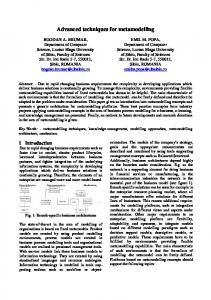
Figure 1: A, B – MRI, axial and coronal views after the first-stage surgery. The residual intradural tumour is noted. The surgical defect is filled with abdominal fat. C, D – MRI, axial and coronal views after the second-stage surgery. After the surgery, there is no residual tumour. T = Intradural tumour; F = fat.
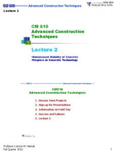
devascularisation of the tumour at the first-stage surgery and subsequent shrinkage of the intradural mass (Figure 1). Another consideration is that in tumours involving LCNs, their sacrifice could cause severe aspiration with continuous cough. The resulting increase in intracranial pressure could give rise to cerebro spinal fluid (CSF) leaks [8]. To try and prevent this, we prefer a staged surgery for tumours with more than 2cm IDE [5]. At the second stage, the approach is determined by the location and size of the residual tumour and the patient’s hearing function. Although petro-occipital-trans-sigmoid approach (POTS) is preferred in most cases, a MTCA or an ELTCA may also be used. Extension to the foramen magnum, clivus or the cavernous sinus – For tumours extending to the foramen magnum and lower clivus, MTCA type D or the ELTCA may be used [9]. In order to prevent tumour recurrence, any part of the clival bone suspected of being infiltrated must be drilled out until healthy bone appears. We had 13 patients; two with tumour involvement of the cavernous sinus, three of foramen magnum and eight of the clivus. In two of them, the tumour involved the cavernous sinus and they were intentionally left intact to avoid compromising cranial nerves III, IV and VI and maintain ocular mobility and after surgery, stereotactic radiotherapy was carried out in both patients. We had three cases involving the foramen magnum and total removal was difficult to achieve in one of them due to persistent bleeding. Figure 2: A, B – Insertion of the stents in the petrous and cervical portions of the ICA. C – Digital x-ray in oblique projection showing the stents fully deployed in the petrous and cervical segments of the ICA. D – Digital subtraction angiography of the ICA after stenting showing resolution of the stenosis.
feature
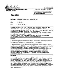
Figure 3: The view of the internal carotid artery. Tumour removal has been completed. Dissection has been carried out down to the stent in an almost bloodless field.
Permanent balloon occlusion (PBO) is performed when the ICA is infiltrated by the tumour and the collateral blood flow is sufficient. In case of insufficient collateral blood flow, we routinely use intraluminal stenting (Figures 2-3). Stenting of the cervical and petrous segments of the ICA was introduced as a pre-operative management protocol by the Gruppo Otologico in the clinical and surgical management of complex HNPs in early 2003 as a method to avoid pre-operative closure of the ICA or high risk bypass procedures and to protect and preserve integrity of the artery during surgery, mainly in cases in which the collateral flow through the circle of Willis is deemed insufficient [11-13]. Twenty-one pre-operative ICA stenting procedures were performed at our centre on 19 patients. Stenting of the ICA allows reinforcement of the artery, reducing the risk of intraoperative injury of its wall while performing a more aggressive carotid dissection in the subadventitial plane. The presence of the stent allows the safe mobilisation of the artery if necessary. This new technique can allow reappraisal of selected cases previously suited only for sub-total resection. To the best of our knowledge, the literature contains only one case of stenting of the intratemporal segment of the ICA for the management of TJPs [7]. A single ICA on the lesion side – As mentioned above, management of the ICA is essential for total tumour removal. For a case with a single carotid artery on the lesion side, possible management options are ‘wait and scan’, partial resection followed by radiotherapy, or total removal subsequent to the pre-operative reinforcement with stents [12]. In a patient with a single carotid artery on the lesion side, bypass surgery can cause severe cerebral ischemic damage. Therefore, the stent insertion may be the best option. We have so far treated two patients with a single ICA without any adverse consequences.
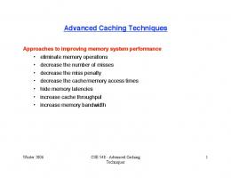
Figure 4: A – A class C4Di2Vi tumour. MRI coronal image showing the tumour attached to the VA. B-F – Surgical sequences of extreme lateral transcondylar approach. B – The transverse process of the atlas (A) is drilled out and the atlanto-occipital joint (J) is removed. (C) = Condyle. C – The tumour (T) is attached to the vertebral and posterior inferior cerebellar arteries infiltrating the clival (Cl) bone, which is partially drilled out. D – The tumour is separated from the PICA. E – CT scan axial view showing the stent in the ICA and the extent of bone removal. F – CT scan coronal view showing absence of the surgically removed occipital condyle (black arrow) compared to non-operated side (white arrow).
Involvement of the ICA – TJPs frequently involve the ICA due to their close anatomical proximity [4]. When indicated, the tumour must be dissected from the arterial wall. This can be achieved by sub-periosteal (or supra-adventitial) dissection of the ICA in the carotid canal (horizontal portion) or
sub-adventitial dissection in the vertical portion [10]. When the artery is completely surrounded by a tumour resulting in severe stenosis on arteriography, manipulation without proper endovascular intervention may give rise to severe bleeding, incomplete removal or a cerebral vascular accident [2].
Vertebral artery (VA) involvement – Although involvement of the VA in TJPs is extremely uncommon, (11 cases reported worldwide, eight of which belong to our series), it represents the pinnacle of difficulty in management of TJPs. Seven out of eight patients underwent surgery. In two patients, pre-operative occlusion was performed. In a previous study [14] we presented the radiological and surgical findings of VA involvement by TJPs to emphasise the importance of VA assessment and propose the addition of the ‘V’ category to the existing Fisch classification [15]. Angiography of the vertebrabasilar system should always be included in the assessment of TJPs planned for surgery. This is to detect anastomotic connections between the external carotid and the VA that are potentially
feature
dangerous during embolisation, such as between branches of the ascending pharyngeal, deep cervical, ascending cervical and occipital arteries with the VA [16], as well as to adequately assess direct involvement of the VA. For the surgical strategy, involvement of the V3 segment requires the addition of an extreme lateral extension to the standard ITFA, in order to ensure its adequate exposure. The V4 segment involvement is inevitably accompanied by a large IDE. In such cases, we prefer a two-stage surgery (Figure 4). Dominant or unilateral sigmoid sinus (SS) on the lesion side – The SS obliteration and the jugular vein often need to be ligated during resection of TJPs. However, ligation SS of the dominant or the presence of unilateral SS on the lesion side may cause intracranial hypertension and venous congestion leading to swelling of the brain [9]. Therefore, pre-operative evaluation of venous drainage of the brain is essential, especially of the ipsilateral mastoid emissary vein or the condylar vein. If their diameters are larger than normal, they should be preserved during surgery. In cases where the collateral venous drainage cannot be preserved or when the patient has no sufficient collateral venous drainage, a more conservative treatment plan such as partial resection with preservation of the SS, gamma knife surgery, or a ‘wait and scan’ approach is recommended. Bilateral or multiple HNPs – In the management of bilateral TJPs, the possibility of bilateral deficits of important LCNs looms large and hence neural preservation is very important to achieve a good quality of life for the patient post-operatively. We had 11 patients with multiple HNPs. According to our management protocol, in patients with LCN deficits on the side of the larger tumour, it is removed first and then the smaller tumour is either followed up or irradiated. On the contrary, if the patients have LCN deficits on the side of the smaller tumour, it is removed first and then the larger tumour is followed up with MRI. During follow-up, if the larger tumour shows evidence of growth, it may be partially removed with the preservation of LCN function or irradiated. In patients with no LCN deficit, the ‘wait and scan’ approach is first applied.
However, if the tumour shows growth, radiotherapy or subtotal removal of the tumour with LCN preservation is performed first. Subsequently, if the tumour continues to grow despite radiotherapy or surgical removal, the other remaining modalities can be applied. Recurrence after previous surgery, radiotherapy or stereotactic radiosurgery – Any revision surgery is a challenge as there are no normal tissue planes and surgical landmarks. Previous surgery or radiation increases the risk of CSF leak and damage to the LCNs and FN [2,9]. The carotid canal is the most common site for recurrence in TJPs and previous dissection increases the risk of injury to the ICA. In such cases, the pre-operative management of the ICA by PBO or stenting is especially important. An ITFA-A with FN rerouting should be performed in all cases with the appropriate extension and extensive bone removal. There is no place for a conservative approach for the FN and external auditory canal in revision surgery. In our present series, 13 cases had undergone previous treatment.
Vagal paragangliomas (VPs) VPs are less common than carotid body tumour (CBT) and TJP, occurring in the fourth and fifth decades of life, and are more common in females [17]. We managed 22 patients with VP from 1988 to 2009 [18]. Our policy is to offer surgery as the treatment choice for patients with VP, irrespective of the existence of vocal cord paralysis, whereas in older patients with no serious complications, a ‘wait and scan’ policy is adopted. For bilateral lesions, if the patient has an abnormal vagus nerve function on one side, surgical removal is considered on that side, and a more conservative management is applied to the contralateral side. The commonly used approaches are the transcervical, transcervical-transmastoid and the ITFA-A.
Conclusion Careful consideration of the complicating factors and thorough pre-operative evaluation and intervention can decrease surgical morbidity in HNPs with a high probability of gross total removal. The application of the abovementioned advanced management techniques will definitely improve prognostic results of this subset of tumours.
References 1.
Moe KS, Li D, Linder TE, Schmid S, Fisch U. An update on the surgical treatment of temporal bone paraganglioma. Skull Base Surgery 1999;9:185–94.
2.
Sanna M, Piazza P, De Donato G, Menozzi R, Falcioni M. Combined endovascular-surgical management of the internal carotid artery in complex tympanojugular paragangliomas. Skull Base 2009;19:26-42.
3.
Lundgren M. Tympanic body tumours in the middle ear — tumours of the carotid body type. Acta Otolaryngol 1949;37:366–79.
4.
Sanna M, Jain Y, De Donato G, Rohit, Lauda L, Taibah A. Management of jugular paragangliomas: the Gruppo Otologico experience. Otol Neurotol 2004;25(5):797-804.
5.
Sanna M, Saleh E, Russo A, et al. Atlas of Lateral Skullbase Surgery. Stuttgart, New York, Georg Thieme Verlag; 1995: 132–45.
6.
Sanna M, Shin SH, De Donato G, Sivalingam S, Lauda L, Vitullo F, Piazza P. Management of complex tympanojugular paragangliomas including endovascular intervention. Laryngoscope 2011;121(7):1372-82.
7.
Al-Mefty O, Teixeira A. Complex tumours of the glomus jugulare: criteria, treatment, and outcome. J Neurosurg 2002;97:1356-66.
8.
Fisch U. Infratemporal fossa approach for glomus tumours of the temporal bone. Ann Otol Rhinol Laryngol 1982;91:474-9.
9.
Patel SJ, Sekhar LN, Cass SP, Hirsch BE. Combined approaches for resection of extensive glomus jugulare tumours. A review of 12 cases. J Neurosurg 1994;80:1026-38.
10. Fisch U, Fagan P, Valavanis A. The infratemporal fossa approach for the lateral skull base. Otolaryngol Clin North Am 1984;17:513-52. 11. Sanna M, Khrais T, Menozzi R, Piazza P. Surgical removal of jugular paragangliomas after stenting of the intratemporal internal carotid artery: a preliminary report. Laryngoscope 2006;116(5):742-6. 12. Piazza P, Di Lella F, Menozzi R, Bacciu A, Sanna M. Absence of the contralateral internal carotid artery: a challenge for management of ipsilateral glomus jugulare and glomus vagale tumours. Laryngoscope 2007;117(8):1333-7. 13. Konishi M, Piazza P, Shin SH, Sivalingam S, Sanna M. The use of internal carotid artery stenting in management of bilateral carotid body tumours. Eur Arch Otorhinolaryngol 2011;268(10):1535-9. 14. Shin SH, Sivalingam S, De Donato G, Falcioni M, Piazza P, Sanna M. Vertebral artery involvement by tympanojugular paragangliomas: management and outcomes with a proposed addition to the fisch classification. Audiol Neurootol 2012;17(2):92-104. 15. Jenkins HA, Fisch U. Glomus tumors of the temporal region technique of surgical resection. Arch Otolaryngol 1981;107(4):209-14. 16. Geibprasert S, Pongpech S, Armstrong D, Krings T. Dangerous extracranial-intracranial anastomoses and supply to the cranial nerves: vessels the neurointerventionalist needs to know. AJNR Am J Neuroradiol 2009;30:1459–68. 17. McNicol AM. Adrenal medulla and paraganglia. In Lioyd RV (Editor), Endocrine Pathology. New York, Springer; 2010: 281–95. 18. Shin SH, Piazza P, De Donato G, Sivalingam S, Lauda L, Vitullo F, Sanna M. Management of vagal paragangliomas including application of internal carotid artery stenting. Audiol Neurootol 2012;17(1):39-53.