Eur Spine J (1998) 7 : 88–94 © Springer-Verlag 1998
J. Y. Margulies Y. Floman G. C. Robin M. G. Neuwirth P. Kuflik M. Weidenbaum J.-P. C. Farcy
Received: 10 May 1997 Accepted: 16 September 1997 J. Y. Margulies (Y) Montefiore Medical Center, III E. 210th St. Bronx, New York 10 467, USA Fax +1-718-920 6373 e-mail:
[email protected] Y. Floman · G. C. Robin Hadassah University, Hospital, Jerusalem, Israel M. G. Neuwirth · P. Kuflik Spine Institute, Beth Israel Medical Center, 10 Union Square East, New York, New York, USA M. Weidenbaum Columbia Presbyterian Medical Center, New York, New York, USA J.-P. C. Farcy Maimonides Orthopaedics, 13th Avenue and 57th Street, Brooklyn, New York, USA
O R I G I N A L A RT I C L E
An algorithm for selection of instrumentation levels in scoliosis
Abstract Appropriate levels for instrumentation and fusion in scoliosis have been a matter of debate among surgeons since the introduction of operative management of this deformity. We set out to examine the hypothesis that the amount of correction achieved in all planes during surgical instrumentation of a curve should be less than, or comparable to, the degree of correction attainable at any non-instrumented adjacent curve. An algorithm was designed to facilitate preoperative planning and intraoperative performance of spinal fusion procedures in the management of scoliosis. To test the validity of the hypothesis and the proposed algorithm, measurements were taken from the preoperative radiographs of 200 patients. The dimensions of the curves were obtained from an initial set of
Introduction Appropriate levels for instrumentation and fusion in idiopathic scoliosis have been a matter of debate among surgeons since the introduction of operative management of this deformity some 80 years ago [7, 18, 23–25, 39, 45]. It is important to fuse the smallest possible number of vertebrae to maintain maximum residual mobility, but end with a well-balanced spine [36, 54]. The longer the instrumentation, the greater the chance to gain control over the various curves and end up with a balanced spine. Shorter instrumentations may yield a well-balanced, mobile spine, with reduction of the scoliotic curves both in the instrumented and non-instrumented portions of the spine.
four X-ray films: (1) standing anteroposterior film of the whole spine, (2) standing lateral film of the whole spine, (3) two properly performed side-bending films including each curve of the spine. With this data, a plan was designed using the algorithm. The results of this plan were compared with the actual results of the surgery, which were revealed only at this stage. All patients in whom actual instrumentation levels fell within those predicted by the proposed algorithm had no imbalance at follow-up. All patients whose actual instrumentation levels were short of those recommended by the algorithm showed obvious imbalance on final postoperative standing radiograph. Key words Scoliosis, surgery · Spine · Spinal fusion
Levels for fusion with Harrington instrumentation were defined according to King’s classification [31, 32]. This approach does not emphasize the sagittal profile of the spine. Loss of lumbar lordosis with sagittal imbalance was quite common and known as the „flat back“ syndrome [1, 8, 17, 34, 35, 52]. Subsequently, the sagittal profile was recognized to be important by Winter, Dickson and Armstrong [2, 11–13, 56–58]. With increased use of CD instrumentation and similar segmental systems, postoperative frontal imbalance has been more frequently noted than that reported with Harrington instrumentation. The newer posterior segmental systems facilitate better operative correction of scoliosis than Harrington instrumentation, and the option of anterior release enables even greater correction to be achieved.
89
films, taken from the radiologic follow-up files of the patients, using the algorithm. This was done before the actual surgical procedure and its results were studied. The proposed instrumentation levels and theoretical magnitude of correction were then compared to the actual results. All final postoperative results were classified as “balanced” or “imbalanced”, according to the criteria for coronal and sagittal imbalance given below.
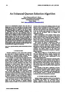
Data acquisition To test the validity of the hypothesis and the algorithm the radiographs of 200 patients were remeasured. The curve dimensions were obtained from an initial set of four plain films: 1. Standing anteroposterior (AP) film of the whole spine 2. Standing lateral film of the whole spine 3. Two properly performed side-bending films including each curve of the spine Fig. 1 The stages of balanced correction: hypothesis
These techniques allow greater potential to straighten curves, but may also lead to imbalance if used in inappropriate ways [39, 44, 55]. It appears that at least some of the imbalance with segmental systems occurred when selection of instrumentation levels was made using King’s criteria. Subsequently, different guidelines were proposed by Dubousset and by Schufflebarger among others, for use with the Cotrel-Dubousset (CD) system [15, 16, 33, 48, 49]. We present an algorithm for selection of levels for segmental instrumentation in scoliosis surgery in the hope that its use will decrease the frequency of imbalance.
Hypothesis The amount of correction achieved in all planes during surgical instrumentation of a curve should be less than, or comparable to, the degree of correction attainable at any non-instrumented adjacent curve. This principle applies to any surgical approach and to any instrumentation applicable to scoliosis (Fig. 1).
Methods An algorithm was designed to facilitate preoperative planning and intraoperative performance of spinal fusion procedures in the management of scoliosis. Patients The radiographs of 200 patients with idiopathic scoliosis were retrospectively reviewed. All patients had been operated on at least 12 months prior to the initiation of the study, and all had reached skeletal maturity. There were 42 males, aged 9–47 years old at surgery, and 158 femalies, aged 9–56. The curve magnitudes ranged from 35° to 95° preoperatively, and from 2° to 60° at follow-up. A proposed plan of instrumentation was made from the preoperative
The neutral vertebra, the stable vertebra, and the end vertebrae were noted, as well as any pelvic and shoulder obliquity. From the lateral films the presence and the degree of the sagittal curve were noted, with particular reference to junctional changes between lordotic and kyphotic regions of the spine. With this data a plan was designed using the algorithm. The results of this plan were compared with the actual results of the surgery, which were only revealed at this stage. Fusion levels were determined by surgeons not involved in the care of these patients.
Definitions Coronal imbalance Coronal imbalance was defined as the occurrence or deterioration of one or more of the following three conditions on standing plain films: 1. Head not centered above the sacrum, but displaced by 1 cm away from the mid-sacral line 2. Shoulders not placed above the hips 3. Existence or progression of trunk shift [19] Sagittal imbalance. Sagittal imbalance was defined as: 1. The first cervical vertebra not above the second sacral segment on the standing lateral film of the spine 2. Kyphosis or lordosis exaggerated or diminished beyond the accepted range of the sagittal vertebral contour [4, 5, 28, 29, 47]. Largest curve. The “largest curve” is that which lies between two end vertebrae on properly performed bending films. Usually, the largest curve on the AP standing film is also the largest on bending films. Occasionally, the largest curve is less structural and is more correctable than an adjacent smaller but more rigid curve when measured on the bending film. Spontaneous curve correction. Spontaneous curve correction is determined by subtracting the Cobb angle values measured on a properly performed bending film from those measured on the standing AP film of the same curve. Surgical curve correction magnitude. Surgical curve correction magnitude is the sum of the range of correction of a curve obtained on bending films and the added correction expected to be achieved by operative release and spinal instrumentation [20]. The following definitions were not modified from their common use.
90
End vertebra. End vertebra delineate the caudal and cephalad limits of the curve; these are the vertebrae from which two curves take opposite directions. They are recognized by opposite-side discopening above and below the vertebral body. One end vertebra is usually common to two adjacent curves.
Stable vertebra. On an AP film, the stable vertebra is that which is most nearly bisected by the mid-sacral line [6, 31]. Neutral vertebra. On an AP film, the neutral vertebra is that which shows the smallest rotation. It is usually next to a disc space that can be made neutral (see below). The neutral disc space and the neutral vertebra are not always contiguous. A neutral disc. A neutral disc is the first one at the end of a curve in which the width on each side opens with bending. Use of the algorithm The use of the algorithm is described in Figs. 2 and 3. The final coordination of the end vertebrae (Fig. 2, step 6) includes verifying that it is not apical on AP or lateral views, that it is neutral and leveled (either on bending films or by surgical maneuver), and that it is within the stable zone. Balance, curve correction, and the number of vertebrae fused must all be taken into consideration. A balanced spine, shorter fusion, and lesser correction are preferable to greater correction that may jeopardize the balance or involve a larger number of vertebrae. It should be kept in mind that the algorithm is a tool to calculate the amount of correction and its influence on the adjacent curves. A full application of the algorithm leads to balanced spine.
Results In the retrospective analysis of 200 operated patients, three groups were identified:
If either end vertebra is apical or junctional on AP or lateral views or out of the stable zone, then include the relevant curve in instrumented fusion*
Fig. 2 Flowchart “The use of the algorithm” Fig. 3 The algorithm “Instrumentation levels for spinal curves”
• Group A. Patients in whom the suggested and actual instrumentation levels matched (n = 178) None of these showed imbalance on follow-up films. • Group B. Patients in whom the algorithm calculations showed that a one-curve instrumentation could have sufficed, but whose actual instrumentation and fusion included an entire adjacent lower curve (n = 6). All of these were also balanced. It is impossible to predict the
91
results had the lower curve been left unfused. No discrepancy of this kind was found regarding an upper curve, proximal to the largest curve. • Group C. A group of patients in whom the actual instrumentation and fusion levels were different from the algorithm “recommendation” (n = 16). All of these patients were found to be unbalanced on follow-up views. Group C was the group of interest in this study. Two subgroups were identified: the first comprised seven patients in whom obvious technical mistakes had been made, such as ending the instrumentation at a level that was either apical or close to an apical vertebra on the coronal or sagittal views. The other subgroup comprised nine patients in whom different errors were made. Examples included failure to include adjacent stiff curves (five patients) or curve overcorrection beyond the flexibility of adjacent curves (four patients). Within this subgroup, there was no uniform mistake pattern, but two common errors can be noted. • A residual low lumbar stiff curve down to S1 was ignored, which caused an oblique lumbosacral take-off and resulted in pelvic obliquity. • The restoration of level shoulders was not taken into account in deciding whether or not to instrument an adjacent upper thoracic curve. In summary, all patients in whom actual instrumentation levels fell within those predicted by the proposed algorithm, or in whom an additional curve was instrumented and fused, possibly unnecessarily, had no imbalance. All other patients whose actual instrumentation levels did not coincide with those recommended by the algorithm showed imbalance on follow-up.
Discussion Classification of idiopathic curves and surgical results In the early history of surgical spine treatment, conclusions regarding which levels should be fused were based on existing methods of preoperative correction. When correction of a scoliotic curve was obtained by lateral angulation of the trunk in a Risser turnbuckle cast, an excessive fusion could fix the trunk in a totally unbalanced position. This problem was so great that, even when localizer casts came into use in the late 1950s, and the problem of fusion of the grossly unbalanced spine was avoided, discussion continued as to whether fusion should cover the curve itself or one or two more vertebrae at each end [20]. Goldstein stressed that the extent of fusion should be decided after considering the specific features of each individual curve [21, 22]. King showed that it was not mandatory to fuse the entire extent of the deformed spine in every case. In fact, in some cases, such as his type II curves, it was preferable to
leave an entire curve unfused to maintain maximum mobility of the spine. Single curves were not a problem in the decision-making process, nor were rigid complicated combined curves. The King classification marked a turning point because it allowed a substantial saving in the number of levels fused in some cases. Postoperative coronal imbalance was relatively uncommon when these curves were treated with Harrington instrumentation. However, it began to appear more frequently with the advent of more powerful means of correction [27, 38, 39, 43, 44, 51, 55]. The imbalance phenomenon emerged in both the sagittal and coronal planes and, as a result, the King classification had to be re-evaluated as a method to determine fusion levels. Sagittal requirements had to be incorporated into the decision making process, since the thoracolumbar contour could be significantly influenced by fusing only the thoracic curve. With the advent of more powerful three-dimensional corrective instrumentation, it appeared that not all the King II curves required the same treatment, since the sagittal contour and the relative flexibility of the different curves made a significant difference. Since no better classification has yet been suggested, it may be more efficient to return to simple morphological descriptions, making individual decisions for each curve on the basis of current knowledge.
Forced curve correction in the coronal plane and rotational spinal release The degree of curve correction attained during surgery can be altered by a variety of measures. At one extreme an in situ fusion can be performed without an attempt to change the curve; at the other a total correction to a straight line may be obtained using extensive anterior and posterior releases and osteotomies. Anterior release, disc excisions, and vertebral body osteotomies may offer a better correction. Posterior instrumentation can then be introduced as a splint, in corrected alignment, without applying additional corrective force. If necessary, even further correction can be contributed by applying force via the posterior instrumentation. The ability to achieve correction beyond that seen on the bending film depends on the surgeon’s technical skills. The evaluation of how much forced correction can be achieved is the function of the surgeon’s art, and is a subjective factor which each surgeon should include in the decision-making process. The ability to correct the rotation at the end of a curve after an anterior release may guarantee balance, as well as permit salvage of the discs below the fusion [46]. In light of all this, it has to be stressed that if the degree of expected surgical correction of the largest curve is greater than the range of spontaneous correction of an adjacent curve, the adjacent curve must be included in the fusion, as otherwise imbalance may occur. In order to
92
avoid a fusion that is too long, the largest curve may be only partially corrected, to match a smaller spontaneous range of correction of the adjacent curve, and the adjacent curve can then be left unfused. If the adjacent curve corrects to 0° or crosses the midline, it should be left uninstrumented. The anticipated correction, which is the surgical curve correction, allows a calculating surgeon some degree of variability in which imbalance can be absorbed. The sagittal plane The sagittal plane must be reconstructed by instrumentation and fusion to prevent sagittal imbalance and to regain “normal posture,” although this “normal posture” has not yet been defined. As a working hypothesis, we can refer to Bridwell, Jackson, and Schultz for basic statements about the normal spine, erect over a neutralized pelvis [5, 28, 47]. Ferguson mentioned the importance of centering the fusion mass over the sacrum as early as in 1930 [18]. The thoracic kyphosis, the thoracolumbar transition area, and the lumbar lordosis must all be carefully considered, as the potential correction of sagittal curves differs according to their level. Forced correction of thoracic kyphosis can be accommodated by the wide range of motion of the adjacent lumbar spine. However, the lumbar range of motion is not unlimited. Fusion should not end at L4–L5 or L5–S1 with extended hips or hypolordosis [30]. The problematic zones are the thoracolumbar junction, where thoracic kyphosis becomes lumbar lordosis over a span of three vertebrae, and the lumbar spine, where inappropriate correction of lordosis may lead to flat-back deformity. The basic, most commonly used instrumentation is a posterior construct. This construct can stabilize a released and mobilized spine in the best possible position and can correct sagittal curves, either by displacing the spine toward a prebent anchored rod, by controlling and correcting malrotation, or by applying compression or distraction forces to the posterior elements. Distraction forces applied to the vertebral arches elongate the posterior column relative to the anterior column and are a kyphosing factor. Kyphosing forces are usually undesirable in the thoracolumbar junction and in the lumbar spine. Compression forces applied to the posterior elements cause relative shortening of the posterior column, and they are a lordosing factor. The rules for instrumentation of the sagittal plane are: 1. Restore the normal contour. 2. Do not stop instrumentation at the apex of a curve (either on an AP or lateral views). i. Always instrument above the apex of the thoracic kyphosis. ii. The thoracolumbar junction should be flat, without lordosis or kyphosis. An attempt should be made to try to
avoid ending the instrumentation in this region, since this may cause imbalance due to the forces applied by the implants. Whenever possible, instrumentation should end in the sagittal plane, at a sagittally neutral vertebra. The anchorage at the ends of a rod become sites for detorsion and control of the sagittal plane contour. Rotation The range of rotation is determined from AP standing and supine bending films in the same way that the range of motion of a curve is determined. The Nash and Moe method was used because it allowed for postoperative evaluation [40]. Since we are dealing with a three-dimensional deformity, each vertebra in the spine may be eventually displaced in all possible axes. We still do not have an efficient tool to express this on a clinical basis. Asher’s approach to spatial description is still too cumbersome for practical use [3]. Consideration of surgical correction of a curve should take into account the rotation of the adjacent curve. Coupling of rotation to bending movement may bias the assessment. One must remember that with most instrumentation systems, including CD, the main correction maneuver is translation rather than derotation. The ensuing angular correction is not always accompanied by rotational correction. Translation rather than derotation was demonstrated in vivo by Farcy [53]. Some systems, such as the Wisconsin segmental spine instrumentation or Cotrel’s short convex compression rod, may increase the rotation of a corrected segment [9, 41, 50]. On the other hand, systems using pedicle screw fixation do permit some control over rotation, and may allow correction using a prebent rod to restore sagittal contour. Rotation should always be considered, so that one may avoid instrumentation to a maximally rotated vertebra. Transfeldt showed that much of the rotation effect in correction occurs out of the fusion mass in the adjacent discs [59]. It becomes evident that forceful derotation without a proper release does not yield any better cosmetic result as far as the rib hump is concerned, and most of the “derotation” occurs outside the fusion region. This occurs mainly below with decompensation as a possible disturbing consequence [37]. Fusion to the sacrum A residual lumbosacral curve must be considered as any other curve. If this curve does not correct itself sufficiently to accommodate correction of the upper curve, it must be included in the fusion area. If the residual curve is not very rigid, fusion to the sacrum can sometimes be avoided at this stage, advising the patient that this may be required in the future. The “price” of fusion to the sacrum
93
is high, and imbalance may be tolerated more easily than fusion to the sacrum [10]. The option of lesser correction of the adjacent lumbar curve also should be considered. It is imperative to remember that a normal spine is built on a normal pelvis. The ability of the pelvis to compensate for spine imbalance is limited in the coronal plane because of the necessity for raising or lowering one hip during gait. The ligamentous complexes and discs at L4–F5–S1 also limit the extent of compensatory rotation, and of AP translation. Compensation is more feasible in the sagittal plane via hip flexion and extension. Scoliosis above a dysplastic spondylolisthesis has a different pathogenesis and must be addressed separately. Fusion to the cervico-thoracic junction Fusion of upper thoracic curves must aim for level shoulders over a balanced spine. If the upper thoracic curve is not flexible enough to balance the shoulders, forceful curve correction may be needed to achieve level shoulders. Occasionally, the higher shoulder is on the concave side of the upper thoracic curve. In this case, correction of the curve may result in greater inequality between shoulder levels [42].
Conclusion In the method described here, the selection of instrumentation and fusion levels is aimed at attaining maximal correction. In certain circumstances, submaximal correction may be selected, in order to reduce the extent of spinal arthrodesis or minimize the danger of neural damage, as long as balance is achieved. In order to leave an adjacent curve without instrumentation, its range of motion must be equal to or greater than the correction of the largest curve. This provides for a balanced spine in the AP plane. If the terms of the algorithm cannot be met, the adjacent curve must be forcibly corrected and fused. Alternatively, avoiding maximal correction of the largest curve, and matching the correction of this curve to the spontaneous correction of the adjacent curve, may yield a reasonable cosmetic result and a balanced spine. Application of this algorithm may help to decrease possibilities for other errors in selection of fusion levels in scoliosis surgery, like fusion to an apex or ignoring residual curves.
References 1. Aaro S, Ohlen G (1983) The effect of Harrington instrumentation on the sagittal configuration and mobility of the spine in scoliosis. Spine 8 : 570– 575 2. Armstrong GWD, Connock SHG (1975) A transverse loading system applied to a modified Harrington instrumentation. Clin Orthop 108 : 70–75 3. Asher M, Cook L (1994) The transverse plane evolution of the major adolescent idiopathic scoliosis deformity. Presented at the 29th Annual Meeting of the Scoliosis Research Society, Portland Oregon 4. Bernhardt M, Bridwell KH (1989) Segmental analysis of the sagittal plane alignment of the normal thoracic and lumbar spines and thoracolumbar junction. Spine 14 : 717–721 5. Bridwell KH, Betz R, Capelli AM, Huss G, Harvey C (1990) Sagittal plane analysis in idiopathic scoliosis patients treated with Cotrel-Dubousset instrumentation. Spine 15 : 921–926 6. Bridwell KH, McAllister JW, Betz RR, Huss G, Clancy M, Schoenecker PL (1991) Coronal decompensation produced by Cotrel-Dubousset “derotation” maneuver for idiopathic right thoracic scoliosis. Spine 16 : 769–777
7. Butte FL (1938) Scoliosis treated by the wedging jacket. Selection of area to be fused. J Bone Joint Surg [Am] 20 : 1–22 8. Cochran T, Irstam L, Nachemson A (1983) Long-term anatomic and functional changes in patients with adolescent idiopathic scoliosis treated by Harrington rod fusion. Spine 8 : 576– 583 9. Crawford AH, Mache J, Caudle R (1988) Sagittal plane correction by contoured Harrington rods and interspinous segmental instrumentation. Orthop Trans 12 : 238 10. Devlin VJ, Boachie-Adjei O, Bradford DS, Ogilvie JW, Transfeldt EE (1991) Treatment of adult spinal deformity with fusion to the sacrum using CD instrumentation. J Spinal Disord 4 : 1–14 11. Dickson RA (1989) Idiopathic scoliosis. BMJ 298 : 906–907 12. Dickson RA (1992) The scientific basis of treatment of idiopathic thoracic scoliosis. Acta Orthop Belg [Suppl] 58 : 107–110 13. Dickson JH, Erwin WD, Rossi D (1990) Harrington instrumentation and arthrodesis for idiopathic scoliosis. A twentyone year follow-up. J Bone Joint Surg [Am] 72 : 678–683
14. Drummond DS, Keene JS (1988) Spinous process segmental spinal instrumentation. Orthopedics 11 : 1403– 1410 15. Dubousset J, Cotrel Y (1991) CD application technique of Cotrel-Dubousset instrumentation for scoliosis deformities. Clin Orthop 264 : 103–110 16. Dubousset J, Cotrel Y (1989) CD instrumentation in the treatment of spinal deformities. Orthopäde 18 : 118–127 17. Fabry G, Van Mekebeek J, Bock E (1989) Back Pain after Harrington rod instrumentation for idiopathic scoliosis. Spine 14 : 620–624 18. Ferguson HB (1930) The study and treatment of scoliosis. South Med J 23 : 116–120 19. Floman Y, Penny JN, Micheli LJ, Riseborough EJ, Hall JE (1982) Osteotomy of the fusion mass in scoliosis. J Bone Joint Surg [Am] 64 : 1307–1316 20. Goldstein JM, Nash CL, Wilham MR (1991) Selection of lumbar fusion levels in adult idiopathic scoliosis patients. Spine 16 : 1150–1154 21. Goldstein LA (1971) The surgical management of scoliosis. Clin Orthop 77 : 32–56
94
22. Goldstein LA (1973) The surgical treatment of idiopathic scoliosis. Clin Orthop 93 : 131–157 23. Harrington PR (1962) Treatment of scoliosis: correction and internal fixation by spinal instrumentation in scoliosis. J Bone Joint Surg [Am] 44 : 591– 610 24. Hibbs RA (1924 A report of fifty-nine cases of scoliosis treated by the fusion operation. J Bone Joint Surg 6 : 3–37 25. Hibbs RA, Risser JC, Ferguson AB (1931) Scoliosis treated by the fusion operation. An end-result study of three hundred and sixty cases. J Bone Joint Surg 13 : 91–104 26. Horton WC, Holt RT, Johnson JR (1988) Zielke instrumentation in idiopathic scoliosis: late effects and minimizing complications. Spine 13 : 1145– 1149 27. Hsu LC, Zucherman J, Tang SC, Leong JC (1982) Dwyer instrumentation in the treatment of adolescent idiopathic scoliosis. J Bone Joint Surg [Br] 64 : 536–541 28. Jackson RP, McManus AC (1994) Radiographic analysis of sagittal plane alignment and balance in standing volunteers and patients with low back pain matched for age, sex, and size: a prospective controlled clinical study. Spine 19 : 1611–1618 29. Jackson RP, McManus AC (1993) Standing segmental lordosis and sagittal plane alignments in asymptomatic volunteers and in patients with low back pain. 28th Annual Meeting of the Scoliosis Research Society, Dublin 30. Jackson RP, Simmons EH, Stripinis D (1989) Coronal and sagittal plane spinal deformities correlating with back pain and pulmonary function in adult idiopathic scoliosis. Spine 14 : 1391–1397 31. King HA, Moe JH, Bradford DS, Winter RB (1983) The selection of fusion levels in thoracic idiopathic scoliosis. J Bone Joint Surg [Am] 56 : 1302– 1313 32. King HA (1988) Selection of fusion levels for posterior instrumentation and fusion in idiopathic scoliosis. Orthop Clin North Am 19 : 247–255 33. Krismer M, Bauer R, Sterzinger W (1992) Scoliosis correction by CotrelDubousset instrumentation. The effect of derotation and three dimensional correction. Spine [Suppl] 17 : 263–269 34. LaGrone MO (1988) Loss of lumbar lordosis. A complication of spinal fusion for scoliosis. Orthop Clin North Am 19 : 383–393
35. LaGrone MO, Bradford DS, Moe JH, Lonstein JE, Winter RB, Ogilvie JW (1988) Treatment of symptomatic flatback after spinal fusion. J Bone Joint Surg [Am] 70 : 569–580 36. Luque ER (1982) Segmental spinal instrumentation for correction of scoliosis. Clin Orthop 163 : 192–198 37. Marchesi DG, Transfeldt EE (1992) Changes in vertebral rotation after Harrington and Luque instrumentation. Spine 17 : 775–780 38. Mason DE, Carango P (1991) Spinal decompensation in Cotrel-Dubousset instrumentation. Spine [Suppl] 16 : 394–403 39. Moe JH, Winter RB, Bradford DS, Lonstein JE (1978) Scoliosis and other spinal deformities. Saunders, Philadelphia 40. Nash CL, Moe JH (1969) A study of vertebral rotation. J Bone Joint Surg Am 51 : 223–229 41. Neuwirth MG, Drummond DS, Casden AS (1993) The results of interspinous segmental instrumentation in the sagittal plane. J Spinal Disord 6 : 1–4 42. O’Brien MF, Lenke LG, Bridwell KH, Baldo C, Blanke K (1993) Recognition and treatment of the proximal thoracic curve. In adolescent idiopathic scoliosis treated with Cotrel-Dubousset instrumentation. Presented at the 28th Annual Meeting of the Scoliosis Research Society, Dublin 43. Puno RM, Johnson JR, Osterman PA, Holt RT (1989) Analysis of the primary and compensatory curvatures following Zielke instrumentation for idiopathic scoliosis. Spine 14 : 738–743 44. Richards BS, Birch JG, Herring JA, Johnston CE, Roach JW (1989) Frontal plane and sagittal plane balance following Cotrel-Dubousset instrumentation for idiopathic scoliosis. Spine 14 : 733–737 45. Risser JC (1964) Scoliosis: past and present. J Bone Joint Surg [Am] 46 : 167–199 46. Roye DP Jr, Farcy JP, Rickert JB, Godfried D (1992) Results of spinal instrumentation of adolescent idiopathic scoliosis by King type. Spine [Suppl] 17 : 270–273 47. Schultz AB, Ashton-Miller JA (1991) Biomechanics of the human spine. In: Mow VC, Hayes WC (eds) Basic orthopaedic biomechanics. Raven Press, New York 48. Shufflebarger HL, Clark CE (1990) Fusion levels and hook patterns in thoracic scoliosis with Cotrel-Dubousset instrumentation. Spine 15 : 916–920
49. Shufflebarger HL, King WF (1987) Composite measurement of scoliosis: a new method of analysis of the deformity. Spine 12 : 228–232 50. Thometz JG, Emans JB (1988) A comparison between spinous process and sublaminar wiring combined with Harrington distraction instrumentation in the management of adolescent idiopathic scoliosis. J Pediatr Orthop 8 : 129–132 51. Thompson JP, Transfeldt EE, Bradford DS, Ogilvie JW, Boachie-Adjei O (1990) Decompensation after CotrelDubousset instrumentation of idiopathic scoliosis. Spine 15 : 927–931 52. Wasylenko M, Skinner SR, Perry J, Antonelli DJ (1983) An analysis of posture and gait following spinal fusion with Harrington instrumentation. Spine 8 : 840–845 53. Weidenbaum M, Farcy J-PC, Higgs GB, Otter M, Ramakrishnan HK, Masiello R, Roye DP Jr, Cochran GVB (1991) An intra-operative technique for measuring changes in spine contour during correction of scoliosis using Cotrel-Dubousset instrumentation. Presented at the 6th Annual Meeting of North American Spine Society, Keystone, Colorado 54. Wenger DR, Carollo JJ, Wilkerson JA Jr (1982) Biomechanics of scoliosis correction by segmental spinal instrumentation. Spine 7 : 260–264 55. West JL, Boachie-Adjei O, Bradford DS, Ogilvie JW (1982) Decompensation following CD instrumentation: a worrisome complication. Orthop Trans 13 : 78–79 56. Winter RB (1975) Scoliosis and other spinal deformities. Acta Orthop Scand 46 : 400–424 57. Winter RB (1978) Combined Dwyer and Harrington instrumentation and fusion in the treatment of selected patients with painful adult idiopathic scoliosis. Spine 3 : 135–141 58. Winter RB (1986) Harrington instrumentation into the lumbar spine. Technique for preservation of normal lumbar lordosis. Spine 11 : 633–635 59. Yamada H, Transfeldt EE, Ogilvie JW, Nagata H, Owashi K (1993) The crankshaft phenomenon in adolescent idiopathic scoliosis – fact or fiction. 28th Annual Meeting of the Scoliosis Research Society, Dublin, Ireland, p 117