This paper presents an efficient method for automatic detection and extraction of ..... On Windows XP, Pentium 4, CPU 1.7 GHz, using MATLAB ver- sion 6.1, the ...
AN EFFICIENT BLOOD VESSEL DETECTION ALGORITHM FOR RETINAL IMAGES USING LOCAL ENTROPY THRESHOLDING Thitiporn Chanwimaluang and Guoliang Fan School of Electrical and Computer Engineering Oklahoma State University, Stillwater, OK 74078 Email: {thitipo,glfan}@okstate.edu ABSTRACT This paper presents an efficient method for automatic detection and extraction of blood vessels in retinal images. Specifically, we also delineate vascular intersections/crossovers. The proposed algorithm is composed of four steps: matched filtering, local entropy thresholding, length filtering, and vascular intersection detection. The purpose of matched filtering is to enhance the blood vessels. Entropy-based thresholding can well keep the spatial structure of vascular tree segments. Length filtering is used to remove misclassified pixels. The algorithm has been tested on twenty ocular fundus images, and experimental results are compared with those obtained from a state-of-the-art method and hand-labeled ground truth segmentations. 1. INTRODUCTION The automatic detection of blood vessels in the retinal images can help physicians for the purposes of diagnosing ocular diseases, patient screening, and clinical study, etc. Information about blood vessels in retinal images can be used in grading disease severity or as part of the process of automated diagnosis of diseases. Blood vessel appearance can provide information on pathological changes caused by some diseases including diabetes, hypertension, and arteriosclerosis. The most effective treatment for many eyerelated diseases is the early detection through regular screenings. Furthermore, a segmentation of the vascular tree seems to be the most appropriate representation for the image registration applications due to three following reasons: 1) it maps the whole retina; 2) it does not move except in few diseases; 3) it contains enough information for the localization of some anchor points. There are many previous works on extracting blood vessels in retinal images. In edge detection-based method [1], since local gradient maxima occur at the boundary of the vessels, the significant edges along these boundaries are extracted. The grouping process searches a partner for each edge which satisfies certain criteria like opposite gradient direction and spatial proximity. In tracking-based method [2], each vessel segment is defined by three attributes, direction, width, and center point. The density distribution of cross section of a blood vessel can be estimated using Gaussian shaped function. Individual segments are identified using a search procedure which keeps track of the center of the vessel and makes some decisions about the future path of the vessel based on certain vessel properties. This method requires that beginning and ending search points are manually selected using cursor. An efficient piecewise threshold probing technique was proposed in [3] where the matched-filter-response (MFR) image is
used for mapping the vascular tree. A set of criteria is tested to determine the threshold of the probe region, and ultimately to decide if the area being probed is a blood vessel. Since the MFR image is probed in a spatially adaptive way, different thresholds can be applied throughout the image for mapping blood vessels. In this paper, we propose a new algorithm to efficiently locate and extract blood vessels in ocular fundus images. The proposed algorithm is composed of four steps, matched filtering, entropybased thresholding, length filtering, and vascular intersection detection. Compare with the method in [2], our proposed algorithm does not involve human intervention. Since our algorithm can automatically estimate one optimal threshold value, it requires less computational complexity compared with the method in [3]. 2. PROPOSED ALGORITHM The proposed algorithm is composed of four steps. Since blood vessels usually have lower reflectance compared with the background, we apply the matched filter to enhance blood vessels with the generation of a MFR image. Secondly, an entropy-based thresholding scheme can be used to distinguish between vessel segments and the background in the MFR image. A length filtering technique is used to remove misclassified pixels. Vascular intersection detection is performed by a window-based probing process. 2.1. Matched Filter In [4], the gray-level profile of the cross section of a blood vessel can be approximated by a Gaussian shaped curve. The concept of matched filter detection is used to detect piecewise linear segments of blood vessels in retinal images. Blood vessels usually have poor local contrast. The two-dimensional matched filter kernel is designed to convolve with the original image in order to enhance the blood vessels. A prototype matched filter kernel is expressed as 2
f (x, y) = − exp( −x ), for |y| ≤ L/2, 2σ 2
(1)
where L is the length of the segment for which the vessel is assumed to have a fixed orientation. Here the direction of the vessel is assumed to be aligned along the y-axis. Because a vessel may be oriented at any angles, the kernel needs to be rotated for all possible angles. A set of twelve 16x15 pixel kernels is applied by convolving to a fundus image and at each pixel only the maximum of their responses is retained. For example, given a retinal image in Fig. 2(a) which has low contrast between blood vessels and background , its MFR version is shown in Fig. 2(b), where we can see blood vessels are significantly enhanced.
2.2. Local Entropy Thresholding
similarly,
Secondly, the MFR image is processed by a proper thresholding scheme in order to extract the vessel segments from the background. An efficient entropy-based thresholding algorithm, which takes into account the spatial distribution of gray levels, is used because an image pixel intensities are not independent of each other. Specifically, we implement a local entropy thresholding technique, described in [5] which can well preserve the spatial structures in the binarized/thresholded image. Two images with identical histograms but different spatial distribution will result in different entropy (also different threshold values). The co-occurrence matrix of the image F is an P × Q dimensional matrix T = [tij ]P ×Q that gives an idea about the transition of intensities between adjacent pixels, indicating spatial structural information of an image. Depending upon the ways in which the gray level i follows gray level j, different definitions of cooccurrence matrix are possible. Here, we made the co-occurrence matrix asymmetric by considering the horizontally right and vertically lower transitions. Thus, tij is defined as follows: tij =
Q P X X
(2)
δ
l=1 k=1
where
δ=1
if
and or and
f (l, k) = i
f (l, k + 1) = j
pij PC
tij
=
L−1 P
L−1 P
i=s+1 j=s+1
tij
for s + 1 ≤ i ≤ L − 1, s + 1 ≤ j ≤ L − 1
(6)
The second-order entropy of the object can be defined as (2)
HA (s) = −
s s 1 XX A Pij log2 PijA 2 i=0 j=0
(7)
Similarly, the second-order entropy of the background can be written as L−1 L−1 1 X X C (2) HC (s) = − Pij log2 PijC (8) 2 i=s+1 j=s+1 Hence, the total second-order local entropy of the object and the background can be written as (2)
(2)
(2)
(9)
HT (s) = HA (s) + HC (s) (2)
f (l, k) = i f (l + 1, k) = j δ = 0 otherwise The probability of co-occurrence pij of gray levels i and j can therefore be written as tij pij = P P (3) tij i
PijC =
j
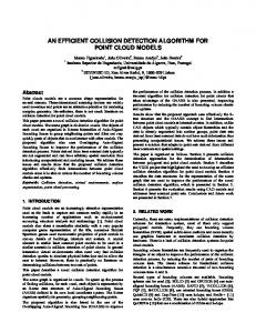
If s, 0 ≤ s ≤ L − 1, is a threshold. Then s can partition the co-occurrence matrix into 4 quadrants, namely A, B, C, and D (Fig. 1).
The gray level corresponding to the maximum of HT (s) gives the optimal threshold for object-background classification. For the MFR image shown in Fig. 2(b), the entropy-based thresholding result is shown in Fig. 2(c) where we can see blood vessels are clearly segmented from the background. 2.3. Length Filtering As seen in Fig. 2(c), there are still some misclassified pixels in the image. Here we want to produce a clean and complete vascular tree structure by removing misclassified pixels. Length filtering is used to remove isolated pixels by using the concept of connected pixels labeling. Connected regions correspond to individual objects. We first need to identify separate connected regions. The length filtering tries to isolate the individual objects by using the eight-connected neighborhood and label propagation. Once the algorithm is completed, only the resulting classes exceed a certain number of pixels, e.g., 250, are labeled as blood vessels. Fig. 2(d) shows the results after length filtering.
Figure 1: Quadrants of co-occurrence matrix [5]. 2.4. Detection of Vascular Intersections/Crossovers
Let us define the following quantities: PA = PC =
s P s P
pij
i=0 j=0 L−1 P P L−1
(4) pij
i=s+1 j=s+1
Normalizing the probabilities within each individual quadrant, such that the sum of the probabilities of each quadrant equals one, we get the following cell probabilities for different quadrants: PijA =
pij PA
tij /(
= =
s P s P
i=0 j=0 tij s P s P i=0 j=0
L−1 P
tij ) i=0 L−1 P L−1 P
tij /
i=0 j=0
tij
tij
for 0 ≤ i ≤ s, 0 ≤ j ≤ s
(5)
Vascular intersections and crossovers are the most appropriate representation in registration process because they exist in every retinal images, and do not move except in some diseases. If a vascular tree is one-pixel wide, the branching points can be detected and characterized efficiently from the vascular tree. Morphological thinning is applied to the vascular tree in order to get one-pixelwide vascular tree as shown in Fig. 2(e). In order to save computational time, a 3×3 neighborhood window is used to probe and find the branching points. If the number of vascular tree in the window is great than 3, it is a branch point. Then a 11×11 neighborhood is applied through a detected branching points in order to eliminate the small intersections [6]. We consider only the boundary pixels of a 11×11 square. If the number of vascular tree on the boundary is greater than 2, we mark it as an intersection/crossover. Fig. 2(f) presents the vascular tree with the intersections and crossovers.
(a)
(b)
(c)
(d)
(e)
(f)
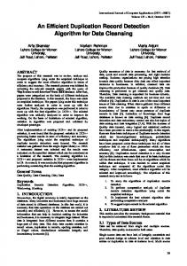
Figure 2: (a) An original retinal image. (b) Matched filtering result. (c) Local entropy thresholding result. (d) Vascular tree. (e) One-pixel wide vascular tree. (f) One-pixel wide vascular tree with intersections and crossovers overlaying on gray-scaled image. 3. SIMULATION RESULTS On Windows XP, Pentium 4, CPU 1.7 GHz, using MATLAB version 6.1, the computational time of the whole process of our algorithm takes approximately 3 minutes for each retinal image. We use the same set of twenty 605 x 700 pixel retinal images (24bpp), as used in [3]. In order to evaluate the performance of our algorithm, we compare our simulation results with the state-of-the-art results obtained from [3] and hand-labeled groundtruth segmentations as shown in Fig. 3. Specifically, we classify retinal images into three categories, normal retinal images, abnormal retinal images with some lesions, and retinal images with obscure blood vessel appearance. From column by column, the first column of Fig. 3 presents the results from normal retinal images. Results from abnormal retinal images with some lesions are shown on the second column. The last column presents results from retinal images with obscure blood vessel appearance. From row by row, the first row shows original retinal images. The second row shows the hand-labeled groundtruth segmentations which were manually labeled by Hoover [3]. The third row presents simulation results from [3]. The last row presents our simulation results. Although, algorithm in [3] performs very well, a significant improvement can be achieved by our algorithm for normal and obscure retinal images. Our method performs very well in extracting blood vessels. Even the smaller blood vessels can be extracted. Matched filtering enhances the contrast of blood vessels against the background. Local entropy thresholding algo-
rithm, which takes into account the spatial distribution of gray levels, performs efficiently in distinguishing between enhanced vessel segments and the background since it can preserve the structure details of an image. However, the presence of lesions in the abnormal retinal image is the major obstacle in extracting blood vessels since they are also mis-enhanced and mis-detected as blood vessels. Our algorithm is sensitive to lesions due to the fact that their boundaries partially match the shape of matched filter kernels, while the algorithm in [3] is more robust to lesions. We expect to improve the robustness of our algorithm by involving color information and additional anatomical constraints for blood vessel detection and extraction. 4. CONCLUSIONS In this paper, we have introduced an efficient algorithm for fully automated blood vessel detection in ocular fundus images using the local entropy thresholding scheme. The proposed method retains the computational simplicity, and at the same time, can achieve accurate segmentation results in the case of normal retinal images and images with obscure blood vessel appearance. In the case of abnormal retinal images with lessons, some lesions are also misdetected in addition to blood vessels. In the future work, we want to improve the robustness of our algorithm by involving in the preprocessing scheme and additional anatomical constraints to separate the lesions in the final vascular tree.
Figure 3: First row: Example images; Second row: Hand-labeled ground truth ([3]); Third row: Results from [3]; Last row: Results from our method. 5. REFERENCES [1] A. Pinz, S. Bernogger, P. Datlinger, and A. Kruger, “Mapping the human retina,” IEEE Trans. Medical imaging, vol. 17, no. 4, August 1998. [2] L. Zhou, M. S. Rzeszotarski, L. Singerman, and J. M. Chokreff, “The detection and quantification of retinopathy using digital angiograms,” IEEE Trans. Medical imaging, vol. 13, no. 4, December 1994. [3] A. Hoover, V. Kouznetsova, and M. Goldbaum, “Locating blood vessels in retinal images by piecewise threshold probing of a matched filter response,” IEEE Trans. Medical imaging, vol. 19, no. 3, March 2000, http://www.ces.clemson.edu/ ahoover. [4] S. Chaudhuri, S. Chatterjee, N. Katz, M. Nelson, and M. Goldbaum, “Detection of blood vessels in retinal images using two-
dimensional matched filters,” IEEE Trans. Medical imaging, vol. 8, no. 3, September 1989. [5] N. R. Pal and S. K. Pal, “Entropic thresholding,” Signal processing, vol. 16, pp. 97–108, 1989. [6] A. Can, H. Shen, J. N. Turner, H. L. Tanenbaum, and B. Roysam, “Rapid automated tracing and feature extraction from retinal fundus images using direct exploratory algorithms,” IEEE Trans. Information Technology in Biomedicine, vol. 3, no. 2, June 1999.