Jun 26, 2008 - (Tld/Xlr) and interact with a conserved modulator (Tsg/xTsg)16â21. Anti-dorsalizing .... model. The model assumes free-ligand diffusion is much smaller than that of .... Admp by Chordin away from their domain of production.
Vol 453 | 26 June 2008 | doi:10.1038/nature07059
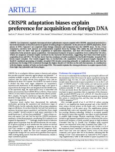
ARTICLES Scaling of the BMP activation gradient in Xenopus embryos Danny Ben-Zvi1, Ben-Zion Shilo1, Abraham Fainsod2 & Naama Barkai1,3 In groundbreaking experiments, Hans Spemann demonstrated that the dorsal part of the amphibian embryo can generate a well-proportioned tadpole, and that a small group of dorsal cells, the ‘organizer’, can induce a complete and well-proportioned twinned axis when transplanted into a host embryo. Key to organizer function is the localized secretion of inhibitors of bone morphogenetic protein (BMP), which defines a graded BMP activation profile. Although the central proteins involved in shaping this gradient are well characterized, their integrated function, and in particular how pattern scales with size, is not understood. Here we present evidence that in Xenopus, the BMP activity gradient is defined by a ‘shuttling-based’ mechanism, whereby the BMP ligands are translocated ventrally through their association with the BMP inhibitor Chordin. This shuttling, with feedback repression of the BMP ligand Admp, offers a quantitative explanation to Spemann’s observations, and accounts naturally for the scaling of embryo pattern with its size. Multicellular organisms develop with a remarkable consistency, maintaining a precise body plan in the face of genetic polymorphism or environmental fluctuations1. Yet, size and shape differ significantly even between closely related species. Developmental processes are thus shaped by seemingly opposing challenges: maintaining robustness at the species level, while allowing sufficient flexibility for evolutionary adaptation2. The interplay between robustness and evolutionary plasticity is poorly understood. In Bilateria, early dorsoventral patterning relies on the graded distribution of BMP activity along the embryo. Two classical experiments performed by Hans Spemann (reviewed in ref. 3) demonstrated the dramatic plasticity of this patterning process in amphibians. First, dorsal halves of bisected embryos develop into well-proportioned tadpoles4. Second, cells taken from the embryonic dorsal blastopore lip and transplanted into the ventral side of a naive embryo induce a complete and well-proportioned secondary axis5. The region responsible for this induction property, ‘Spemann’s organizer’, was identified later in other vertebrates, and its inductive capacity is attributed primarily to the secretion of BMP inhibitors6. However, the mechanism underlying the ability of dorsal-half embryos to grow into well-scaled tadpoles, and the ability to generate two complete and properly scaled tadpoles upon organizer transplantation, remained unknown. Experiments by Cooke further demonstrated the precision of scaling, and verified that compensation is not due to overgrowth of the remaining cells, but to their proportionate assignment to the different tissues7. Despite large differences in shape and size, the molecular network that generates the BMP gradient is remarkably conserved across evolution8–11. In flies and vertebrates, BMP ligands are initially expressed in broad domains, with the localized secretion of a conserved BMP inhibitor (Sog/Chordin, respectively12,13) providing the key organizing dorsoventral asymmetry14,15 (Supplementary Fig. 1a– d). The inhibitors diffuse, undergo cleavage by a conserved protease (Tld/Xlr) and interact with a conserved modulator (Tsg/xTsg)16–21. Anti-dorsalizing morphogenic protein (Admp) is a BMP ligand found in many Bilateria but is missing in Drosophila. In contrast to other BMP ligands, it is expressed dorsally with BMP inhibitors, and is subject to autoregulatory transcriptional repression by the BMP
pathway22,23. A role for Admp in providing scaling was recently suggested, following the observation that depletion of Admp abolishes patterning in dorsal-half embryos24. Theoretical analysis of the BMP gradient formation in the Drosophila embryo distinguished two qualitatively different patterning mechanisms25–28 (Supplementary Fig. 1e, f). In the ‘inhibitionbased’ mechanism, patterning is governed by the creation of an inhibition gradient over a uniform field of activators. In the ‘shuttling-based’ mechanism, patterning relies on the physical translocation of the activator to the midline, mediated by its binding to the inhibitor. Both mechanisms can generate a graded profile of BMP activation26,29, but the finding that the shuttling mechanism generates a sharp and robust gradient led to the proposal that it is in use. This prediction was subsequently verified experimentally26,30–32 (reviewed in refs 33 and 34). In this study, we show that shuttling is used also in the Xenopus embryo, and that, with the auto-repression of Admp, it ensures the scaling of the BMP activation profile with embryo size. Shuttling is required for scaling A key question is whether the conservation of network constituents implies the conservation of their integrated function, and if so, how an increased functional complexity can evolve. To examine whether shuttling plays a part in establishing the BMP activation gradient in Xenopus, we focused first on the ability of dorsal-half embryos to generate a well-proportioned embryo. This scaling property is difficult to explain by most models of morphogen gradients, and was proposed as evidence that patterning does not involve morphogens7. To assess the constraints imposed by this scaling property rigorously, we formulated a mathematical model that is based on the conserved core of this patterning network. The model includes two BMP ligands, Admp and Bmp (where Bmp stands for the three ligands BMP2/4/7), a BMP inhibitor (Chordin) and the protease Xlr (Fig. 1a). We allowed for the diffusion of all components, the binding of the BMP ligands to Chordin, and the degradation of Chordin by Xlr. We also considered the production of Chordin and Admp at the dorsal pole and the auto-repression of Admp by BMP signalling24 (see Methods and Supplementary Information for equations and further details of the screen).
1 Department of Molecular Genetics, Weizmann Institute of Science, Rehovot 76100, Israel. 2Department of Cellular Biochemistry and Human Genetics, Faculty of Medicine, Hebrew University, Jerusalem 91120, Israel. 3Department of Physics of Complex Systems, Weizmann Institute of Science, Rehovot 76100, Israel.
1205 ©2008 Macmillan Publishers Limited. All rights reserved
ARTICLES
NATURE | Vol 453 | 26 June 2008
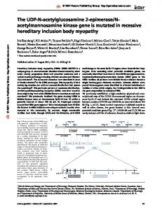
To examine more generally whether shuttling is required for scaling, we defined a rigorous measure that quantified the extent of shuttling, namely the translocation of the total ligand to the ventral pole, in each of the networks tested (Methods and Supplementary Information). Shuttling was observed in all networks that were capable of robust scaling (Fig. 1f). Moreover, the parameters of the consistent networks obeyed the molecular requirement for shuttling, as described previously in Drosophila26. First, the binding of Chordin to the BMP ligands largely facilitated the diffusion of the BMP ligands (Fig. 1g–j). Second, Chordin was degraded primarily when complexed with BMP ligands (Fig. 1g, h). Additionally, we found that scaling requires that Chordin binds to Bmp with a significantly higher affinity than to Admp (Fig. 1i, j).
Diffusion
Xlr
+
Chd
Bmp
Chd–Bmp
Diffusion
* *
Xlr
l
Lig lChd /lChd
BMP signalling
g
Wild type d
Wild type d
v
l
0.4 0.2
v
e Shuttling
BMP signalling
Inhibition
Wild type
l
i
Wild type
Dorsal half (scaled axis) d
0.5 0.7 Shuttling coefficient
0.9
102
h 102 101
100
100
10–1
10–1
10–2
10–2 101
102
10–2 10–1 100
101
102
10–2 10–1 100 101 DComp/DLig
102
Dorsal half
kAdmp/kBmp
d
0.3
101
10–2 10–1 100 Dorsal half
Shuttling
0.6
0.1
c Shuttling
Inhibition
0.8 Inhibition
Chd–Admp
Admp
b
f Probability
*
Mechanism by which scaling is achieved Our numerical analysis confirmed that the known patterning network can support the scaling of pattern with size, and suggested that shuttling plays an important part in providing this ability. To
Diffusion
a
Diffusion
Diffusion
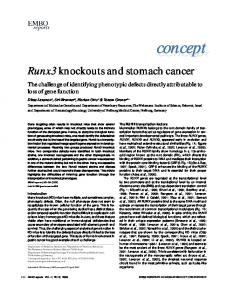
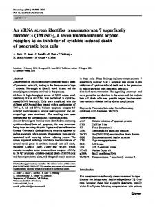
We screened systematically for parameters (rate constants and diffusion coefficients) for which the activation gradient is robust and scales with embryo size. To this end, we assigned each of the nine parameters three possible values, ranging over at least two orders of magnitude. The BMP activation gradient was solved numerically for all the networks defined in this nine-dimensional cube. Of the 26,000 networks examined, approximately 1,100 displayed a proper polarity, but only 21 were also capable of scaling in a dorsal-half embryo. An example for a gradient that did not scale is shown in Fig. 1b, d whereas a scaled gradient is shown in Fig. 1c, e. Examining the solutions, we noted that the respective gradients were established by distinct mechanisms. In the network that did not scale, the overall level of the ligands remained approximately uniform, and the activation gradient reflected the gradient of the inhibitor Chordin. In contrast, in the network that did scale, the ligands were physically concentrated at the ventral pole. The activation gradient was thus generated by the shuttling of the ligands to the ventral pole.
Dorsal half (scaled axis) v
d
l
102
j 102
101
101
100
100
10–1
10–1
10–2
10–2 10–2 10–1 100 101 DComp/DLig
102
v
Figure 1 | Numerical evidence for shuttling. a, Model used in the screen. See Methods and Supplementary Information for equations and parameters. b, d, Activation profiles defined by the inhibition-based model. The model assumes that all components diffuse at the same rate. The model was solved numerically for the whole embryos (black) and the dorsal-half embryos (grey). The unscaled profile, measured in absolute length, is shown in b whereas the scaled profile measured in relative lengths, scaled by the size of the field, is shown in d. d, l, v, dorsal, lateral and ventral regions of the embryo, respectively. c, e, Activation profiles defined by the shuttling-based model. The model assumes free-ligand diffusion is much smaller than that of Chordin and the complex. Chordin is degraded primarily when complexed with a BMP ligand (parameters marked with * in Fig. 1a are small). The unscaled profile is shown in c whereas the scaled profile is shown in e. f, Consistent networks establish pattern by shuttling. Distribution of the shuttling coefficient Sh in consistent networks (black) and in all the networks
that establish a dorsoventral gradient (grey). Sh < 1 indicates a shuttling mechanism, whereas Sh < 0 an inhibition mechanism. g, h, Relative diffusion and degradation in networks that establish dorsoventral polarity. The x axis displays the ratio between diffusion coefficients of the inhibitorthe ratio bound and free BMP ligand (DComp/DLig). The y axis displays � � Lig between degradation of BMP-bound and free inhibitor lChd =lChd . Networks that were sampled in the screen are in light grey. Because this plot is a projection from a nine-dimensional space, each point symbolizes many networks where the respective ratios were held fixed, but parameters were changed systematically. g, Grey circles correspond to networks establishing proper dorsoventral polarity. h, Black circles correspond to networks establishing proper dorsoventral polarity, support scaling and robustness. i, j, Affinity of Chordin to Admp versus Bmp. Same as g–h with the y axis denoting the ratio between of binding rates of Chordin to Admp and Bmp (kAdmp/kBmp).
1206 ©2008 Macmillan Publishers Limited. All rights reserved
ARTICLES
NATURE | Vol 453 | 26 June 2008
ð1Þ
where DChd is the Chordin diffusion coefficient and kLig is the binding rate of the BMP ligand to Chordin. This profile is valid in most places, (x . e with e / 1/[BMP]tot R 0). It is robust to changes in the levels of network components but does not scale with embryo size; indeed, embryo size does not appear in equation (1), and thus does not influence the shape of the gradient. Similarly, solving the model numerically under conditions of secondary-axis induction, we find that two axes ensue, but these two axes decay at the same rate as the original axis and do not scale to half-embryo size (Fig. 2a–c). Thus, shuttling of a single ligand is not sufficient for scaling. We extended the model to account for the additional ligand Admp, and its feedback-mediated repression. This model can also be solved analytically (Box 1), predicting the activation profile: S(x)