Brief Communications Diagnosis of ruptured pulmonary hydatid cyst by means of flexible fiberoptic bronchoscopy: A report of three cases Abdul Salam Yaseen Taha, MB, ChB, FICMS, Kurdistan, Iraq
T
here are 3 radiologic signs considered diagnostic of ruptured pulmonary hydatid cyst (PHC): perivesicular pneumocyst, double-domed arch, and water lily.1 Apart from these, every localized Dr Taha radiologic density seen in any patient more than 3 years of age in an endemic area should be looked on as a possible ruptured hydatid cyst (HC).1 Nevertheless, situations in which the diagnosis of ruptured PHC is difficult are far from being rare in countries of high endemicity.1-3 Thus a preliminary bronchoscopy is a perfectly justifiable step in the diagnostic work-up.1 Herein, we report 3 selected cases of Iraqi patients with ruptured PHC in whom definitive diagnoses were made with the use of the flexible fiberoptic bronchoscope (FOB).
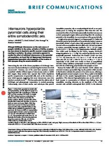
Clinical Summaries PATIENT 1. A 36-year-old man presented with a 1-week history of cough, expectoration of greenish sputum, fever, right-sided pleuritic chest pain, and hemoptysis. Physical examination showed no abnormalities. The chest films (Figures 1 and 2) revealed a rounded opacity in the superior segment of the right lower lobe. FOB revealed a whitish material (laminated membrane of ruptured PHC) in a subsegmental orifice of the apical lower segmental bronchus.
From the Department of Surgery, College of Medicine, University of Sulaimani, Kurdistan, Iraq. Received for publication May 17, 2004; revisions accepted March 1, 2005; accepted for publication March 14, 2005. Address for reprints: Dr Abdul Salam Y. Taha, Assistant Professor and Head of the Department of Thoracic and Cardiovascular Surgery, College of Medicine, University of Sulaimani, Kurdistan, Iraq (E-mail:
[email protected]). J Thorac Cardiovasc Surg 2005;30:1196-7 0022-5223/$30.00 Copyright © 2005 by The American Association for Thoracic Surgery doi:10.1016/j.jtcvs.2005.03.024
1196
Figure 1. Anteroposterior chest film showing a rounded opacity in the superior segment of the right lower lobe.
The Journal of Thoracic and Cardiovascular Surgery ● October 2005
Brief Communications
Figure 2. Lateral chest film showing a rounded opacity in the superior segment of the right lower lobe.
PATIENT 2. A 36-year-old man presented with a 3-month history of intermittent fever and dry cough. Chest examination revealed right-sided wheeze. The chest films revealed a persistent, homogeneous, diamond-shaped opacity in the right upper zone with a smooth inferior border: the interlobar fissure. The initial impression was either ruptured PHC or encysted pleural effusion. FOB revealed whitish material (fragment of laminated membrane) in the anterior and posterior segmental bronchi. The patient underwent right thoracotomy, which confirmed the diagnosis. PATIENT 3. A 26-year-old housewife presented with a 2-week history of cough and massive hemoptysis. Her chest film showed an ovoid opacity in the left upper lobe. During bronchoscopy, fresh bleeding from the lingular bronchus, as well as the laminated membrane, was seen. A left thoracotomy was performed. Infected disintegrated membrane was found and removed.
Discussion Bronchoscopy is unnecessary in cases of ruptured PHC with a pathognomonic clinical picture, radiologic picture, or both.1,4
However, it is indispensable when there is suspicion of tumor or when the radiologic picture is atypical.1,2 The source of hemoptysis (if present) can be traced (as in case 3). The finding of laminated membrane in a bronchus draining the cyst cavity (be it lobar, segmental, or subsegmental) is diagnostic.1,5 A piece of the membrane can be obtained and examined with a hand lens1 or microscopically to confirm its laminated nature. The bronchial aspirate can be examined for the presence of hooklets. The reported patients had atypical chest radiographic signs. FOB enabled a definite diagnosis in all of them. Moreover, it ruled out the possibility of encysted pleural effusion (case 2), which requires conservative rather than surgical therapy. In a place in which pulmonary tuberculosis is endemic, like Iraq, the combination of hemoptysis and persistent pulmonary opacities is quite suspicious. FOB rules out this diagnosis by visualizing the membrane of the HC. This is another example of the superiority of FOB because of its characteristics (safety, small size, flexibility, and better vision). It is worthy to mention that all of our patients had laminated membranes in either the segmental or subsegmental bronchus, which otherwise could be missed by using the rigid scope. The use of a bronchoscope in the diagnosis of doubtful cases of ruptured PHC is not new.1,5 Professor F. Saidi1 reported a case of a 6-year-old girl with left lower lobe ruptured HC diagnosed bronchoscopically by visualizing an intrabronchial laminated membrane. In his opinion, such a finding can be observed by some surgeons with extensive experience in dealing with PHC.1 Two other cases were reported in Tunisian children aged 5 and 10 years with chronic pulmonary opacities posing a diagnostic problem.5 It is worthy of mention that both workers used the rigid bronchoscope. A review of the computerized medical literature from 1966 through 1998 revealed no single article on FOB and HC. Along with Henry and colleagues5 and Saidi,1 this report strongly shows the advantage of bronchoscopy (and FOB in particular) in the diagnosis of puzzling cases of ruptured HCs.
References 1. Saidi F. Surgery of hydatid disease. London: W.B. Saunders Co, Ltd; 1976. 2. Elhasani NB. Pulmonary hydatid disease (part one). Postgrad Doctor. 1985;8:44. 3. Elhasani NB. Pulmonary hydatid disease (part two). Postgrad Doctor. 1985;8:84. 4. Xanthasis D, Efthimiadis M, Papadakis G, et al. Hydatid disease of the chest. Report of 91 patients surgically treated. Thorax. 1972;27:517-28. 5. Henry P, Khalfallah A, Lakhal A, et al. Bronchoscopy in the diagnosis of complicated pulmonary hydatid cyst in children. Rev Mal Respir. 1984;1:313-7.
The Journal of Thoracic and Cardiovascular Surgery ● Volume 30, Number 4
1197