Recent studies have demonstrated that histone methylation can be dynamically regulated through active demethylation. However, no demethylase specific to histone H3 trimethylLys4 (H3K4me3) has been identified. Here we report that the Drosophila melanogaster protein ‘little imaginal discs’ (Lid), a JmjC domain–containing trithorax group protein, can demethylate H3K4me3. Consistent with its genetic classification, Lid positively regulates Hox gene expression in S2 cells. Histone methylation contributes to diverse biological processes1. Recent studies indicate that histone methylation, like other histone modifications, is dynamically regulated through active demethylation
a
b
Size M F-Lid (kDa)
170 130 95 72 56
400
300
200
100
Lid
0
– + H3K4 SET7
c
H3K4me3 peptide (1–21) Me 14 Da +Lid
d Relative intensity
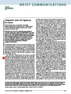
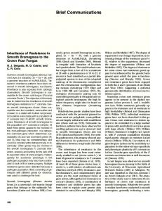
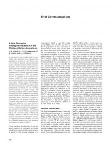
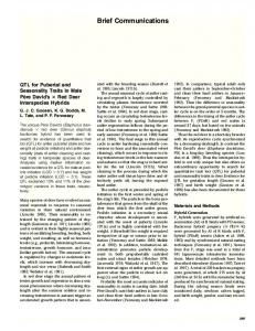
Figure 1 Lid is a histone demethylase with specificity for H3K4me3. (a) Coomassie-stained SDS-PAGE gel of Flag-tagged Lid protein purified from baculovirus-infected Sf9 cells. M, marker; F-Lid, Flag-tagged Lid. (b) Recombinant Lid can demethylate substrates generated by a SET7 mutant. Various radiolabeled methylhistone substrates were generated and used in demethylation assays with recombinant Lid. The release of radioactively labeled formaldehyde was used to measure enzymatic activity. Owing to steric constrains of the SET7 catalytic site, the wildtype SET7 can only monomethylate H3K4, whereas the Y245A mutant can di- and trimethylate H3K4. (c,d) Lid demethylates both di- and trimethylated H3K4 peptides. Histone peptides (residues 1–21) containing either tri- (c) or dimethylated (d) H3K4 were subjected to demethylation reactions in the presence or absence of Lid followed by mass spectrometric analysis. (e) Quantification of the mass spectrometry results in c and d. Relative percentage of peptide substrates at different methylation states after demethylation reaction is shown. Lid has similar efficiency in converting H3K4me3 to H3K4me2 and H3K4me2 to H3K4me1 in vitro.
2,764.6
2,750.6
–Lid
– +
– +
– +
H3K4me2 peptide (1–21) Me 14 Da +Lid
– +
– +
– +
H3K4 H3K9 H3K27 H3K36 H3K79 H4K20 SET7 DIM5 EZH2 SET2 DOT1 SUV4Y245A 20 H1
2,863.2
2,849.2
–Lid m/z
m/z
e
Substrate % after HDM assay
Nara Lee1,2, Junyu Zhang3, Robert J Klose1,2, Hediye ErdjumentBromage4, Paul Tempst4, Richard S Jones3 & Yi Zhang1,2
by two distinct classes of enzymes. The first class of histone demethylase, exemplified by LSD1, catalyzes demethylation of H3K4me2 and H3K4me1 in a flavin adenine dinucleotide (FAD)-dependent manner2. The second class of histone demethylase encompasses a large protein family3 and uses a conserved JmjC domain to catalyze demethylation in an Fe(II)- and a-ketoglutarate–dependent hydroxylation reaction4. Several JmjC domain–containing proteins with specificity toward various methylation states of H3 Lys9 and Lys36 (H3K9 and H3K36) have been characterized4–9. However, no H3K4me3-specific demethylase has been identified thus far, leaving open the possibility that H3K4me3 might not be a reversible modification. To facilitate identification of novel histone demethylases, we performed a phylogenetic analysis of the JmjC domain–containing proteins in six model organisms3. This analysis allowed us to divide the JmjC domain–containing proteins into seven subfamilies on the basis of conservation in the JmjC domain and overall protein domain architecture3. Members from three of the seven subfamilies have been shown to encode active histone demethylases. Of the remaining four subfamilies, the JARID1 subfamily is of particular interest because members of this subfamily contain multiple conserved functional domains (Supplementary Fig. 1a online), some of which are reported to participate in transcriptional regulation10,11. Notably, the sole
Formaldehyde release (c.p.m.)
The trithorax-group protein Lid is a histone H3 trimethyl-Lys4 demethylase
Relative intensity
© 2007 Nature Publishing Group http://www.nature.com/nsmb
B R I E F C O M M U N I C AT I O N S
K4me3 substrate K4me2 substrate 100 80 60 40 20 0 me3 me2 me1 me0
1Howard
Hughes Medical Institute and 2Department of Biochemistry and Biophysics, Lineberger Comprehensive Cancer Center, University of North Carolina, Chapel Hill, North Carolina 27599-7295, USA. 3Department of Biological Sciences, Southern Methodist University, Dallas, Texas 75275, USA. 4Molecular Biology Program, Memorial Sloan Kettering Cancer Center, 1275 York Avenue, New York, New York 10021, USA. Correspondence should be addressed to Y.Z. (
[email protected]). Received 21 December 2006; accepted 16 February 2007; published online 11 March 2007; doi:10.1038/nsmb1216
NATURE STRUCTURAL & MOLECULAR BIOLOGY
VOLUME 14
NUMBER 4
APRIL 2007
341
B R I E F C O M M U N I C AT I O N S
a
Anti-Flag
Anti-H3K4me3
DAPI
Anti-Flag
Anti-H3K4me3
DAPI
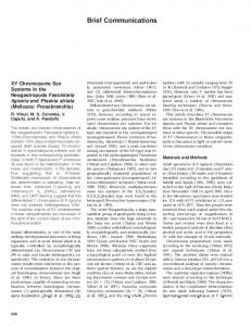
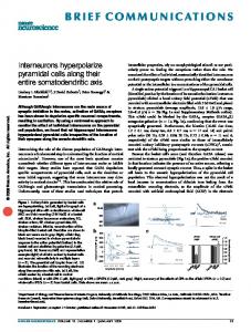
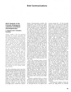
Figure 2 Multiple domains contribute to Lid enzymatic activity. (a) Overexpression of wild-type Lid (top images), but not the H637A mutant (bottom images) results in a global decrease of H3K4me3 levels. Drosophila S2 cells were transfected with Flag-tagged Lid and costained with antibodies against Flag tag and H3K4me3. (b) Demethylase activity of Lid mutants was monitored by H3K4me3 staining in transfected cells. Deletion of the JmjN, Arid and the zinc-finger domains abolished the enzymatic activity, whereas deletion of the C-terminal PHD fingers did not affect Lid’s enzymatic activity.
Lid WT
H637A
© 2007 Nature Publishing Group http://www.nature.com/nsmb
b
Drosophila homolog of this subfamily, Lid, has been identified genetically as a trithorax group (trxG) protein12. The Lid protein contains all the functional domains found in the mammalian JARID1 proteins, and its JmjC domain is also highly conserved (Supplementary Fig. 1b). To test whether Lid is an active demethylase, we expressed an N-terminally Flag-tagged Lid protein in Sf 9 cells and purified it to near homogeneity (Fig. 1a). Incubation of the recombinant Lid protein with various radiolabeled histone substrates in a demethylase assay resulted in release of radioactive formaldehyde only when substrates were generated with a mutant SET7 (Fig. 1b and Supplementary Methods online), which specifically di- and trimethylates (me2/3) H3K4 (ref. 13). Notably, Lid did not demethylate substrates
generated by the wild-type SET7, which only monomethylates H3K4 (ref. 13), nor did it demethylate other known histone methylation sites (Fig. 1b). These results indicate that Lid is an H3K4me2/3-specific demethylase. To confirm the substrate specificity of Lid, synthetic peptides containing di- or trimethylated H3K4 were incubated with Lid and analyzed by mass spectrometry. In agreement with the formaldehyde-release assay, Lid was able to demethylate both H3K4me3 and H3K4me2 peptides (Fig. 1c,d). Quantification of the demethylated peptide product indicated that Lid has similar efficiency toward di- and trimethylated peptides (Fig. 1e). Collectively, the above results indicate that Lid is an H3K4me2/3-specific demethylase in vitro. To evaluate whether Lid functions as an active H3K4 demethylase in vivo, we transfected a Flag-tagged Lid expression plasmid into Drosophila S2 cells and analyzed the effect on H3K4 methylation by indirect immunofluorescence using H3K4 methylation–specific antibodies. Overexpression of Lid greatly reduced H3K4me3 levels (Fig. 2a, top images). The reduction was dependent on the demethylase activity of Lid, as overexpression of a Lid mutant (H637A) predicted to disrupt Fe(II) binding had no effect on H3K4me3 levels (bottom images). Although Lid is capable of demethylating an H3K4me2 peptide in vitro (Fig. 1b), overexpression of Lid did not cause a noticeable change in the H3K4me2 or H3K4me1 levels (Supplementary Fig. 2a online). Similarly, trimethylation on H3K9, H3K27 and H3K36 were also unaffected (Supplementary Fig. 2b). Collectively, these data indicate that Lid can specifically demethylate H3K4me3 in vivo. The Lid protein contains several other functional domains in addition to the JmjC domain, including JmjN, Arid, PHD and a zinc finger (Supplementary Fig. 1a). We have previously demonstrated that, in addition to the JmjC domain, the zinc finger of
a
c
∆jmjN (∆1–218)
∆Arid (∆214–441)
∆PHD (∆893–1838)
∆ZF (∆766–1838)
Size (kDa)
M
NE
b
Lid
Ser5
Merge
d –RT
170
dsRNA GFP lid
130
+RT GFP
lid
lid 95 Ubx
72
Lid
Ser2
56
Merge rp49
43 WB anti-Lid
Lid
DAPI
Merge
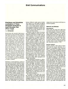
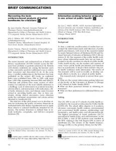
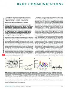
Figure 3 Lid localizes to interbands but does not colocalize with active RNA polymerase II. (a) Western blotting analysis of Drosophila embryo nuclear extracts (125 mg) indicates that the Lid antibody is specific for Lid. M, protein marker; NE, nuclear extract. (b) Distribution of Lid on larval polytene chromosomes. Immunostaining reveals that Lid is localized to interbands but excluded from the chromocenter (arrow) and heavy DAPI bands. (c) Lid does not colocalize with active RNA polymerase II. Costaining of Lid with the Ser5- or Ser3-phosphorylated form of RNA polymerase II is shown. (d) lid knockdown in S2 cells results in decreased Ubx expression. S2 cells were treated with double-stranded RNAs (dsRNAs) that target lid or GFP. Knockdown was verified by reverse-transcription PCR. GFP dsRNA serves as a control for the RNA interference treatment. rp49 serves as a control for equal input. The numbers of PCR cycles for lid, Ubx and rp49 were 32, 40 and 25, respectively.
342
VOLUME 14
NUMBER 4
APRIL 2007
NATURE STRUCTURAL & MOLECULAR BIOLOGY
© 2007 Nature Publishing Group http://www.nature.com/nsmb
B R I E F C O M M U N I C AT I O N S JHDM2A (ref. 9) and the JmjN domain of JHDM3A (ref. 7) are essential for enzymatic activity. To define the domain requirements for Lid enzymatic activity, we generated four expression constructs with deletions of the JmjN, Arid, the zinc finger and the two C-terminal PHD fingers (Supplementary Fig. 3 online). In contrast to expression of the wild-type Lid (Fig. 2a), expression of the deletion mutants had no effect on H3K4me3 levels, with the exception of the PHD-finger deletion mutant, which retained demethylase activity (Fig. 2b). On the basis of these results, we conclude that although the C-terminal PHD fingers are dispensable for activity, all other domains contribute to the demethylase activity of Lid. It is likely that these domains contribute either to substrate recognition or folding of the JmjC domain. To gain insight into the potential function of Lid, we generated a polyclonal antibody against Lid protein. After demonstration of the antibody specificity by western blotting of Drosophila embryo nuclear extracts (Fig. 3a), we investigated the distribution of Lid on Drosophila polytene chromosomes. Lid is broadly distributed on these chromosomes (Fig. 3b), suggesting that Lid may regulate methylation at many genomic loci. However, Lid is excluded from the chromocenter (left image, arrow) and 4¢,6-diamidino-2-phenylindole (DAPI)-heavy regions (Fig. 3b, right image and Supplementary Fig. 4 online). The fact that Lid is excluded from DAPI-heavy regions prompted us to investigate whether Lid is enriched in actively transcribed regions of chromatin. To this end, we costained Lid with RNA polymerase II phosphorylated at Ser5 and Ser2 in the C-terminal domain, modifications that mark transcriptional initiation or elongation, respectively. The strongest bands for active RNAPII do not show concomitant Lid staining (Fig. 3c). This is in direct contrast to other TrxG proteins such as Brm and Kis, which show extensive colocalization with active RNAPII14. The polytene staining data presented above suggest that Lid may not directly participate in active transcription because it does not overlap with active RNA polymerase II. However, the genetic classification of the lid gene as a trxG gene indicates that Lid positively regulates Hox gene expression. Available lid mutant alleles seem to be hypomorphic and show highly variable phenotypes12. Some lid homozygotes die as embryos, whereas others survive to late larval or pupal stages. Those with more severe postembryonic phenotypes have extremely small imaginal discs, which makes molecular analysis of the mutants technically difficult. Therefore, we analyzed the effect of Lid on Hox gene expression in S2 cells using double stranded RNA–mediated knockdown. Consistent with the genetic classification of lid as a trxG gene, Lid seems to positively regulate the Hox gene Ubx, as knockdown of lid decreased the abundance of Ubx transcripts (Fig. 3d). H3K4me3 is enriched in promoters of active genes15,16; consequently, H3K4me3 is regarded as a mark of active transcription. Consistent with this notion, several trxG proteins involved in maintaining the ‘on’ state of Hox genes have H3K4 methyltransferase
NATURE STRUCTURAL & MOLECULAR BIOLOGY
VOLUME 14
activity17,18. Therefore, it was surprising that the biochemical activity of Lid antagonizes those of other trxG proteins. Classification of lid as a trxG gene is based on the observation that lid mutant alleles respectively suppress and enhance homeotic phenotypes produced by PcG and trxG mutant alleles12. Consistent with its genetic classification, Lid positively regulates the Ubx gene in S2 cells. To reconcile its biochemical activity with its genetic classification, we favor the possibility that Lid indirectly regulates Hox gene expression. However, we cannot rule out the possibility that an H3K4 methylationdemethylation cycle may be an integral part of the gene activation process. A similar situation has been reported for H2B ubiquitination, where both ubiquitination and deubiquitination are required for efficient transcription19,20. Identification of direct target genes of Lid will allow us to differentiate between these two possibilities in the future. Note: Supplementary information is available on the Nature Structural & Molecular Biology website. ACKNOWLEDGMENTS We thank L. Fabrizio for help with mass spectrometry and S. Rogers and G. Rogers (University of North Carolina, Chapel Hill) for sharing fly reagents. This work was supported by US National Institutes of Health grants GM68804 (to Y.Z.), GM46567 (to R.S.J.) and P30 CA08748 (to P.T.). Y.Z. is an Investigator of the Howard Hughes Medical Institute. R.J.K. is funded by the Canadian Institutes of Heath Research. AUTHOR CONTRIBUTIONS N.L. and Y.Z. designed the experiments. N.L., J.Z., R.J.K., H.E.-B. and P.T. performed the experiments. Y.Z., N.L. and R.S.J. wrote the paper. COMPETING INTERESTS STATEMENT The authors declare no competing financial interests. Published online at http://www.nature.com/nsmb Reprints and permissions information is available online at http://npg.nature.com/ reprintsandpermissions 1. Martin, C. & Zhang, Y. Nat. Rev. Mol. Cell Biol. 6, 838–849 (2005). 2. Shi, Y. et al. Cell 119, 941–953 (2004). 3. Klose, R.J., Kallin, E.M. & Zhang, Y. Nat. Rev. Genet. 7, 715–727 (2006). 4. Tsukada, Y. et al. Nature 439, 811–816 (2006). 5. Cloos, P.A. et al. Nature 442, 307–311 (2006). 6. Fodor, B.D. et al. Genes Dev. 20, 1557–1562 (2006). 7. Klose, R.J. et al. Nature 442, 312–316 (2006). 8. Whetstine, J.R. et al. Cell 125, 467–481 (2006). 9. Yamane, K. et al. Cell 125, 483–495 (2006). 10. Benevolenskaya, E.V., Murray, H.L., Branton, P., Young, R.A. & Kaelin, W.G., Jr. Mol. Cell 18, 623–635 (2005). 11. Lu, P.J. et al. J. Biol. Chem. 274, 15633–15645 (1999). 12. Gildea, J.J., Lopez, R. & Shearn, A. Genetics 156, 645–663 (2000). 13. Xiao, B. et al. Nature 421, 652–656 (2003). 14. Srinivasan, S. et al. Development 132, 1623–1635 (2005). 15. Ng, H.H., Robert, F., Young, R.A. & Struhl, K. Mol. Cell 11, 709–719 (2003). 16. Santos-Rosa, H. et al. Nature 419, 407–411 (2002). 17. Beisel, C., Imhof, A., Greene, J., Kremmer, E. & Sauer, F. Nature 419, 857–862 (2002). 18. Sedkov, Y. et al. Nature 426, 78–83 (2003). 19. Henry, K.W. et al. Genes Dev. 17, 2648–2663 (2003). 20. Kao, C.F. et al. Genes Dev. 18, 184–195 (2004).
NUMBER 4
APRIL 2007
343