Behavioral Neuroscience 2007, Vol. 121, No. 1, 218 –223
Copyright 2007 by the American Psychological Association 0735-7044/07/$12.00 DOI: 10.1037/0735-7044.121.1.218
Context- But Not Familiarity-Dependent Forms of Object Recognition Are Impaired Following Excitotoxic Hippocampal Lesions in Rats M. A. Good, P. Barnes, V. Staal, A. McGregor, and R. C. Honey Cardiff University
Dual-process models of recognition memory in animals propose that recognition memory is supported by two independent processes that reflect the operation of distinct brain structures: a familiarity process that operates independently of the hippocampus and a context-dependent (episodic) memory process that is dependent on the hippocampus. A novel variant of an object recognition procedure was used to examine this proposal. Healthy rats showed a preference for exploring a novel object rather than a familiar object: a familiarity-dependent recognition effect. They also showed a preference for exploring a familiar object that was presented in a different spatiotemporal context rather than a familiar object that was presented either in a different spatial or temporal context: a context-dependent form of recognition that is sensitive to “what” object has been presented “where” and “when.” Rats with excitotoxic hippocampal lesions showed the familiarity-dependent but not the context-dependent form of recognition. The results provide support for dual-process theories of recognition memory. Keywords: episodic, recognition, objects, hippocampus, memory
of memory was that hippocampal lesions would have no effect on preferences that can be supported by the operation of a familiarity process but would disrupt context-dependent recognition.
Recent psychobiological models of memory propose that successful recognition can be supported by two different processes: a familiarity process that does not rely on the hippocampus and a context-dependent episodic process that is reliant on the hippocampus (e.g., Aggleton & Brown, 1999; see also Fortin, Wright, & Eichenbaum, 2004; Sutherland & Rudy, 1989; Tulving, 2002; Yonelinas, Otten, Shaw, & Rugg, 2005; but see Wais, Wixted, Hopkins, & Squire, 2006; Wixted & Squire, 2004). Recognition memory in rodents can be assessed with the spontaneous object recognition paradigm (e.g., Mumby, 2001). In this paradigm, animals are initially exposed to an object for a brief period of time and then subsequently are given a choice test in which the familiar object and a novel object are presented. Healthy animals show a preference for exploring a novel object rather than the familiar object—a preference that could be based solely on the operation of the familiarity process. There is also some evidence from a variant of the same paradigm that is consistent with the operation of a context-dependent episodic process. Thus, when healthy animals are presented with an array of equally familiar objects, they show a preference for exploring the object that has been presented least recently and in a different position (see Kart-Teke, De Souza Silva, Huston, & Dere, 2006; see also Eacott & Norman, 2004). These two basic procedures provide behavioral support for dual-process accounts of recognition memory. Here we investigated the effect of hippocampal lesions on different components of object recognition memory. The clear prediction from the dual-process models
Method Subjects and Surgery Ten male Lister hooded rats, approximately 7– 8 months of age at the time of testing, received bilateral ibotenic acid (Biosearch Technologies, San Rafael, CA; 63 mM solution, pH 7.4) lesions of the hippocampus via methods and at stereotaxic coordinates described previously (McGregor, Hayward, Pearce, & Good, 2004). Ten sham-operated control rats received a similar treatment except that no toxin was injected into the hippocampus. Once the rats had recovered from the surgery, they were transferred to their home cages and underwent behavioral testing after a minimum of 14 days of postoperative recovery.
Apparatus and Objects A wooden arena was painted matte gray (W ! L ! H: 100 cm ! 100 cm ! 50 cm) and was filled with wood shavings to a depth of approximately 1.5 cm. Objects were obtained from a variety of commercial sources. They were constructed from materials that could not be easily gnawed by the rats and were nonporous (e.g., glass, glazed ceramic, metal). The shape and size of the objects were approximately matched, and each item was free standing and weighted to withstand the investigative behavior of rats. The objects included glass bottles, tin cans, ceramic ornaments, and glassware. These objects were wiped down with a solution of 70% ethanol in distilled water before they were placed into the maze to reduce the influence of odor cues, and the experimenter handled the objects using surgical gloves. A camera was suspended from the ceiling, 90 cm above the center point of
M. A. Good, V. Staal, A. McGregor, and R. C. Honey, School of Psychology, Cardiff University, Cardiff, United Kingdom; P. Barnes, Department of Biosciences, University of Wales, Cardiff. Correspondence concerning this article should be addressed to M. A. Good, School of Psychology, Cardiff University, Cardiff, Tower Building, Park Place, Cardiff CF1O 3AT, United Kingdom. E-mail:
[email protected] 218
BRIEF COMMUNICATIONS
the arena, and was attached to a video recorder (model NV-MV20, Panasonic, Bracknell, England), monitor, and a reduced instruction set computer (RISC PC). We used EthoVision video tracking system (Noldus, Wageningen, Holland) to track and record the movement of the animals in the maze.
Behavioral Procedures Object exploration was indexed by the amount of time rats spent attending to (actively sniffing or interacting with) the object at a distance no greater than 2 cm (Ennaceur & Delacour, 1988). In addition to analyzing raw contact times, we also converted these times into discrimination ratios (DRs) of the following form: time spent exploring mismatched object(s)/time spent exploring all objects. DRs greater than .5 indicated that the rat was exploring the mismatched object(s) more than the other object(s). Analyses of these DRs were less subject to influence of variability in the duration of contacts by the individual rats with the objects. Habituation. The rats received 2 days of acclimatization to the test arena for 10 min each day. The rats then received 2 consecutive days of testing on each of the tasks depicted in Figure 1. The tasks were presented in the order shown in Figure 1 (proceeding from top to bottom) and were each separated by a 2-day rest period. In each test and task, we used a novel set of objects, and the nature of the target items was counterbalanced. Novelty. Each rat was initially presented with two identical sample objects (B and B) that were placed approximately 30 cm apart at the center of the arena. The rats were allowed to explore the objects for 5 min and were required to accumulate a minimum of 40 s of object exploration. All rats in each stage of testing met this criterion. The rats were then removed from the arena for 2 min and were placed in an opaque holding cage located on a table in the experimental room. In this interval, both objects were then removed from the arena, and the arena was cleaned with a fresh cloth containing a solution of 70% alcohol in distilled water. A novel object and duplicate of the sample object were then cleaned with a fresh cloth and placed approximately 30 cm apart in the center of the arena (i.e., A and B). The types of the objects that served as the novel (A) and familiar (B) objects were counterbalanced across rats, as was the position in which they were presented during the tests (i.e., on the left or the right). Each rat was then placed in the arena with the pair of test objects for an additional 5 min and was then returned to the holding room. We supposed that control rats would be more likely to explore the novel object, A, than the familiar object, B. Temporal context. There were two sample stages, separated by a 2-min interval during which rats were placed in a holding cage adjacent to the arena. In each sample stage, rats were presented with a different pair of identical objects for a fixed period of 5 min (i.e., A and A followed by B and B). After the second sample stage, rats were placed in the holding cage for 2 min, during which time the arena was cleaned. One object from each of the pairs was placed in the middle of the maze, approximately 30 cm apart (i.e., A & B), and the resulting pair was presented to each rat for 5 min. The types of the objects presented in the two stages and the position (left or right) in which they were presented during the test were counterbalanced. We anticipated that control rats would be more likely to explore the object for which there was a greater
219
temporal mismatch between training and test (i.e., the remote Object A). Spatial context. Four different objects were placed in the corners of the arena, approximately 5 cm from the side walls (A, B, A", and B"). Each rat was released from the center of the arena and allowed to explore the objects for 5 min, whereupon the rat was removed and was placed in a holding cage for 2 min. The arena and all of the objects were cleaned, and the four objects repositioned in the maze. Two objects (B & B") occupied the same positions as they had during the sample phase, and the remaining two objects (A & A") were presented in different locations. The objects selected and the spatial planes along which the switch took place (i.e., top left to bottom right versus top right to bottom left) were counterbalanced. After a 2-min retention interval, the rat was then placed in the center of the arena and was allowed to explore the new arrangement of objects for 5 min. We predicted that control rats would be more likely to explore the objects that had been moved to a new spatial location. Spatiotemporal context. Each rat received two (5-min) sample stages (see Figure 1) that were separated by a 2-min interval during which the rat was placed in a holding cage. In the first sample stage, half of the rats in each group were presented with two different novel objects (A and B) located in each of the top two corners of the arena (their position, left or right, was counterbalanced within groups), and the remaining rats received presentations of two different novel objects, one in each of the bottom two corners of the arena (again, with the positions counterbalanced). During the interval, when the rat was placed in a holding cage, the first set of objects was removed, the maze was cleaned, and the objects were replaced with two different novel objects (C and D) located in the corners that had been unoccupied in the first sample trial. The rat was then released from the center of the arena and was allowed to explore these objects for an additional 5 min. Each rat was then placed in a holding cage for 2 min, during which time copies of each of the four objects (A, B, C, and D) were placed in the arena. Two of the objects (B and C), one from the first sample array and one from the second, were placed in the same locations used during the sample stage. We switched the positions of the remaining two objects (A and D) relative to their positions during the sample stages. We counterbalanced the pairs of objects selected for switched positions with those that were not selected as we did with the objects that served as either B or C and A and D. The rat was then reintroduced into the arena and was allowed to explore the object array for an additional 5 min. We predicted that an object that had been presented recently and had been placed in the same spatial location (i.e., Object C; see Figure 1) would elicit the least exploratory activity and that the object that had been presented earlier in a sequence and had been placed in a different spatial location during the test stage (i.e., Object A; see Figure 1) would generate the most exploratory activity. Histology. After completion of behavioral testing, we deeply anaesthetized the hippocampal lesioned and sham-operated rats in Experiment 1 using Euthatal (200 mg/kg sodium pentobarbital, Merial Animal Health, Harlow, Essex, England), and we perfused their hearts using physiological saline and 10% formol saline. The brains were then removed and were stored in 10% formol saline solution for at least 4 hr. The formol saline solution was then replaced with 25% sucrose solution, and the brains were allowed to become saturated in the solution for between 24 and 48 hr
220
BRIEF COMMUNICATIONS
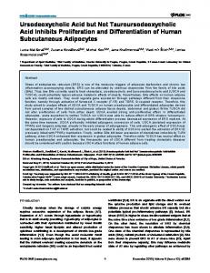
A
Task
B B
Novelty
Temporal Context
A
A
Spatial Context
A Spatio-temporal Context
Test
Exposure
A B
B
B
A
B
A
B
A′
B
B′
A′
B′
A
D
B
C
A
B C
D
B
Figure 1. A: Summary of the behavioral procedures for each of the object recognition tasks. With this design, we first assessed rats’ tendency to explore a novel object more than a familiar object. We then separately examined the principal components of episodic-like memory: first, by assessing rats’ memory for the temporal order (temporal context) in which two sets of objects were presented and then, by assessing rats’ sensitivity to a change in the spatial position of objects (spatial context). Finally, we assessed the rats’ tendency to identify and explore a test object that was presented both earlier in a sequence and in a different location relative to other objects that differed along only one of these dimensions. B, left side: Reconstructions of the maximum and minimum extent of the lesion (from left to right section taken at –3.1, #3.6, #4.28, #5.32, #6.38, #6.82, and – 8.4 mm from bregma (adapted from The Rat Brain in Stereotaxic Coordinates [pp. 1–7], by G. Paxinos and C. Watson, 1986, Orlando: FL: Academic Press. Copyright 1986 by Elsevier. Adapted with permission). B, right side: An example of a horizontal section of an intact hippocampus in sham-operated rats (upper left and right panels) and the maximum and minimum extent of cell loss in hippocampal lesioned rats (lower left and right panels) taken at a mid-dorsoventral level (2.5! magnification; scale bar $ 1 mm).
221
BRIEF COMMUNICATIONS
A 60 Mean Contact Time (sec)
Results
Behavioral Testing Novelty. Figure 2A (left columns) shows the level of exploratory activity elicited by a novel object (A) and a familiar object (B; see Figure 1). This figure indicates that sham-operated rats and rats with hippocampal lesions explored the test objects to a similar extent and that both groups showed a similar preference for exploring the novel object (A) rather than the familiar (B; see Figure 1) object. An analysis of variance (ANOVA) confirmed that there was no effect of group, F & 1; there was an effect of object type, F(1, 18) $ 16.23, p & .001; and there was no interaction between these factors, F & 1. The data were also converted into a DR (of the form: time spent with novel object/time spent with both the novel and familiar objects). The mean DR for rats with hippocampal lesions was .60 (SE $ ' .02) and mean DR for controls was .63 (SE $ ' .04). There was no significant difference between these means, t(18) $ 0.47, p ( .50, and both groups differed significantly from 0.5, ts(9) $ 3.81 and 2.68, ps & .05, for hippocampal lesioned and sham-operated rats, respectively. Temporal context. Figure 2a (middle columns) shows the level of exploratory activity directed toward test objects that were either presented recently (B) or more remotely (A; see Figure 1A). This figure shows that both groups displayed a preference for the objects presented least recently, although the magnitude of the effect appears to be smaller in rats with hippocampal lesions. An ANOVA revealed that there was no effect of group, F(1, 18) $ 1.30, p ( .26; there was an effect of object type, F(1, 18) $ 7.64, p & .05; and there was no interaction between these factors, F & 1. Nevertheless, an analysis of the DRs revealed a significant difference between sham-operated and hippocampal lesioned rats: the mean DR for the control rats was .69 (SE $ ' .03), and the mean DR for the hippocampal lesioned rats was .56 (SE $ ' .03), t(18) $ 2.98, p & .01. In addition, the performance of the sham-
Sham
40
*
30 20 10
B Mean Contact Time (sec)
Temporal Context
Hippocampal
50
Different Position
Same Position
Remote
Familiar Novelty
Recent
0
Histology Figure 1B shows the maximum and minimum extent of the lesion throughout horizontal sections obtained from the dorsal and ventral portions of the hippocampus and a representative example of the extent of cell loss at a mid-dorsoventral level (approximately –5.5 mm from bregma). Inspection of the cell loss in the rats with hippocampal lesions revealed that all the rats sustained a minimum of 70% cell loss in the hippocampus, and therefore no rats were excluded from this group. The lesion extended throughout the cornu ammonis subfields and the dentate gyrus in both the dorsal and ventral portions of the hippocampus. There was little or no damage to adjacent structures such as the subiculum, where it was evident that damage was confined to the more ventral aspects of this brain region, and there was no damage to the entorhinal cortex.
Hippocampal
50
Novel
before being sectioned (40 %m), mounted on slides, and stained with cresyl violet. The extent of the lesion in each rat was estimated by projecting an image of the section onto histological references images adapted from The Rat Brain in Stereotaxic Coordinates by Paxinos and Watson (1986) and transcribing the area of cell loss onto graph paper to calculate the proportion of area damaged.
Spatial Location
Sham
40 30 20 10 0 Object C
Object B
Object D
Object A Different Spatiotemporal Context
Figure 2. A: The mean contact time (in seconds) with objects during the object novelty, temporal context, and spatial context tests in rats with hippocampal lesions (n $ 10) and in control (sham-operated) rats (n $ 10). Asterisk indicates nonsignificant difference between object contact times. B: The mean contact time (in seconds) with each object (A–D) during the spatiotemporal context test in rats with hippocampal or sham lesions. Error bars show the standard error of the mean.
operated rats, but not that of the rats with hippocampal lesions, differed significantly from 0.5, ts(9) $ 2.98, p & .05, and 1.65, p ( .13, respectively. Spatial context. Figure 2A (right columns) shows the exploratory activity directed toward the test objects that were either located in the same positions as during exposure (B and B") or in different positions (A and A"; see Figure 1). Sham-operated rats were more likely to explore the objects presented in a different position than objects presented in the same position, but this preference was not apparent in rats with hippocampal lesions. An ANOVA revealed that there was no significant effect of group, F(1, 18) $ 3.56, p ( .05, and no significant effect of object type, F(1, 18) $ 1.74, p ( .20, but that there was a significant interaction between these factors, F(1, 18) $ 23.08, p & .001. Tests of simple main effects revealed a significant preference in shamoperated rats for objects presented in a different position, F(1, 18) $ 18.27, p & .001, but no such effect in the rats with hippocampal lesions. The mean DRs for the sham-operated control and hippocampal lesioned rats were .67 (SE $ ' .02) and .43 (SE $ ' .03), respectively, t(18) $ 5.66, p & .01. In addition, the performance of the control rats, but not that of the rats with hippocampal lesions, differed significantly from 0.5, ts(9) $ 8.14, p & .01, and 1.98, p ( .05, respectively.
222
BRIEF COMMUNICATIONS
Spatiotemporal context. Sham-operated rats spent more time exploring Object A (see Figure 2B), which was presented in both different spatial and different temporal contexts, than they did exploring the other objects (B, C, and D). In contrast, rats with hippocampal lesions spent a similar amount of time with each of the four objects (A–D). An ANOVA revealed no significant main effect of group, F(1, 18) $ 2.00, p ( .17; however, it did reveal a main effect of object type, F(3, 54) $ 3.35, p & .05, and a significant interaction between these factors, F(3, 54) $ 3.30, p & .05. Simple main effects analysis revealed that the mean time that rats spent exploring Object A differed between the groups, F(1, 69) $ 4.60, p & .05, and that there was a main effect of object type in control rats, F(3, 54) $ 6.52, p & .01, but not in lesioned rats (F & 1). Further analysis revealed that the main effect of object type in sham-operated rats reflected a significant difference between the exploration of Object A and exploration of the three remaining objects (Duncan’s pairwise comparisons: p & .05) and that explorations of the remaining Objects B, C, and D did not differ ( ps ( .05). This analysis confirms that sham-operated rats, but not rats with hippocampal lesions, showed a marked preference for exploring the object that was presented in a different spatiotemporal context relative to other objects that differed in one of these properties. This was further confirmed by analysis of the DR data that revealed a significant difference between the groups (sham-operated rats: DR $ .65, SE $ ' .04; hippocampal lesioned rats: DR $ .47, SE $ ' .04; t(18) $ 2.85, p & .05). Furthermore, the performance of the sham-operated rats, but not that of the hippocampal-lesioned rats, differed significantly from 0.5, ts(9) $ 3.23, p & .05, and 0.79, p ( .44, respectively.
Discussion The exploration of objects by control rats was determined by a number of factors: familiarity and both temporal and spatial contexts. When two test objects differed in familiarity, control rats explored the novel object; when the test objects were equally familiar, rats explored the least recently presented object; and when the test objects were equally familiar and had been presented equally recently, control rats explored objects whose positions had changed between exposure and test. The final observation for control rats was that they were more inclined to explore an object that had been presented less recently and in a different position than objects that had either been presented less recently or presented in a changed position (see also Dere, Huston, & De Souza Silva., 2005a, 2005b; Eacott & Norman, 2004; Kart-Teke et al., 2006). This latter result indicates that the influence of changes in temporal and spatial contexts may combine in an additive manner. The rats with hippocampal lesions only showed a clear influence of one factor on their exploratory behavior: familiarity. This pattern of findings is clearly consistent with the suggestion that the hippocampus plays a role in the context-dependent forms of recognition but not in context-independent forms of recognition (e.g., Aggleton & Brown, 1999). There are, however, a variety of possible accounts of the context-dependent forms of recognition that inform our interpretation of the failure to find such effects in hippocampal rats. One type of account of context-dependent object recognition suggests that during the exposure stage, control rats form an integrated or episodic memory of the temporal and spatial contexts
in which an object has been presented (e.g., Norman & O’Reilly, 2003; O’Keefe & Nadel, 1978; O’Reilly & Rudy, 2001). During the test stage, rats explore an object to the extent that there is a mismatch between the object that is currently being inspected and the stored integrated memories formed during the exposure stage. This analysis is consistent with earlier accounts of habituation (e.g., Konorski, 1967; Sokolov, 1963) and with the psychobiological accounts of recognition memory that have been the focus of interest in this article (e.g., Aggleton & Brown, 1999; cf. Ergorul & Eichenbaum, 2004). One obvious interpretation of the finding that hippocampal lesions disrupt context-specific forms of recognition is that rats with such lesions fail to form such memories. A closely related dual-process account suggests that the hippocampus contributes specifically to recollection-based, as opposed to familiarity-based, forms of recognition memory (Fortin et al., 2004). The principle evidence in support of this hypothesis stems from an analysis of the receiver operating characteristics (ROC) curves generated by healthy rats and rats with hippocampal lesions in an odor recognition task. The ROC curve of healthy rats displayed both an asymmetrical and curvilinear component, suggesting the operation of both recollection- and familiarity-based forms of recognition memory. In contrast, only the curvilinear component of the ROC curve was evident in rats with hippocampal lesions. On the basis of these results, Fortin et al. (2004) concluded that hippocampal damage in rats disrupted a recollective component of recognition memory. Of course, this interpretation relies on one’s accepting a specific analysis of the ROC curves (cf. Rotello, Macmillan, Reeder, & Wong, 2006; Slotnick & Dodson, 2005; Yonelinas, 1999). An alternative account is predicated on the assumptions that an object’s memory has many elements and that if any of these have been recently activated (either by the object itself or associatively by the spatial context) then they will be less likely to provoke exploration of a recently presented object (see Wagner, 1981). According to this account the temporal and spatial influences on object exploration are (relatively) independent and although the memory for an object is associated with its spatial context, this account does not include an explicit temporal tag. The influence of relative recency is a product of the decay of elements from the refractory state, in which elements are less able to affect exploration, to an inactive state in which they become available to initiate exploration. Within this account, one plausible locus for the effect of hippocampal lesions on context-dependent recognition is in changing the rate at which the elements that have recently been activated decay into their inactive state (see Honey & Good, 2000). Like the theories outlined in the previous paragraph, this account would also need to be supplemented by a second process (of habituation) that could result in preferences for a novel object over a familiar object in hippocampal lesioned rats (see, for example, Groves & Thomson, 1970; Hawkins & Kandel, 1984; Horn & Hill, 1964). The results of this study provide evidence for dual-process accounts of object recognition and for the specific suggestion that the hippocampus is involved in context-dependent recognition, but not in context-independent recognition. Further work will be required to clarify both the specific nature of the dual processes and the contribution of the hippocampus to these processes.
BRIEF COMMUNICATIONS
References Aggleton, J. P., & Brown, M. W. (1999). Episodic memory, amnesia, and the hippocampal-anterior thalamic axis. Behavioural and Brain Sciences, 22, 425– 444. Dere, E., Huston, J. P., & De Souza Silva, M. A. (2005a). Episodic-like memory in mice: Simultaneous assessment of object, place and temporal order memory. Brain Research and Brain Research Protocols, 16, 10 –19. Dere, E., Huston, J. P., & De Souza Silva, M. A. (2005b). Integrated memory for objects, places, and temporal order: Evidence for episodiclike memory in mice. Neurobiology of Learning and Memory, 84, 214 –221. Eacott, M. J., & Norman, G. (2004). Integrated memory for object, place, and context in rats: A possible model of episodic-like memory? Journal of Neuroscience, 24, 1948 –1953. Ennaceur, A., & Delacour, J. (1988). A new one-trial test for neurobiological studies of memory in rats: 1. Behavioral data. Behavioural Brain Research, 31, 47–59. Ergorul, C., & Eichenbaum, H. (2004). The hippocampus and memory for “what,” “where,” and “when.” Learning & Memory, 11, 397– 405. Fortin, N. J., Wright, S. P., & Eichenbaum, H. (2004, September 9). Recollection-like memory retrieval in rats is dependent on the hippocampus. Nature, 431, 188 –191. Groves, P. M., & Thompson, R. F. (1970). Habituation: A dual-process theory. Psychological Review, 77, 419 – 450. Hawkins, R. D., & Kandel, E. R. (1984). Is there a cell biological alphabet for simple forms of learning? Psychological Review, 91, 375–391. Honey, R. C., & Good, M. (2000). Associative components of recognition memory. Current Opinion in Neurobiology, 10, 200 –204. Horn, G., & Hill, R. M. (1964, April 18). Habituation of the response to sensory stimuli of neurones in the brain stem of rabbits. Nature, 202, 296 –298. Kart-Teke, E., De Souza Silva, M. A., Huston, J. P., & Dere, E. (2006). Wistar rats show episodic-like memory for unique experiences. Neurobiology of Learning and Memory, 85, 173–182. Konorski, J. (1967). Integrative activity of the brain. Chicago: University of Chicago. McGregor, A., Hayward, A. J., Pearce, J. M., & Good, M. A. (2004). Hippocampal lesions disrupt navigation based on the shape of the environment. Behavioral Neuroscience, 118, 1011–1021. Mumby, D. G. (2001). Perspectives on object-recognition memory following hippocampal damage: Lessons from studies in rats. Behavioural Brain Research, 127, 159 –181. Norman, K. A., & O’Reilly, R. C. (2003). Modeling hippocampal and
223
neocortical contributions to recognition memory: A complementarylearning-systems approach. Psychological Review, 110, 611– 646. O’Keefe, J., & Nadel, L. (1978). The hippocampus as a cognitive map. Oxford, England: Clarendon Press. O’Reilly, R. C., & Rudy, J. W. (2001). Conjunctive representations in learning and memory: Principles of cortical and hippocampal function. Psychological Review, 108, 311–345. Paxinos, G., & Watson, C. (1986). The rat brain in stereotaxic coordinates (compact 3rd ed.). Orlando, FL: Academic Press. Rotello, C. M., Macmillan, N. A., Reeder, J. A., & Wong, M. (2006). The remember response: Subject to bias, graded, and not a process-pure indicator of recollection. Psychonomic Bulletin & Review, 12, 865– 873. Slotnick, S. D., & Dodson, C. S. (2005). Support for a continuous (singleprocess) model of recognition memory and source memory. Memory & Cognition, 33, 151–170. Sokolov, E. N. (1963). Perception and the conditioned reflex. London: Pergamon. Sutherland, R. J., & Rudy, J. W. (1989). Configural association theory: The contribution of the hippocampus to learning, memory, and amnesia. Psychobiology, 17, 129 –144. Tulving, E. (2002). Episodic memory: From mind to brain. Annual Review of Psychology, 53, 1–25. Wagner, A. R. (1981). SOP: A model of automatic memory processing in animal behavior. In N. E. Spear & R. R. Miller (Eds.), Information processing in animals: Memory mechanisms (pp. 5– 48). Hillsdale, NJ: Erlbaum. Wais, P. E., Wixted, J. T., Hopkins, R. O., & Squire, L. R. (2006). The hippocampus supports both the recollection and the familiarity components of recognition memory. Neuron, 49, 459 – 466. Wixted, J. T., & Squire, L. R. (2004). Recall and recognition are equally impaired in patients with selective hippocampal damage. Cognitive, Affective, & Behavioral Neuroscience, 4, 58 – 66. Yonelinas, A. P. (1999). The contribution of recollection and familiarity to recognition and source-memory judgments: A formal dual-process model and an analysis of receiver operating characteristics. Journal of Experimental Psychology: Learning, Memory, and Cognition, 25, 1415– 1434. Yonelinas, A. P., Otten, L. J., Shaw, K. N., & Rugg, M. D. (2005). Separating the brain regions involved in recollection and familiarity in recognition memory. Journal of Neuroscience, 25, 3002–3008.
Received August 7, 2006 Revision received September 22, 2006 Accepted September 29, 2006 !