PROGRESS Circuit-breakers: optical technologies for probing neural signals and systems Feng Zhang, Alexander M. Aravanis, Antoine Adamantidis, Luis de Lecea and Karl Deisseroth
Abstract | Neuropsychiatric disorders, which arise from a combination of genetic, epigenetic and environmental influences, epitomize the challenges faced in understanding the mammalian brain. Elucidation and treatment of these diseases will benefit from understanding how specific brain cell types are interconnected and signal in neural circuits. Newly developed neuroengineering tools based on two microbial opsins, channelrhodopsin-2 (ChR2) and halorhodopsin (NpHR), enable the investigation of neural circuit function with cell-type-specific, temporally accurate and reversible neuromodulation. These tools could lead to the development of precise neuromodulation technologies for animal models of disease and clinical neuropsychiatry. Treatments for neuropsychiatric disorders include drug therapies, surgical interventions, electrode-based deep brain stimulation (DBS), transcranial magnetic stimulation (TMS), vagus nerve stimulation (VNS) and psychotherapy. These methods represent different modalities for interfacing with the diseased brain, from molecules to cognition. Although seemingly disparate, the effects of these treatments converge at the level of neural circuits, altering the way neurons individually communicate and collectively compute. The brain consists of numerous subtypes of excitatory, inhibitory and modulatory neurons, and it has become increasingly clear that specific cell types have crucial roles in many neuropsychiatric diseases. For example, the death of dopaminergic neurons in the substantia nigra pars compacta leads to Parkinson’s disease in humans and related symptoms in animal models1, the generation of new neurons in the hippocampus has been linked to the therapeutic efficacy of antidepressants2, and dysfunction of parvalbumin (PV)-positive γ-aminobutyric acid (GABA)-releasing interneurons in the prefrontal cortex (PFC) has been implicated in schizophrenia3–5.
To better understand the contribution of specific cell types to the physiology of neuropsychiatric disease, a pair of microbial light-sensitive proteins have been developed that, when used togther, can optically interrogate intact neural circuits and bidirectionally control animal behaviour6. As described below, co-expression of these proteins enables bidirectional modulation of electrical signals (activation or inhibition) with millisecond precision and thus allows probing of the downstream circuit-level effects of turning specific cells on and off. Moreover, the ability to control the activity of specific neural populations may make it possible to generate new models of neuropsychiatric diseases by directly mimicking errant electrical signals in the brain with millisecond precision. Finally, therapies using cell-specific modulation might eventually be employed to selectively regulate malfunctioning neurons, thereby optimizing treatment efficacy and reducing the side-effects associated with less specific therapeutic interventions. Many other elegant approaches have been developed recently for cell-type-specific control7–11; in this Progress article, we discuss the recent advances in genetically targeted
NATURE REVIEWS | NEUROSCIENCE
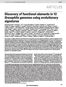
optical neuromodulation with microbial opsins, and give some broad general perspectives relevant to both basic science research and clinical application. Optogenetics: two microbial opsins Neuropsychiatric diseases may result from circuit-level effects of a group of malfunctioning neurons; for instance, the PV-positive interneurons in the dorsolateral PFC of schizophrenic patients have reduced GABA immunoreactivity relative to controls, suggesting reduced inhibition of cortical pyramidal neurons4, 5. To best understand the role of a specific neuron subtype in living animals, we must be able to bidirectionally turn these neurons on and off with cell-type specificity, high temporal precision and rapid reversibility. To satisfy these requirements, the microbial light-sensitive proteins Chlamydomonas reinhardtii Channelrhodopsin-2 (ChR2) and Natronomonas pharaonis halorhodopsin (NpHR) have been introduced into neurons6,12–22,25. ChR2 (initially cloned in REF. 23) is a monovalent cation channel that allows Na+ ions to enter the cell following exposure to ~470 nm blue light, whereas the NpHR (described in REF. 24) is a chloride pump that activates upon illumination with ~580 nm yellow light (FIG. 1a). As the activation maxima of these two proteins are over 100 nm apart (FIG. 1b), they can be controlled independently to either drive action potential firing or suppress neural activity in intact tissue6 (FIG. 1c shows implementation of this protocol in hippocampal neurons using cell-attached and whole-cell patch clamp), and together may modulate neuronal synchrony17. Both proteins have fast temporal kinetics, on the scale of milliseconds, making it possible to drive reliable trains of high frequency action potentials in vivo using ChR2 (REF. 15) and suppress single action potentials within high frequency spike trains using NpHR6. Because NpHR remains active for many minutes when exposed to continuous light6 and deactivates quickly when light is turned off, it can be used to mimic lesions in a rapid, stable and reversible manner. Stability in general is particularly important; it has long been
VOLUME 8 | AUGUST 2007 | 577 © 2007 Nature Publishing Group
PROGRESS a
Blue light
Yellow light
ChR2
NpHR
K+
Na+
b
c
1
Cl–
ChR2 NpHR
Activation
0.8 0.6 0.4 0.2 0 325
425
525
625
725
Wavelength (nm)
Figure 1 | Optogenetic tools: ChR2 and NpHR. a | Schematic of channelrhodopsin-2 (ChR2) and the halorhodopsin (NpHR) pump. Following illumination with blue light (activation maximum ~470 nm, REF.23), ChR2 allows the entry of cations (mostly Na+ and very low levels of Ca2+) into the cell. NpHR is activated by yellow light illumination (activation maximum ~580 nm, REF. 6) and allows the entry of Cl – anions. b | Action spectra for ChR2 and NpHR. The excitation maxima for ChR2 and NpHR are separated by ~100 nm, making it possible to activate each opsin independently with light. c | Cell-attached (top) and whole-cell current-clamp (bottom) traces from hippocampal neurons showing all-optical neural activation and inhibition. Blue pulses represent the blue light flashes used to drive ChR2-mediated activation and the yellow bar denotes NpHR-mediated inactivation. Part a was modified with permission from Nature REF.52 © (2007) Macmillan Publishers Ltd. Part c was modified with permission from Nature REF.6 © (2007) Macmillan Publishers Ltd.
known that many archaebacterial species express red-shifted light-activated halorhodopsins that can pump chloride ions and modulate membrane potential (for example, H. salinarum, H. halobium, and N. pharaonis), but we selected the Natronomonas halorhodopsin to experimentally develop for neural control, as NpHR has enhanced stability and chloride affinity6. Moreover, halorhodopsin does not require the addition of the cofactor all-trans-retinal (ATR) to function in intact mammalian tissue6, a result that is consistent with previous findings with the homologous opsin ChR2 (REFS 13,25). The ability to functionally express ChR2 and NpHR without the addition of any exogenous cofactor is crucial for applications to basic science investigations, preclinical disease models and possible clinical translation. Light delivery into the brain To modulate the activity of ChR2- and NpHR-expressing neurons, light must be delivered to the brain region of interest. Recently a fibreoptic-based system was
developed that is suitable for delivering light in vivo16 to both superficial and deep brain structures. In this approach, a thin optical fibre (~0.1 to 0.2 mm in diameter) can be inserted through a cannula guide (FIG. 2a) that is targeted precisely to the lightsensitized neurons, to deliver effective photostimulation to specific neuronal subtypes in the brain of freely moving rats (for example, expressing ChR2 or NpHR; Fig. 2b). The optical fibre can be coupled to a bright light source such as a diode laser16. Alternative solutions may involve digital light processing (DLP) micromirrors for projection, or high intensity light-emitting diodes (LEDs) directly mounted on the head of mice or rats; moreover, in larger animals such as primates, millimetre-scale LEDs or LED arrays could be directly implanted into deeper brain structures. In animals, the LEDs can be either tethered to an external power source or powered by a battery pack carried by the animal to allow normal behaviour; the LEDs could even be powered wirelessly by radio frequency or magnetic induction.
578 | AUGUST 2007 | VOLUME 8
Protein expression in neuron subtypes Different types of neurons are characterized by unique gene expression patterns26. Many of the neuron types implicated in neuropsychiatric disorders can be identified based on their immunoreactivity to antibodies that recognize neurotransmitters (for example, dopamine, cholecystokinin or neuropeptide Y) or neurochemical markers (for example PV, calretinin or calbindin), suggesting possible targets for experimental or therapeutic control. Similar principles extend to disorders lying outside conventional neuropsychiatric disease categories. For example, animals lacking melaninconcentrating hormone (MCH) are reported to be hypophagic and lean27, and optical inhibition of the MCH-secreting neurons in the lateral hypothalamus using NpHR might therefore be used to modify food intake or obesity. A wide array of techniques is now available for genetically specifying the expression pattern of probes like ChR2 and NpHR in mouse models (for thorough discussions on strategies for making transgenic mouse lines see REFS 28,29) or in other experimental systems7, 11. For potential preclinical animal models and potential clinical applications, viral gene delivery also may provide a convenient and quick approach for mediating ChR2 and NpHR expression. Viral vectors carrying the genes encoding ChR2 or NpHR can be delivered stereotactically into discrete brain regions6,16,30 (for detailed discussions on viral gene transfer into the nervous system see REF. 31). Recombinant lentiviral and adeno-associated viral (AAV) vectors32, 32 have been popular choices for gene transfer into patients. Lentiviral vectors are integrated into the genome of the target cell and confer permanent gene expression. AAV-mediated expression may be less stable because a much smaller percentage (less than 1%) of the virus is integrated. These vectors can also be rendered cell-type-specific by the choice of promoter34–36, viral receptor37 or spatial targeting strategy38. However, the limited packaging capacity of many viral vectors restricts the size of transgene cassettes that can be used (10 kb or less for lentivirus and 5kb or less for AAV31). Identification of relevant neural circuits The cell types pertinent to most neuropsychiatric diseases remain to be identified and in most cases even compelling hypotheses for candidate cell types are lacking. Therefore, to generate hypotheses that will elucidate the cellular basis of disease mechanisms, it will be advantageous to identify
www.nature.com/reviews/neuro © 2007 Nature Publishing Group
PROGRESS relevant neurons based on their activity or functional connectivity during the manifestation of disease symptoms (for example, during states of elevated stress or anxiety). One possible method is to take advantage of promoters from immediate-early genes (IEGs; for example, FOS and ARC) to drive expression39, 40 of fluorescent-protein-tagged ChR2 and NpHR in neurons that are active during particular behaviours relevant to diseases. ChR2 or NpHR expression can also be restricted to cells that are synaptically connected to a previously-identified relevant population, in order to determine which connections are most important to disease symptoms. For example, it is possible to use viral delivery to retrogradely label neurons with ChR2 or NpHR based on their projection patterns38, 41. A recombinant, glycoprotein-deleted form of rabies virus has been shown to mediate strong protein expression not only in the neurons that project to the site of viral delivery, but also in their presynaptic partners that may be located in distant neural circuits41. With this approach, it may be possible to stimulate only the cells in a particular brain region that connect to cells in another brain region of interest, and to determine how these connections are altered in a disease state. This approach may be especially useful in diseases such as schizophrenia and depression, where the implicated cell types receive input from and make functional connections with neurons in heterogeneous regions of the brain (such as the hippocampus and neocortex). Analogous development of anterograde labelling methods will complement this approach and enable more powerful analysis of relevant circuits. Integration with output measurements To be informative in an experimental setting, optical neuromodulation must be tightly coupled with an appropriate quantitative readout. Stimulation of neurons using light makes it easier to perform simultaneous electrical recordings in vivo without electrical artefact interference from stimulation. This unique aspect of optical neuromodulation allows electrical monitoring of activity changes in brain regions within, or downstream from, the site of stimulation with no stimulus artefact-induced delay. Therefore, it is now possible to map out the functional connectivity of disease circuits in real time. It also will be important to combine optical stimulation/inhibition with noninvasive forms of output measurement, including optical imaging, positron emission
a
Optical fibre
b Cannula
Cranioplastic cement
Skull Cortex Illumination
c Electrical stimulation Depth electrode (1.27 mm diameter)
Targeted neuron type Modulation
Intended effect
Modulation
Side effect
Adjacent non-targeted neuron
Optical stimulation Implanted optical fibre (0.2 mm diameter) ChR2 or NpHR
Targeted neuron type expressing ChR2 or NpHR Modulation
Intended effect
Unaffected
No side effect
Adjacent non-targeted neuron
Figure 2 | In vivo optical neuromodulation in animal models of neuropsychiatric disease. a | A cannula is implanted into the head of the experimental animal to guide an optical fibre to the targeted brain region. b | The optical fibre is coupled to a strong light source (here a 488 nm laser diode16) to bring blue or yellow light into the brain. c | Genetic targeting of channelrhodopsin (ChR2) or halorhodopsin (NpHR) into defined classes of disease-model-relevant neurons may allow cell-specific neuromodulation and avoid inadvertent stimulation of disease-model-irrelevant neurons as occurs with electrical stimulation. Figure modified with permission from REF. 16 © (2007) Institute of Physics Pub.
tomography (PET) and functional magnetic resonance imaging (fMRI). In particular, optical recording of neural activity in vivo42,43 or in brain slices holds great potential for monitoring effects on the activity of large neural populations. Ultimately, as genetically encoded activity probes44,45 mature, it will be possible to perturb a specific population of neurons and examine their influence on another genetically defined population of neurons. Finally, particularly for the study of the cellular and circuit-level mechanisms of neuropsychiatric diseases, the effects of modulating specific cell types in vivo needs to be quantified through behavioural assays. Light delivery with long, flexible and lightweight fibreoptics16 or
NATURE REVIEWS | NEUROSCIENCE
LED leads will allow experimental animals to behave freely during targeted photostimulation. It also may be possible to establish novel, reversible behavioural models of neuropsychiatric diseases that are linked to specific cell types as the ability to control the electrical activity of specific populations of neurons in discrete regions of the brain has the potential to reversibly mimic underactivity or overactivity of candidate neurons. Additionally, when applied to existing behavioural models used as output measures, cell-specific neuromodulation will be useful to either exacerbate or rescue the behavioural phenotypes and thereby provide insights for future clinical translation.
VOLUME 8 | AUGUST 2007 | 579 © 2007 Nature Publishing Group
PROGRESS Box 1 | Designing possible optogenetic neuromodulation therapies Below are several brain diseases that could, in principle, be treated or studied in animal models using optogenetic neuromodulation. Parkinson’s disease. Deep brain stimulation devices have been efficacious in correcting movement disorders in patients with advanced stage Parkinson’s disease. High frequency stimulation is thought to suppress firing of neurons in the subthalamic nucleus (STN). Optical neuromodulation could be used to shut down excitatory glutamatergic neurons in the STN that express halorhodopsin (NpHR) under the control of an excitatory neuron-specific promoter; this cell type specificity will result in a more direct inhibition, and thus less side-effects, compared with electrode-based DBS. Depression. DBS to the subgenual cingulate area 25 (Cg25) appears to alleviate depression symptoms47. NpHR could be introduced into Cg25 to specifically and potently reduce neuronal activity in this area. Alternatively, channelrhodopsin-2 (ChR2) could be expressed in the nucleus accumbens to model proposed DBS-mediated excitation48. Obsessive-compulsive disorder (OCD). Irreversible lesions to the anterior capsule and analogous DBS to that region are established means for treating severe and intractable OCD49. Such approaches could be emulated using NpHR-mediated inhibition of the anterior capsule, or of regions such as BA32, which show metabolic decreases following OCD remission. Chronic pain. Electrical stimulation methods include local peripheral nerve stimulation, local cranial nerve stimulation50 and ‘subthreshold’ motor cortex stimulation51. Reasonable optogenic approaches might include NpHR-mediated inhibition of specific pain fibres or foci, sparing other fibre types. ChR2 could also be used to provide effective pain relief by driving inhibitory or analgesic neurons. Development of tolerance and dependence might be less of an issue with this method of selectively driving analgesic neurons compared with pharmacological treatments. Epilepsy. Quenching or blocking epileptogenic activity is an exciting prospect. Many epilepsy patients have a stereotyped pattern of activity spread resulting from an epileptogenic focus. Brief activation of NpHR could be used to suppress the abnormal activity before it spreads, or to truncate the abnormal activity early in its course. Activation of ChR2 in γ-aminobutyric acid (GABA)-releasing interneurons could provide a similar result.
Possible clinical applications The ability to control the activity of a defined class of neurons has the potential to advance clinical neuromodulation. Existing electrode-based DBS methods indiscriminately stimulate all neurons within a given volume, including cells that are not implicated in the disease state, thus leading to unwanted side-effects and even reduced efficacy as opposing excitatory and inhibitory cell types are affected by the electrodes. Genetic control makes it possible to develop more precise therapies by restricting the excited or inhibited neurons to the diseaserelevant population (FIG. 2c). Moreover, the ability to simultaneously record electrical activity during optical stimulation without electrical artefacts makes it possible to engage in responsive neuromodulation by dynamically adjusting the stimulation or inhibition intensity based on feedback from the activity state in the brain. This feature may be especially useful for diseases characterized by sporadic fluctuations in electrical activity such as epilepsy. The combination of recording and optical control could also be used to bridge severed connections, for example to relay information from the brain to distal limbs in the case of severed spinal cord. BOX 1 describes several potential scenarios for optical neuromodulation therapies.
Although the direct use of optical neuromodulation in humans has tremendous potential to control disease circuits with a precision unparalleled by drugs or electrode-based techniques, two key issues require additional research and development. First, the genes encoding ChR2 and NpHR must be safely and stably transferred into the patient’s neurons. Progress is being made on this front as several viral and non-viral-based gene delivery methods are currently in late stages of clinical testing32. Second, a lesser, but still significant, problem is the engineering of an appropriate optical device. To achieve effective optical control, it will be necessary to deliver sufficient intensities of light to the correct local circuit, perhaps using analogous methods to those developed for light delivery in animal models. Recent advances in microelectronics, high-capacity batteries and optoelectronics have made the development of clinical-quality optical brain stimulators a tractable engineering problem. Most importantly, long before optical neuromodulation is ready for use in neuropsychiatric clinics, the knowledge gained from the use of these tools in animal models is likely to have an impact on our understanding of neuropsychiatric circuit pathology. Moreover, further understanding
580 | AUGUST 2007 | VOLUME 8
of brain diseases in this way is likely to influence future development of existing pharmaceutical agents and medical devices; for example, knowing the cell populations responsible for the efficacy of DBS treatments could lead to better electrode placement and design. Future perspectives The convergence of genetic, optical and engineering advances holds substantial potential for mechanistic investigation of disease aetiology and treatment. The ability to restrict neuromodulation to genetically specified neuronal subtypes will enable more precise analysis of brain function. However, for these optogenetic methods to reach their full potential it is still necessary to improve the tools of their application. Achieving more precise genetic control through the identification of novel promoters together with enhanced understanding of transcriptional regulation, and developing less invasive modulation (for example, by harnessing infrared light) may lead to new breakthrough technologies. A final perspective arises from the viewpoint of the practising psychiatrist. We do not yet know the precise neural ‘code’ that is relevant to symptoms of psychiatric disease, so in most cases we can not test hypotheses by delivering precisely timed pulses despite having the tools to do so. However, we predict that key psychiatric symptoms like those associated with depression (for example, Glossary Deep brain stimulation (DBS). A surgical procedure involving the implantation of electrodes to deliver electrical pulses to specific brain regions; used to treat neuropsychiatric disorders such as Parkinson’s disease, and currently under evaluation for other disorders, including clinical depression and Tourette’s syndrome.
Retrograde label A method used to examine neuronal connectivity in which an injected label travels within neurons in the opposite direction of neural information flow, thereby labelling the cell bodies that project to the injected location. An anterograde label will track axons projecting from the injection site.
Transcranial magnetic stimulation (TMS). A non-invasive method for exciting neurons by inducing electrical currents in the brain with rapidly changing external magnetic fields; currently under evaluation for diagnostic and therapeutic use in diseases, including depression and Parkinson’s disease.
Vagus nerve stimulation (VNS). A device-based neural stimulation method that uses an electrical lead to stimulate the vagus nerve in the neck, which may then influence central neurons. It is used to treat intractable epilepsy and has recently been applied to clinical depression.
www.nature.com/reviews/neuro © 2007 Nature Publishing Group
PROGRESS hopelessness, anhedonia or low energy) may have less to do with coding or computation, and more to do with alterations in the spatiotemporal spread (also referred to as percolation) of activity through bottlenecks in neural circuits controlled by specific cell types46. Indeed many aetiologies and treatments could be classified as positive or negative modulators of activity propagation that do not affect coding per se46. By contrast, disease states with borderline and schizophrenic features in which aspects of the self can be experienced as alien (as with auditory hallucinations or dissociative symptoms) might be due to a high-speed coding problem in which neurons reporting self-generated stimuli are ‘mis-tagged’ with a non-self millisecond-scale timing tag (for example, oscillating at the wrong frequency or without correct synchronization). Both the ‘tagging model’ and the ‘percolation model’, which are based on, and fit well with, clinical psychiatry experience, correspond to theoretical extremes in the range of psychiatric disease mechanisms. Ultimately both models may be susceptible to testing with optical6,46 and other high-speed neuroengineering technologies. Feng Zhang, Alexander M. Aravanis and Karl Deisseroth are in the Department of Bioengineering, W083 Clark Center, 318 Campus Drive West, Stanford University, California, USA. Antoine Adamantidis and Luis de Lecea are in the Department of Psychiatry and Behavioural Sciences, 401 Quarry Road, Stanford University, California, USA. Correspondence to K.D. e-mail:
[email protected] doi:10.1038/nr n2192
13.
14.
15.
16.
17.
18.
19.
20.
21.
22.
23.
24.
25.
26. 27.
1.
Beal, M. F. Experimental models of Parkinson’s disease. Nature Rev. Neurosci. 2, 325–334 (2001). 2. Santarelli, L. et al. Requirement of hippocampal neurogenesis for the behavioural effects of antidepressants. Science 301, 805–809 (2003). 3. Lewis, D. A. GABAergic local circuit neurons and prefrontal cortical dysfunction in schizophrenia. Brain Res. Brain Res. Rev. 31, 270–276 (2000). 4. Hashimoto, T., et al. Gene expression deficits in a subclass of GABA neurons in the prefrontal cortex of subjects with schizophrenia. J. Neurosci. 23, 6315–6326 (2003). 5. Volk, D., Austin, M., Pierri, J., Sampson, A. & Lewis, D. GABA transporter-1 mRNA in the prefrontal cortex in schizophrenia: decreased expression in a subset of neurons. Am. J. Psychiatry 158, 256–265 (2001). 6. Zhang, F. et al. Multimodal fast optical interrogation of neural circuitry. Nature 446, 633–639 (2007). 7. Lima, S. Q. & Miesenbock, G. Remote control of behaviour through genetically targeted photostimulation of neurons. Cell 121, 141–152 (2005). 8. Karpova, A.Y., Tervo, D.G., Gray, N.W. & Svoboda, K. Rapid and reversible chemical inactivation of synaptic transmission in genetically targeted neurons. Neuron 48, 727–735 (2005). 9. Tan, E.M., et al. Selective and quickly reversible inactivation of mammalian neurons in vivo using the Drosophila allatostatin receptor. Neuron 51, 157–170 (2006). 10. Lerchner, W., et al. Reversible silencing of neuronal excitability in behaving mice by a genetically targeted, ivermectin-gated Cl– channel. Neuron 54, 35–49 (2007). 11. Szobota, S. et al. Remote control of neuronal activity with a light-gated glutamate receptor. Neuron 54, 535–545 (2007). 12. Boyden, E. S., Zhang, F., Bamberg, E., Nagel, G. & Deisseroth, K. Millisecond-timescale, genetically
28.
29.
30.
31.
32.
33.
34.
35.
36.
37.
targeted optical control of neural activity. Nature Neurosci. 8, 1263–1268 (2005). Zhang, F., Wang, L. P., Boyden, E. S. & Deisseroth, K. Channelrhodopsin-2 and optical control of excitable cells. Nature Methods 3, 785–792 (2006). Wang, H., et al. High-speed mapping of synaptic connectivity using photostimulation in Channelrhodopsin-2 transgenic mice. Proc. Natl Acad. Sci. USA 104, 8143–8148 (2007). Arenkiel, B. R. et al. In vivo light-induced activation of neural circuitry in transgenic mice expressing channelrhodopsin-2. Neuron 54, 205–218 (2007). Aravanis, A. M. et al. An optical neural inteface: in vivo control of rodent motor cortex with integrated fiberoptic and optogenetic technology. J. Neural Eng. 4, S143–S156 (2007). Han, X. & Boyden, E. S. Multiple-color optical activation, silencing and desynchronization of neural activity with single-spike temporal resolution. PLoS ONE 2, e299 (2007). Petreanu, L., Huber, D., Sobczyk, A. & Svoboda, K. Channelrhodopsin-2-assisted circuit mapping of longrange callosal projections. Nature Neurosci. 10, 663–668 (2007). Li, X., et al. Fast noninvasive activation and inhibition of neural and network activity by vertebrate rhodopsin and green algae channelrhodopsin. Proc. Natl Acad. Sci. USA102, 17816–17821 (2005). Nagel, G., et al. Light activation of channelrhodopsin-2 in excitable cells of Caenorhabditis elegans triggers rapid behavioral responses. Curr. Biol. 15, 2279–2284 (2005). Ishizuka, T., Kakuda, M., Araki, R. & Yawo, H. Kinetic evaluation of photosensitivity in genetically engineered neurons expressing green algae light-gated channels. Neurosci. Res. 54, 85–94 (2006). Schroll, C., et al. Light-induced activation of distinct modulatory neurons triggers appetitive or aversive learning in Drosophila larvae. Curr. Biol. 16, 1741–1747 (2006). Nagel, G., et al. Channelrhodopsin-2, a directly lightgated cation-selective membrane channel. Proc. Natl Acad. Sci. USA 100, 13940–13945 (2003). Soliman, G. S. H. & Truper, H. G. Halobacterium pharaonis: a new, extremely haloalkaliphilic archaebacterium with low magnesium requirement. Zentralblatt für Bakteriologie Mikrobiologie und Hygiene erste Abteilung Originale C, Allgemeine, angewandte und ökologische Mikrobiologie 3, 318–329 (1982). Bi, A. et al. Ectopic expression of a microbial-type rhodopsin restores visual responses in mice with photoreceptor degeneration. Neuron 50, 23–33 (2006). Lein, E. S. et al. Genome-wide atlas of gene expression in the adult mouse brain. Nature 445, 168–176 (2007). Shimada, M., Tritos, N. A., Lowell, B. B., Flier, J. S. & Maratos-Flier, E. Mice lacking melanin-concentrating hormone are hypophagic and lean. Nature 396, 670–674 (1998). Tecott, L. H. & Wehner, J. M. Mouse molecular genetic technologies: promise for psychiatric research. Arch. Gen. Psychiatry 58, 995–1004 (2001). Miyoshi, G. & Fishell, G. Directing neuron-specific transgene expression in the mouse CNS. Curr. Opin Neurobiol. 16, 577–584 (2006). Cetin, A., Komai, S., Eliava, M., Seeburg, P. H. & Osten, P. Stereotaxic gene delivery in the rodent brain. Nature Protoc. 1, 3166–3173 (2006). Davidson, B. L. & Breakefield, X. O. Viral vectors for gene delivery to the nervous system. Nature Rev. Neurosci. 4, 353–364 (2003). Kaplitt, M. G., et al. Safety and tolerability of gene therapy with an adeno-associated virus (AAV) borne GAD gene for Parkinson’s disease: an open label, phase I trial. Lancet 369, 2097–2105 (2007). de Boer, A. G. & Gaillard, P. J. Drug targeting to the brain. Annu. Rev. Pharmacol. Toxicol. 47, 323–355 (2007). Chhatwal, J. P., Hammack, S. E., Jasnow, A. M., Rainnie, D. G. & Ressler, K. J. Identification of cell-type-specific promoters within the brain using lentiviral vectors. Gene Ther. 14, 575–583 (2007). Osten, P., Grinevich, V. & Cetin, A. Viral vectors: a wide range of choices and high levels of service. Handb. Exp. Pharmacol., 177–202 (2007). van den Pol, A. N., Acuna-Goycolea, C., Clark, K. R. & Ghosh, P. K. Physiological properties of hypothalamic MCH neurons identified with selective expression of reporter gene after recombinant virus infection. Neuron 42, 635–652 (2004). Taymans, J. M., et al. Comparative analysis of adenoassociated viral vector serotypes 1, 2, 5, 7, and 8 in mouse brain. Hum. Gene Ther. 18, 195–206 (2007).
NATURE REVIEWS | NEUROSCIENCE
38. Wickersham, I. R., Finke, S., Conzelmann, K. K. & Callaway, E. M. Retrograde neuronal tracing with a deletion-mutant rabies virus. Nature Methods 4, 47–49 (2007). 39. Barth, A. L., Gerkin, R. C. & Dean, K. L. Alteration of neuronal firing properties after in vivo experience in a FosGFP transgenic mouse. J. Neurosci. 24, 6466–6475 (2004). 40. Wang, K. H., et al. In vivo two-photon imaging reveals a role of arc in enhancing orientation specificity in visual cortex. Cell 126, 389–402 (2006). 41. Wickersham, I. R., et al. Monosynaptic restriction of transsynaptic tracing from single, genetically targeted neurons. Neuron 53, 639–647 (2007). 42. Ferezou, I., Bolea, S. & Petersen, C. C. Visualizing the cortical representation of whisker touch: voltagesensitive dye imaging in freely moving mice. Neuron 50, 617–629 (2006). 43. Piyawattanametha, W., et al. Fast-scanning two-photon fluorescence imaging based on a microelectromechanical systems two-dimensional scanning mirror. Opt. Lett. 31, 2018–2020 (2006). 44. Miesenbock, G. & Kevrekidis, I. G. Optical imaging and control of genetically designated neurons in functioning circuits. Annu. Rev. Neurosci. 28, 533–563 (2005). 45. Kotlikoff, M. I. Genetically encoded Ca2+ indicators: using genetics and molecular design to understand complex physiology. J. Physiol. 578, 55–67 (2007). 46. Airan R. D. et al. High-speed imaging reveals neurophysiological links to behaviour in an animal model of depression. Science 5 July 2007 (doi: 10.1126/science.1144400). 47. Mayberg, H. S. et al. Deep brain stimulation for treatment-resistant depression. Neuron 45, 651–660 (2005). 48. Schlaepfer, T. E. et al. Deep brain stimulation to reward circuitry alleviates anhedonia in refractory major depression. Neuropsychopharmacology (2007). 49. Rauch, S. L. et al. A functional neuroimaging investigation of deep brain stimulation in patients with obsessive-compulsive disorder. J. Neurosurg. 104, 558–565 (2006). 50. Machado, A., Ogrin, M., Rosenow, J. M. & Henderson, J. M. A 12-month prospective study of gasserian ganglion stimulation for trigeminal neuropathic pain. Stereotact. Funct. Neurosurg. 85, 216–224 (2007). 51. Rasche, D., Ruppolt, M., Stippich, C., Unterberg, A. & Tronnier, V. M. Motor cortex stimulation for long-term relief of chronic neuropathic pain: a 10 year experience. Pain 121, 43–52 (2006). 52. Hausser, M. & Smith, S. L. Neuroscience: controlling neural circuits with light. Nature 446, 617–619 (2007).
Acknowledgements We acknowledge the collective effort of the entire Deisseroth laboratory over the years and our many outstanding coauthors, including R.D. Airan, G.J. Augustine, E. Bamberg, E.S. Boyden, M. Ehlers, G. Feng, A. Gottschalk, V. Gradinaru, J. Henderson, L.A. Meltzer, M. Mogri, G. Nagel, M. Roy, M.B. Schneider and L.-P. Wang. We also acknowledge our colleagues and mentors including R. Altman, P.S. Buckmaster, S. Delp, J. Huguenard, R.C. Malenka, S. Quake, M. Scott, R.W. Tsien and P. Yock. F.Z. is supported by a Ruth L. Kirschstein NRSA. A.M.A. is supported by the Walter and Idun Berry Foundation. A.A. is supported by the Belgian American Educational Foundation and the Fondation Leon Fredericq. L.L. is supported by the NIMH and NIDA. K.D. is supported by NARSAD, APIRE and the Snyder, Culpeper, Coulter, Klingenstein, Whitehall, McKnight, and Albert Yu and Mary Bechmann Foundations, as well as by NIMH, NIDA and the NIH Director’s Pioneer Award Program. We apologize to those authors whose work we could not cite or discuss due to space limitations. The materials and methods described herein are freely distributed and supported by the authors.
Competing interests statement The authors declare no competing financial interests.
DATABASES The following terms in this article are linked online to: Entrez Gene: http://www.ncbi.nlm.nih.gov/entrez/query. fcgi?db=gene ARC | cholecystokinin | FOS | MCH | neuropeptide Y UniProtKB: http://ca.expasy.org/sprot calbindin | calretinin | PV
FURTHER INFORMATION Karl Deisseroth’s homepage: http://www.stanford.edu/group/dlab/ Access to this links box is available online.
VOLUME 8 | AUGUST 2007 | 581 © 2007 Nature Publishing Group