Journal of General Virology (2004), 85, 1401–1412
DOI 10.1099/vir.0.79980-0
A new member of the interleukin 10-related cytokine family encoded by a poxvirus Nathan W. Bartlett,1 Laure Dumoutier,2 Jean-Christophe Renauld,2 Sergei V. Kotenko,3 Colin E. McVey,1 Han-Joo Lee4 and Geoffrey L. Smith1 1
Department of Virology, Faculty of Medicine, Imperial College London, St Mary’s Campus, Norfolk Place, London W2 1PG, UK
Correspondence Geoffrey L. Smith
2
[email protected]
LICR, Experimental Medicine Unit, Universite´ Catholique de Louvain, Brussels, Belgium
3
Department of Biochemistry & Molecular Biology, New Jersey Medical School, Newark, USA
4
Department of Genetics, Harvard Medical School, Department of Molecular Biology, Massachusetts General Hospital, Boston, MA 02114, USA
Received 15 January 2004 Accepted 18 February 2004
Poxviruses express numerous proteins involved in manipulating the host immune response. Analysis of the primary sequence and predicted structure of the 134R protein of Yaba-like disease virus (Y134R) indicated that it is similar to cellular proteins of the IL-10 family, specifically IL-19, IL-20 and IL-24. A flag-tagged Y134R was expressed from mammalian cells and identified as a secreted, monomeric glycoprotein that stimulated signal transduction from class II cytokine receptors IL-20Ra/IL-20Rb (IL-20R type1) and IL-22R/IL-20Rb (IL-20R type 2). Y134R induced phosphorylation of signal transducers and activators of transcription, their translocation to the nucleus and the induction of reporter gene expression. In contrast, Y134R was unable to induce similar responses from either the IL-22 or IFN-l (IL-28A, IL-28B, IL-29) class II cytokine receptors. To examine the role Y134R plays during a poxvirus infection, a vaccinia virus recombinant expressing Y134R was constructed and tested in a murine intranasal infection model. Compared with control viruses, the virus expressing Y134R had a reduced virulence, manifested by reduced weight loss, signs of illness and virus titres in infected organs. These results demonstrate that Y134R is a new viral member of the IL-10-related cytokine family and that its activity in vivo affects virus virulence.
INTRODUCTION The Poxviridae is a group of complex DNA viruses that replicate in the cytoplasm and express many proteins that modulate the host immune system (Alcami, 2003; Seet et al., 2003). Many of these immunomodulatory proteins were identified by bioinformatic analysis that revealed similarity with cellular proteins. Yaba-like disease virus (YLDV), Tanapox virus and Yaba monkey tumour virus (YMTV) are members of the genus Yatapoxvirus of the Chordopoxvirinae. These viruses infect simians; however, transmission to humans may cause an acute febrile illness associated with a few localized skin lesions (deHarven & Yohn, 1966; Espan˜a et al., 1971). The YLDV genome has been sequenced and gene Y134R was predicted to encode a protein (Y134R) with similarity to members of the IL-10-related cytokine family (Lee et al., 2001), specifically the human melanoma differentiation-associated 7 (mda-7) gene product, now designated IL-24 (Jiang et al., 1995; Caudell et al., 2002). The IL-10 family of proteins includes IL-10 and other cytokines with structural similarity such as IL-19 (Gallagher et al., 2000), IL-20 (Blumberg et al., 2001), IL-22 0007-9980 G 2004 SGM
Printed in Great Britain
(Dumoutier et al., 2000), IL-24 (Jiang et al., 1995; Caudell et al., 2002) and IL-26 (Knappe et al., 2000). Several herpesviruses and poxviruses encode proteins related to the IL-10 family. Human herpesvirus 4 (Epstein–Barr virus) and Orf virus express proteins that are closely related to IL-10 from their human or ovine host (Moore et al., 1990; Fleming et al., 1997; Kotenko et al., 2000) and viral proteins that are more distantly related to IL-10 are encoded by human and primate cytomegalovirus (Kotenko et al., 2000; Lockridge et al., 2000) and YLDV (Lee et al., 2001). Collectively, these mammalian and virus proteins form the IL-10-related cytokine family (Kotenko, 2002; Renauld, 2003). The functions of IL-10-like cytokines are only partially understood. Human IL-24 expression diminishes in melanocytes with tumorigenesis and melanoma progression (Jiang et al., 1995), but is restored during induction of growth arrest and terminal differentiation (Huang et al., 2001). Overexpression of IL-24 can induce apoptosis in several human cancer cell lines (Jiang et al., 1996; Su et al., 1998, 2003; Kawabe et al., 2002; Lebedeva et al., 2002) and may be linked with p38 mitogen-activated protein kinase 1401
N. W. Bartlett and others
activity and upregulation of growth arrest and DNA damage-inducible genes (Sarkar et al., 2002; Sauane et al., 2003). IL-24 is a secreted glycoprotein (Wang et al., 2002) expressed by cells of the immune system (Schaefer et al., 2001; Garn et al., 2002). The receptors for IL-24 and other IL-10-related cytokines are members of the class II cytokine receptor family, a group of heterodimeric, transmembrane proteins (Renauld, 2003). IL-19, IL-20 and IL-24 utilize the IL-20R type 1 that consists of IL-20Ra (long-chain) and IL20Rb (short-chain) subunits. IL-24 and IL-20 also bind to the IL-20Rb subunit complexed with the long-chain IL-22R subunit, designated IL-20R type 2 (Dumoutier et al., 2001b; Wang et al., 2002). Upon receptor engagement these cytokines activate the Janus kinase (JAK)/signal transducers and activators of transcription (STAT) signal transduction pathway, resulting in phosphorylation, nuclear translocation and transcriptional activity of STAT3 and STAT1 (Dumoutier et al., 2001b; Parrish-Novak et al., 2002). The immunological functions of IL-10-related cytokines are unclear. Transgenic mice overexpressing either human or murine IL-20 showed hyperproliferation and abnormal differentiation of skin cells. Furthermore, STAT-driven luciferase expression in response to IL-20 was enhanced by the proinflammatory cytokines IL-1b and tumour necrosis factor (TNF)-a, indicating a role for IL-20 in the inflammatory response in skin (Blumberg et al., 2001). The murine counterpart of IL-24, designated IL-4-induced secreted protein, is secreted by CD4+ T cells under Th2 differentiation conditions (Schaefer et al., 2001), whilst the rat counterpart (mob-5 or C49a) affects Ras-mediated neoplasia (Zhang et al., 1997) and proliferation of fibroblasts during wound healing (Soo et al., 1999). Human PBMC can secrete IL-19, IL-20 and IL-24; however, the distribution of receptors for these cytokines suggested that immune cells may not be their primary target (Wolk et al., 2002). Recently, IL-24 was reported to inhibit angiogenesis and endothelial cell differentiation via the STAT3 signal transduction pathway following activation of the IL-22R (Ramesh et al., 2003). This study has characterized the IL-10-related cytokine Y134R encoded by YLDV. Bioinformatic analyses and protein modelling predicted Y134R to be an a-helical glycoprotein most closely related to IL-24. Data presented show that Y134R is secreted from cells and stimulates the class II cytokine receptors IL-20R type 1 and IL-20R type 2 to induce the JAK/STAT signal transduction pathway, as measured by STAT phosphorylation, nuclear translocation and reporter gene activation. Expression of this viral protein reduced vaccinia virus (VACV) virulence in a murine infection model.
METHODS Cells and viruses. Human embryonic kidney (HEK) 293T,
HEK293-EBNA and keratinocyte HaCaT cells and monkey CV-1, 1402
BS-C-1 and OMK cells were grown in Dulbecco’s modified Eagle’s medium (DMEM) supplemented with 10 % foetal bovine serum (FBS). Baby hamster kidney (BHK)-21 fibroblasts were grown in Glasgow minimum essential medium supplemented with 5 % tryptose phosphate broth and 10 % FBS. Human HT-29 intestinal epithelial cells were grown in Iscove–Dulbecco’s medium supplemented with 10 % FBS, 0?55 mM L-arginine, 0?24 mM L-asparagine and 1?25 mM L-glutamine. BW5147, a murine lymphoma cell line (ATCC) was cultured in Iscove–Dulbecco’s medium supplemented with 10 % FBS, 50 mM 2-mercaptoethanol, 0?55 mM L-arginine, 0?24 mM L-asparagine and 1?25 mM L-glutamine. BW-LICR2 cells were obtained by stable transfection of the BW5147 mouse T cell lymphoma with the human LICR2/IFNlR cDNA cloned into the pDisplay vector (Invitrogen). YLDV was obtained from the ATCC, propagated in OMK cells and purified as described (Lee et al., 2001). The VACV strain Western Reserve (WR) lacking the B8R gene (vDB8R) (Symons et al., 2002) and recombinants thereof were grown and titrated on BS-C-1 cells. Transfections and protein expression by mammalian cells.
Y134Rflag was generated by PCR using purified YLDV genomic DNA as template and oligonucleotides (59-GCACGAATTCAATATATAACAAAATG-39 and 59-ATATTCTAGATTACTTATCGTCGTCATCCTTGTAATCACCTCCACCATTTTTGCCATAACCCATG-39) that contained EcoRI and XbaI restriction sites (underlined) and a flag tag Asp-Tyr-Lys-Asp-Asp-Asp-Asp-Lys (bold) followed by a stop codon. The PCR product was digested with EcoRI and XbaI and cloned into mammalian expression plasmid pCI (Promega) forming pCI-Y134Rflag. Recombinant pCEP4 plasmids encoding human C-terminal flag-tagged IL-19, IL-20, IL-22 and IL-24 (Dumoutier et al., 2001b) and expression plasmids encoding class II cytokine receptor subunits IL-20Ra, IL-20Rb, IL-22R and IL-10Rb (Wang et al., 2002) have been described. For analysis of STAT3 phosphorylation and nuclear translocation, Y134R and IL-10-related cytokines were expressed transiently in HEK293T cells. IL-20 and Y134R were expressed transiently in HEK293-EBNA cells as described (Dumoutier et al., 2001b). Recombinant human IL-22 was produced in Escherichia coli (Dumoutier et al., 2001a) and recombinant IFN-l1/IL-29 was produced as described (Kotenko et al., 2003). Production of rabbit polyclonal antiserum against Y134R.
For expression in E. coli the 134R gene encoding an N-terminal (6)His tag was generated by PCR using pCI-Y134Rflag plasmid DNA as template and primers (59-GTAACACATATGTTAAATTGTGGAATAGAACAC-39 and 59-TTTTAAGCTTTTAAATTTTTGCCATAACCC-39) that contained NdeI and HindIII sites (underlined). The product was digested with these enzymes and cloned into pET28a (Novagen) to form pET28-HisY134R. This plasmid was introduced into B834(DE3) cells (Novagen) and HisY134R was expressed in inclusion bodies by addition of 1?0 mM IPTG for 3 h at 37 uC. These were released in BugBuster protein extraction reagent, isolated as instructed by the manufacturer (Novagen) and solubilized in 20 mM Tris/HCl, 0?5 M NaCl, 5 mM imidazole, 6 M guanidine hydrochloride, 1 mM 2-mercaptoethanol. HisY134R was purified by Ni2+ affinity chromatography (HiTrap chelating HP column; Amersham Pharmacia Biotech) under denaturing conditions according to the manufacturer’s instructions. The purified protein (100–200 mg) was injected into New Zealand White rabbits to produce anti-Y134R serum (Harlan Sera Labs) from which the Ig fraction was purified by affinity chromatography on a HiTrap protein A–Sepharose column (APB) as instructed by the manufacturer. Immunoblotting. Cells were infected or mock-infected with virus at 10 p.f.u. per cell in the presence, where indicated, of 40 mg b-Darabinofuranoside (araC) ml21, 1 mg tunicamycin ml21 or 1 mM
monensin (all from Sigma). Alternatively, cells were stimulated with 10 % conditioned supernatant from HEK293T cells transfected with Journal of General Virology 85
Poxvirus-encoded cytokine empty vector or plasmid encoding Y134R or other cytokines. At various times post-infection/stimulation cells were harvested directly into reducing SDS-PAGE loading buffer. Supernatants were centrifuged (2500 g, 10 min) to remove cellular debris, filtered through a 0?1 mm filter and then concentrated in a centrifugal filter device (Amicon). Where indicated, deglycosylation of N-linked carbohydrate from proteins in supernatants was achieved using peptide-Nglycosidase F (PNGase F) (Prozyme) according to the manufacturer’s instructions. Proteins in cell extracts and supernatants were resolved by SDS-PAGE and analysed by immunoblotting with either mouse a-flag mAb (Sigma), rabbit a-Y134R IgG, mouse aphosphoSTAT3 mAb or rabbit a-phosphoSTAT1 IgG (each from Upstate Biotechnology and diluted 1 : 1000) as described (Bartlett et al., 2002). Construction of a recombinant VACV expressing Y134R. A
recombinant VACV expressing Y134R under the control of a strong synthetic early and late promoter (pSEL) from the B8R locus of the vDB8R virus was constructed by transient dominant selection using the Ecogpt selectable marker (Boyle & Coupar, 1988; Falkner & Moss, 1990). The Y134R ORF was amplified by PCR using pCI-Y134flag as DNA template and oligonucleotides 59-CATTCTCGAGGCCACCATGAAATTATACTTTTATTGTATT-39 and 59-TTACATCGATTTAAATTTTTGCCATAACCCATG-39, which contained XhoI and ClaI sites (underlined). The product was digested with these enzymes and cloned into the pDB8R plasmid (Symons et al., 2002) downstream of the VACV pSEL forming the plasmid pDB8RSEL-Y134R. This was transfected into vDB8R-infected CV-1 cells and, following resolution of a mycophenolic acid-resistant intermediate virus, Y134R-expressing (vDB8R-Y134R) and parental control viruses were isolated. A revertant virus (vDB8R-Rev) was created by transfecting pDB8R into vDB8R-Y134R-infected cells. Generation and characterization of vDB8R-Y134R were carried out under the biological containment conditions and procedures required by the UK Health and Safety Commission’s Advisory Committee on Genetic Modification and with their notification. Immunofluorescence. BHK-21 cells on 13 mm glass coverslips
were transfected with plasmids encoding class II cytokine receptor subunits. At 16 h post-transfection cells were starved in serum-free medium for 6 h before being stimulated for 20 min with conditioned HEK293T supernatants (10 % of overlay volume) containing Y134R or IL-10-related cytokines. Cells were fixed in 4 % paraformaldehyde (PFA) in 250 mM HEPES for 20 min on ice. Following quenching for 15 min on ice with 20 mM glycine in PBS, cells were permeabilized and blocked with 0?1 % Triton X-100 in PBS containing 10 % FBS for 20 min at room temperature. Cells were washed with PBS and then incubated with a-STAT3 rabbit IgG diluted 1 : 200 in PBS containing 10 % FBS for 1 h at room temperature followed by Alexa Fluor 488-conjugated goat anti-rabbit IgG (both from Cellomics). Cells were washed three times in PBS containing 10 % FBS and once in water before mounting in Mowiol as described previously (Sanderson et al., 1996). Samples were examined with an LSM 5 Pascal attached to an Axioplan 2 imaging microscope (Zeiss) and images were captured and prepared using Zeiss LSM image browser software version 3.2. Luciferase assays. HT-29 cells were electroporated (107 cells in 400 ml, 250 V, 192 V, 1200 mF) with 15 mg of pGRR5, pRL-TK and IL-20Rb cDNA cloned as described (Dumoutier et al., 2001b). BW-LICR2 cells were electroporated (107 cells in 800 ml, 270 V, 74 V, 1200 mF) with 15 mg of pGRR5 and pRL-TK. Transfected cells
were seeded in 24-well plates and incubated for 5 h at 37 uC to allow the expression of IL-20Rb. The cells were stimulated with the indicated cytokine for 2 h before being collected by centrifugation and lysed. Both luciferase activities were monitored with the Dual Luciferase Reporter Assay System kit (Promega) according to the manufacturer’s instructions. http://vir.sgmjournals.org
Assay for virus virulence. The virulence of vDB8R-Y134R com-
pared with control viruses was assessed in female BALB/c mice (6–8 weeks old) as described (Alcami & Smith, 1992). To assess virus titres in organs, mice were sacrificed at the indicated times post-infection. Lungs and brains were removed and homogenized by forcing through a 70 mm nylon cell strainer. Organ homogenates were freeze–thawed three times and sonicated before being assayed for infectious virus by plaque assay on BS-C-1 cells. Bioinformatic analyses and protein modelling. Alignments
were done using CLUSTAL W (Thompson et al., 1994) and formatted using GeneDoc (Nicholas, 1997). The hydrophobicity profile of the protein sequence was created using the program ProtScale (Kyte & Doolittle, 1982). Secondary and three-dimensional structure models were predicted from amino acid sequences by local structure PsiPred (Jones, 1999) and 3D-PSSM fold recognition methods (Kelley et al., 1999, 2000). A structural model of Y134R was based on sequence alignments and recognized folds, using the Swiss-PdbViewer program for alignment (version 3.7; www.expasy.ch/spdbv/) and the optimize mode of the SWISS-MODEL program server (version 3.5; www.expasy.ch/swissmod/SWISS-MODEL.html) for automatic model building (Peitsch, 1995; Guex & Peitsch, 1997; Schwede et al., 2003). The Y134R structure was modelled using IL-19 (pdb i.d. 1n1f) as a template (Chang et al., 2003). Several cycles of energy minimization were performed to optimize bond geometry and to improve the stereochemistry of the final model using the optimize mode using the Swiss-PdbViewer implementation of the GROMOS96 43B1 force field, together with the SWISS-MODEL server minimization protocol. The quality of the models was accessed using the WhatCheck program (Hooft et al., 1996). Identification of secondary structure elements, calculation of the electrostatic surface potential and visualization were made using the program PyMol (DeLano, 2002).
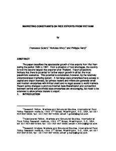
RESULTS Y134R is related to members of the IL-10 cytokine family Previously, a comparison of Y134R with protein databases established that Y134R is related to the family of IL-10related proteins (Lee et al., 2001). This was reinvestigated by comparison with a non-redundant protein sequence database available at http://www.ncbi.nlm.nih.gov/ using the BLASTP 2.2.6 (PSI-BLAST) program. An alignment of Y134R (without signal peptide) with human cytokines IL-24, IL-20 and IL-19 is shown in Fig. 1(a). Of these, Y134R was most closely related to IL-24 with 28 % amino acid identity and 55 % similarity. These proteins have only two conserved cysteines, whereas IL-19 and IL-20 have six, and Y134R contains a single motif for the addition of N-linked carbohydrate that is conserved in IL-24 (Fig. 1a). The hydrophobicity profile of Y134R (Fig. 1b) shows the hydrophobic signal peptide which, using the SignalP program at http://us.expasy.org/, was predicted to be cleaved between S18 and L19. Fig. 1(c) shows a model of Y134R based on the X-ray crystal structure of IL-19 (Chang et al., 2003). The model suggests a possible disulphide bond between C3 and C50. Although the distance separating these residues is 4?8 A˚, 2?8 A˚ greater than the ideal distance of 2 A˚ for a disulphide bond, the N-terminal cysteine is predicted to be located in a flexible loop, and hence these cysteines might be close enough to form a disulphide 1403
N. W. Bartlett and others
Fig. 1. Y134R is related to members of the IL-10-like cytokine family. (a) Amino acid alignment of Y134R, without the putative signal peptide, with human IL-19, IL-20 and IL-24. The protein accession numbers are: Y134R (CAC21372), IL-24 (NP 006841), IL-20 (NP 061194), IL-19 (NP 037503). Bars above the Y134R sequence indicate regions predicted to be ahelical and correspond to those illustrated in (c). Asterisks denote conserved cysteine residues. Identical amino acids or those with similar biochemical properties are shaded in black; identities across two or three of the sequences are highlighted in grey. The N-glycosylation site conserved in Y134R and IL-24 is boxed. (b) Hydrophobicity plot of Y134R showing the hydrophobic signal peptide. (c) Predicted three-dimensional structure of Y134R based on the structure of IL-19. The numbers of the different a-helices and the positions of cysteine residues are indicated.
bridge. This region of the model also contains another loop that is made up of a string of positively charged lysine and arginine residues that may be involved in receptor interactions. The Y134R gene encodes a secreted protein To characterize Y134R, a C-terminal flag-tagged copy of the Y134R gene was transfected into HEK293T cells. For comparison, flag-tagged cDNAs encoding IL-19, IL-20, IL-22 and IL-24 were expressed in parallel. Supernatants from all transfections were analysed by immunoblotting with a mAb that recognizes the flag epitope (Fig. 2a). HEK293T cells secreted Y134Rflag as two different forms, which probably represent differences in glycosylation, whilst the other cytokines were expressed as reported previously 1404
(Dumoutier et al., 2001b). To compare Y134Rflag with the untagged Y134R made by YLDV- or vDB8R-Y134R-infected cells, the rabbit anti-Y134R polyclonal IgG was used in immunoblotting (Fig. 2b). Y134R was secreted from infected cells as two different forms that were approximately 13 and 20–22 kDa, slightly smaller than those observed for Y134Rflag. The predicted size of mature Y134R is 16?1 kDa; thus it is likely that the diffuse 20–22 kDa band is glycosylated. The smaller form did not exactly match the predicted size for unknown reasons. Expression from VACV yielded significantly higher levels of Y134R than from YLDV, consistent with the use of a strong VACV promoter. To assess when Y134R is expressed during YLDV infection, supernatants were collected from cells infected in the presence or absence of araC at various times post-infection Journal of General Virology 85
Poxvirus-encoded cytokine
Fig. 2. Y134R is a secreted protein that is produced late in YLDV infection. (a) Transient expression of flag-tagged IL-19, IL-20, IL-22, IL-24, Y134R or empty vector (pCI) from HEK293T cells. Tissue culture supernatants were harvested from cells 3 days post-transfection and proteins were resolved by SDS-PAGE and analysed by immunoblotting with a mouse a-flag mAb. In the case of pCI and Y134Rflag, supernatants were concentrated 10-fold before analysis. (b) Detection of Y134R in supernatants from pCI-Y134Rflag (pCI134)-transfected HEK293T cells 3 days post-transfection (concentrated 10-fold), YLDV-infected OMK cells 3 days post-infection (concentrated 10-fold) and vDB8R-Y134R (v134R)-infected BS-C-1 cells 2 days postinfection (concentrated fourfold) by immunoblotting using rabbit a-Y134R polyclonal IgG. (c) Time-course analysis of Y134R expression from YLDV-infected CV-1 cells. Cells were mock infected or infected in the presence or absence of araC. Supernatants were collected at various times post-infection and concentrated equally for all samples. Proteins were analysed by SDS-PAGE and immunoblotting using a-Y134R rabbit IgG.
and were analysed by immunoblotting (Fig. 2c). In the presence of araC, no Y134R was detected, indicating that the gene is expressed only late in infection. Y134R is a monomer that requires glycosylation for secretion To determine whether Y134R is a monomer, dimer or oligomer, supernatants from vDB8R-Y134R-infected cells were analysed by size-exclusion chromatography as http://vir.sgmjournals.org
Fig. 3. Y134R is a glycosylated monomer. (a) A concentrated supernatant from cells infected with vDB8R-Y134R was mixed with gel filtration calibration proteins [BSA (67 kDa), hen egg ovalbumin (43 kDa), bovine pancreatic chymotrypsin (25 kDa) (all from APB) and cytochrome c (13?6 kDa) (Sigma)] and fractionated by size-exclusion chromatography on a Superdex 75 10/300 GL column (APB). Proteins present in fractions 13–21 were analysed by SDS-PAGE and immunoblotting with aY134R IgG. (b) Cells were infected with vDB8R-Y134R for 40 h in the presence or absence of tunicamycin or monensin. Supernatants were concentrated fivefold. Protein in supernatant from cells infected in the absence of drugs was either untreated or treated with PNGase F to remove N-linked carbohydrate. Proteins in all samples were resolved by SDS-PAGE and immunoblotted with either a-Y134R IgG or a-A41L rabbit serum.
described previously (Bartlett et al., 2002). Y134R eluted from the column with a size consistent with it being monomeric under physiological conditions (Fig. 3a). To investigate whether Y134R is glycosylated, Y134R in the supernatant of vDB8R-Y134R-infected cells was analysed by immunoblotting before or after treatment with PNGase F to remove N-linked carbohydrate (Fig. 3b). PNGase F treatment reduced the size of Y134R to that of a smaller form found predominantly inside cells. Consistent with this observation, Y134R synthesized in the presence of tunicamycin, an inhibitor of N-linked glycosylation, was not secreted and was present only as intracellular forms, the 1405
N. W. Bartlett and others
smaller of which co-migrated with the PNGase F-treated sample. In contrast, monensin, an inhibitor of O-linked glycosylation, neither inhibited the secretion of Y134R nor the size of the mature protein. Parallel analysis of the VACV glycoprotein A41L, which also contains N-linked carbohydrate (Ng et al., 2001), showed that tunicamycin reduced its size but did not prevent secretion. Identification of the class II cytokine receptors utilized by Y134R Given the predicted structural similarity shared by Y134R and IL-10-related cytokines, we investigated whether Y134R could signal via receptors used by these cytokines. The human HaCaT keratinocyte cell line (Boukamp et al., 1988) expresses both IL-20 receptors that signal via STAT3 upon ligand binding (Blumberg et al., 2001). To assess whether Y134R could activate this signal transduction pathway, HaCaT cells were treated with conditioned supernatants with or without Y134Rflag and the level of STAT3 phosphorylation was compared with that induced by treatment with supernatants containing different human cytokines (Fig. 4a). After 20 min incubation with Y134R or each other cytokine tested, there was a significant increase in the level of phosphorylated STAT3 above that induced by incubation with supernatants taken from cells transfected with empty vector (pCI). To elucidate which receptor(s) Y134R utilized, BHK-21 cells were transfected with different receptor subunit combinations to yield functional heterodimeric receptors. These cells were stimulated with different cytokines for 20 min and then cell extracts were prepared and analysed for STAT3 (Fig. 4b) or STAT1 (Fig. 4c) phosphorylation. Y134R, IL-24 and IL-20 signalled via both the IL-20R type 1 (IL-20Ra/IL20Rb) and IL-20R type 2 (IL-22R/IL-20Rb) and induced both STAT3 and STAT1 activation in cells expressing these receptors. In contrast, IL-24, IL-20 and Y134R were unable to induce STAT3 phosphorylation in cells expressing the IL22 receptor (IL-22R/IL-10Rb). The observation that IL-19 could only activate STATs in cells expressing the IL-20R type 1 and not in cells expressing IL-20R type 2, as previously reported (Dumoutier et al., 2001b), confirmed the specificity of receptor expression. Y134R activates the JAK/STAT signal transduction pathway Following specific ligand–receptor interaction at the cell surface, STATs become phosphorylated on tyrosine residues, dimerize and enter the nucleus to induce transcription of responsive genes (Darnell, 1997). Therefore, the location of STAT3 was investigated in cells expressing the IL-20R type 2 (Fig. 5a–c) or the IL-22R (Fig. 5d, e). BHK-21 cells expressing transiently the IL-20R type 2 were stimulated with conditioned HEK293T supernatants containing IL-19, IL-24 or 134R (Fig. 5a–c). In agreement with the immunoblotting data (Fig. 4), both IL-24 and Y134R activated STAT3 in cells expressing this IL-20 receptor, resulting in 1406
Fig. 4. Y134R induces STAT activation via type II cytokine receptors. (a) HaCaT keratinocytes were stimulated with conditioned HEK293T supernatant (10 % of overlay volume) from transfection with empty vector (pCI) or cDNAs encoding IL-19, IL-20, IL-22, IL-24 or Y134R for the time indicated. Cell extracts were analysed by immunoblotting with a-phospho-STAT3 mAb. Equal protein loading was evaluated by immunoblotting for atubulin (aTub). (b, c) BHK-21 cells were transfected with expression plasmids encoding the following class II cytokine receptor subunits: IL-20Ra and IL-20Rb, IL-22R and IL-20Rb or IL-22R and IL-10Rb. Cells were stimulated with conditioned HEK293T supernatants containing the indicated cytokine or Y134R for 20 min and the cell extracts were analysed by immunoblotting for STAT3 (b) or STAT1 (c) phosphorylation.
translocation of STAT3 to the nucleus. In contrast, IL-19 was unable to activate STAT3 via the IL-20R type 2 and no nuclear localization of STAT3 was apparent. To analyse further whether Y134R could induce STAT3 activation via the IL-22R, cells expressing this receptor were stimulated with either IL-22 or Y134R (Fig. 5d, e). As expected, IL-22 induced nuclear localization of STAT3 in these cells whereas Y134R did not. To characterize receptor specificity and STAT activation by Y134R further, HT-29 cells, which express the IL-22 receptor (IL-22R/IL-10Rb), were transfected with a STATluciferase reporter gene and were then incubated with Journal of General Virology 85
Poxvirus-encoded cytokine
Fig. 5. Y134R induces nuclear translocation of STAT3. BHK21 cells were transfected with cDNAs encoding IL-22R and IL20Rb, or IL-22R and IL-10Rb. Cells were stimulated for 20 min with conditioned HEK293T supernatant containing the indicated cytokine and then were processed for immunofluorescence to assess nuclear translocation of STAT3. Arrows indicate cells showing punctuate nuclear fluorescence.
different ligands (Fig. 6a). In agreement with STAT phosphorylation and nuclear translocation results, these cells did not respond to either Y134R or IL-20. However, HT-29 cells transfected with plasmids encoding IL-20Rb and the STAT–luciferase reporter gene, so that they expressed the IL-20R type 2 (IL-22R/IL-20Rb), produced luciferase in response to IL-22, Y134Rflag and IL-20. This confirmed the receptor specificity of Y134R (Fig. 6b). Another class II cytokine receptor has been described recently and binds IL-28A, IL-28B and IL-29. These cytokines are also called IFN-ls and belong to the same family of small a-helical cytokines as IL-10, IL-19, IL-20, IL24 and Y134R. The receptor consists of the IL-10Rb (also called CRF2-4 and which is shared by the IL-22R) and IFNlR1 (also called LICR2, IL-28Ra and CRF2-12) (Kotenko et al., 2003; Sheppard et al., 2003). To determine whether Y134R could signal via this receptor, the BWIL-28R-LICR2 cell line (which stably expresses the IFN-l receptor) was transfected with the STAT–luciferase gene and then treated with IFN-l1/IL-29, Y134Rflag or negative control transfection supernatant (pCI) (Fig. 6c). Luciferase production induced by Y134Rflag was no greater than that observed for cells incubated with the supernatant from transfection with the empty vector (pCI), indicating that Y134R does not signal through this class II cytokine receptor. Expression of Y134R reduces VACV virulence Having demonstrated a biological activity of 134R in vitro, we next examined the contribution of Y134R to the http://vir.sgmjournals.org
Fig. 6. STAT-regulated gene expression is induced by Y134R. HT-29 cells were transfected with (a) pGRR5 and pRLTK luciferase or (b) IL-20Rb cDNA, pGRR5 and pRLTK. Five hours later transfected cells were stimulated with recombinant IL-22 protein (10 ng ml”1) or supernatants from HEK293T cells transfected with empty vector (pCI), Y134R or IL-20. Luciferase activity was monitored after 2 h. Results are expressed in arbitrary units including standardization using Renilla luciferase as an internal control, and correspond to the mean of duplicate cultures. (c) BW-LICR2 cells were transfected with pGRR5 and then stimulated with 20 ng IFN-l1/IL-29 protein ml”1 or HEK293T supernatants (10 % overlay volume) from transfections with empty vector (pCI) or Y134R. Luciferase activity was monitored after 2 h. Results are expressed in arbitrary units including standardization using Renilla luciferase as an internal control, and correspond to the mean of duplicate cultures.
virulence of a poxvirus. Because YLDV does not induce disease in mice and no primate model was available, we sought to examine a role for Y134R via its expression in VACV. Y134R was expressed from a VACV strain WR mutant engineered to lack the VACV IFN-c binding protein, B8R. This protein does not affect VACV virulence in a murine intranasal model because it does not inhibit mouse IFN-c (Symons et al., 2002), but this virus would be predicted to be attenuated in humans because the VACV 1407
N. W. Bartlett and others
B8R protein does inhibit human IFN-c (Alcami & Smith, 1995; Mossman et al., 1995). Therefore, this strain was selected as a potentially safer parent virus from which Y134R could be expressed. The parent virus vDB8R, the virus expressing Y134R (vDB8R-Y134R) and a revertant virus from which the Y134R gene was removed (vDB8R-rev) were used to infect BALB/c mice intranasally. The outcome was assessed by monitoring body weight and signs of illness as described previously (Alcami & Smith, 1992). Mice infected with vDB8R-Y134R lost less weight and presented milder disease symptoms than mice infected with control viruses (Fig. 7a, b). The differences were significant for weight loss (P