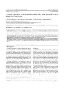
0013-7227/97/$03.00/0 Endocrinology Copyright © 1997 by The Endocrine Society
Vol. 138, No. 6 Printed in U.S.A.
Cloning and Functional Expression of the Luteinizing Hormone Receptor Complementary Deoxyribonucleic Acid from the Marmoset Monkey Testis: Absence of Sequences Encoding Exon 10 in Other Species* FU-PING ZHANG, ANTTI S. RANNIKKO, PULAK R. MANNA, HAMISH M. FRASER, AND ILPO T. HUHTANIEMI Department of Physiology, University of Turku, Kiinamyllynkatu 10, FIN-20520 Turku, Finland; and the Medical Research Council Reproductive Biology Unit, Center for Reproductive Biology (H.M.F.), Edinburgh, United Kingdom EH3 9 EW ABSTRACT Based on sequence homologies among the human, porcine, rat, and mouse genes for the LH receptor (LHR), overlapping partial fragments of LHR complementary DNAs (cDNAs) were multiplied from marmoset monkey testicular RNA using reverse transcription-PCR. Ligations of the individual cDNA fragments generated a full-length monkey LHR cDNA (2031 bp) containing the complete amino acidcoding sequence (676 amino acids). Northern hybridization analysis of monkey testicular RNA, using a complementary RNA probe corresponding to the full-length cDNA, demonstrated major transcripts of 5.5 and 1.4 kilobases and minor ones of 4.0, 2.7, and 1.9 kilobases. Sequence analysis of the monkey LHR cDNA revealed a striking feature, i.e. the absence of an 81-bp nucleotide sequence corresponding to exon 10, present in the LHR cDNAs of all other species studied to date. The monkey LHR cDNA displayed 83–94% overall sequence homology with the other mammalian LHR cDNAs. Reverse transcription-PCR with human exon 10-specific primers demonstrated the total absence of this sequence from the monkey LHR messenger RNA. Southern hybridization of monkey genomic DNA using a human
exon 10 probe demonstrated its presence in the monkey gene and that it is totally spliced out from the primary transcript. COS cells transfected with the monkey LHR cDNA showed similar high affinity (Kd 5 0.25 nmol/liter) of [125I]iodo-hCG binding as those transfected with human LHR cDNA (Kd 5 0.20 nmol/liter). The cells expressing the recombinant monkey and human LHR displayed similar responses of extracellular cAMP and inositol trisphosphate to hCG. In conclusion, marmoset monkey LHR seems to lack the sequence corresponding to exon 10 of the LHR gene in other mammalian species. The truncation does not alter LHR function, as the monkey receptor protein bound hCG and evoked cAMP and inositol trisphosphate responses comparable to those of the human LHR containing the exon 10-encoded structure. As the sequence homologous to exon 10 is missing in the other two glycoprotein receptors, i.e. those of FSH and TSH, this extra exon is apparently inserted into the LHR messenger RNA of some species during evolution from intronic sequences by a change in alternative splicing. (Endocrinology 138: 2481–2490, 1997)
T
N-terminal half and a plasma membrane-associated C-terminal half of the full-length protein (1). The extracellular domain is encoded by the first 10 and a part of the 11th exon, and it is capable of high affinity hormone binding. The rest of exon 11 encodes the transmembrane and intracellular parts of the receptor, which are capable of low affinity hormone binding and signal transduction (6 –9). Recent studies on serial mutagenesis of the LHR complementary DNA (cDNA) emphasize that the structural integrity of the receptor protein is important for its functions. Several point mutations or deletion of Lys583, located in the third extracellular loop, maintain the affinity of the receptor to ligand, but greatly diminish the ligand-mediated signaling (10, 11). Two point mutations of the LHR in the ionizable amino acid residues, i.e. Glu332 3 Lys332 and Asp3333 Lys333, displayed nearly the same binding affinity for hCG as wildtype receptor, but they also failed to evoke increased cAMP production (12). Deletion of amino acids 1–11 or the leucinerich repeats 1, 2, 3, 4, 5, and 6 individually, by contrast, resulted in a loss of detectable binding activity (13). Numerous mutations of the LHR gene have also been found to be involved in pathophysiological conditions. The syndrome of familial male-limited precocious puberty is
HE LH RECEPTOR (LHR) plays an important role in reproductive physiology. LHR is primarily expressed in the gonads, and it targets the action of LH to Leydig cells in the testis and to thecal, granulosa, and luteal cells in the ovary. The LH-receptor interaction activates two intracellular signaling pathways: adenylyl cyclase-stimulated cAMP production and phospholipase C-stimulated formation of inositol phosphates and elevation of intracellular free Ca21 (1, 2). The structure and function of LHR have been extensively studied, and this receptor, together with those of FSH and TSH, belongs to a subfamily of glycoprotein hormone receptors among the G protein-associated seven-transmembrane-domain receptors (1, 3–5). Unlike many members of the 7-transmembrane domain receptor family, which are intronless (1), the 3 glycoprotein hormone receptor genes contain 9 (TSH and FSH receptors) or 10 (LHR) introns. LHR is composed of an extracellular Received December 23, 1996. Address all correspondence and requests for reprints to: Dr. Ilpo Huhtaniemi, Department of Physiology, University of Turku, FIN-20520 Turku Finland. E-mail:
[email protected]. * This work was supported by grants from the Academy of Finland and the Sigrid Juse´lius Foundation.
2481
2482
caused by mutations mainly located in the third intracellular loop or in the sixth transmembrane segment of the LHR, resulting in its constitutive action (14 –16). Inactivating mutations of the human LHR lead to Leydig cell hypoplasia, a form of male pseudohermaphroditism resulting from the failure of fetal testicular Leydig cells to differentiate and produce testosterone due to defective LH/hCG signal transduction (17–20). To further understand the structure-function relationships of the LHR gene and to determine whether there are structural differences in the LHR between different species, we undertook the cloning of the complete nucleotide sequence as well as functional expression of marmoset monkey LHR cDNA. The intriguing finding was that in the monkey LHR cDNA, the DNA sequence corresponding to exon 10 of the LHR gene in all other species studied to date is missing in all splice variants. The truncated monkey LHR cDNA was successfully expressed in COS cells, allowing high hCG binding and hCG-mediated cAMP and inositol trisphosphate (IP3) production. Materials and Methods Isolation of RNA and DNA Adult monkey testes and ovaries were obtained at autopsy from adult marmoset monkeys (Callithrix jacchus), a New World primate, frozen in liquid nitrogen, and stored at 270 C until isolation of RNA and DNA. The isolation of total RNA was started by homogenizing the tissues in 10 ml/g tissue of 4 m guanidinium thiocyanate solution, followed by the CsCl2 centrifugation method (21). After centrifugation, the supernatant was discarded, and the pelleted RNA was extracted with phenol-chloroform and precipitated with ethanol. Genomic DNA was isolated from monkey testis, and human placenta and muscle were isolated by the proteinase K method. Briefly, the tissues were digested with proteinase K in lysis buffer (5 ml/g tissue) at 42 C overnight. Sequentially, the same volume of phenol-chloroform-isomyl alcohol (50:49:1) was added to the digestion mixture. After centrifugation, the supernatant was removed to a new tube and precipitated with ethanol.
Reverse transcription-PCR (RT-PCR) and PCR analyses For RT-PC and PCR reactions, several primers were designed based on the conserved sequences of the human, porcine, rat, and mouse LHR cDNAs. These primers were named human oligonucleotide (oh; Fig. 1 and Table 1). Subsequently, several other primers specific for the monkey LHR cDNA were designed according to the known partial monkey LHR cDNA sequences generated by RT-PCR; these primers were named monkey oligonucleotide (om; Fig. 1 and Table 1). The RT and PCR reactions were carried out sequentially in the same
FIG. 1. Cloning strategy of the monkey LHR cDNA. The thick lines represent the partial and holoreceptor of monkey LHR cDNAs. The blank box indicates the position of the missing exon 10 that is present in the other known LHR cDNAs. The oligonucleotide primers are related to the diagram of the monkey LHR cDNA. The primer pairs used for RT-PCR were oh 2–3, oh 4 –5, and oh 6 –7 to produce fragments H1, H2, and H3. Primer pairs oh1-om1, oh2-om2, om3–5, and om4-oh8 generated fragments M1, M2, M3, and M4.
Endo • 1997 Vol 138 • No 6
MONKEY LH RECEPTOR
assay tube (22). Two to 5 mg total RNA from testis or ovary were reverse transcribed using the avian myeloblastosis virus reverse transcriptase (Promega Corp., Madison, WI) and one of the antisense primers (see Fig. 1). The reverse transcribed, single strand cDNAs were further amplified by PCR using the various primer pairs (see Fig. 1); in each case, the antisense primer was the same as the primer used in the RT reaction. Fifty microliters of the RT-PCR mixture contained 1 nm of each oligo primer, 200 mm deoxy-NTPs, 1.5 mm MgCl2, 20 U RNasin (Promega), 12.5 U avian myeloblastosis virus reverse transcriptase, and 2.5 U Taq DNA polymerase (Promega). The reaction was started at 50 C for 10 min (at room temperature), followed by a period of 3 min at 97 C, and then run for 35– 40 PCR cycles (96 C, 1 min; 57 C, 1 min; 72 C, 2 min), and final extension for 10 min at 72 C. The PCR reaction from monkey genomic DNA was performed with the hot start method. The reaction was started at 97 C for 5 min, followed by 52 C for 2 min, and 72 C for 3 min, and then run 30 PCR cycles (97 C, 1 min; 52 C, 1, 72 C, 2 min) and final extension for 10 min at 72 C. The RT-PCR and PCR products were resolved in 1–1.5% agarose gels, and the cDNA fragments with expected sizes were cut from the gels and isolated using the QIAEX II gel extraction kit (Qiagen, Chatsworth, CA). The DNA fragments were subcloned into the T vector (Promega) according to the manufacturer’s instruction for sequencing and construction of the full-length LHR cDNA (see below).
Construction of the monkey and human LHR cDNAs for expression studies The overlapping fragments of the monkey LHR cDNA were digested by restriction endonucleases (Promega) at appropriate sites (see Fig. 1) TABLE 1. Primer structures Primers were designed according to the human LHR cDNA Oligo h1 59-CAGATGGTAGGGGACAACA-39 Oligo h2 59-GCCGCCGCTGCCACGAG-39 Oligo h3 59-AGGAGGTTGTCAAAGGCATTAG-39 Oligo h4 59-CCCGATTAAAATACTTGAGCA-39 Oligo h5 59-ATTAGCCTCTGAATGGACTCT-39 Oligo h6 59-CGATGTGCTCCTGAACCAG-39 Oligo h7 59-GCGAGTCTTGTCTAGGAGAG-39 Oligo h8 59-GTTAAAATTACTGGTACAGGTA-39 Oligo h9 59-ACAGAATTTTCACATT-39 Oligo h10 59-AGTGTTTGTTAATTCAC-39 Primers were designed according to the monkey LHR cDNA Oligo Oligo Oligo Oligo Oligo Oligo Oligo Oligo
m1 m2 m3 m4 m5 m6 m7 m8
59-AGTCGAGTGAGACCGGCCC-39 59-GCCGGTCTCACTCGACTATCAC-39 59-GTACAAAGTCATGCGTTAAA-39 59-TGCTGCTCATAGCCTCGGTTG-39 59-TCCAGGTGAATAGCATAGGT-39 59-GGATGCCACGCTGACTTACCC-39 59-TCAGCAAATATGGCAGGATA-39 59-TTAATTCAGCCAAATCAGGA-39
MONKEY LH RECEPTOR and ligated by the T4 DNA ligase (Promega) using standard molecular biology methods. The complete monkey LHR cDNA was excised from the T vector by digestion with SacII and SalI, rendered blunt ended using the Klenow and T4 DNA polymerase enzymes (Promega), and subcloned into the eukaryotic expression vector pZeoSV21 (Invitrogen, San Diego, CA), prepared by digestion with EcoRV and dephosphorylation to yield pZeoSV21/monkey LHR (sense) and pZeoSV21/monkey LHR (antisense). As control, human LHR cDNA was excised from pBS-hLHR by digestion with EcoRV and BamHI and subcloned into pZeoSV21 at the HindIII and BamHI sites to yield pZeoSV21/human LHR.
Sequencing The partial and complete monkey LHR cDNAs inserted into the T vector were sequenced from both strands using an automatic sequencing machine (Perkin-Elmer, Foster City, CA). The nucleotide and amino acid sequences were further analyzed by DNASTAR (DNASTAR, Madison, WI) and BLAST Search (23) computer programs.
Northern hybridization analysis Twenty micrograms of total RNA from monkey testis were resolved on 1.2% formaldehyde denaturing agarose gel and transferred onto nylon membrane (Hybond-N, Amersham, Aylesbury, UK). Prehybridization and hybridization were performed as previously described (24). Briefly, the filters were prehybridized for at least 4 h at 65 C in a solution containing 50% formamide, 3 3 SSC (1 3 SSC 5 150 mm NaCl and 15 mm sodium citrate, pH 7.0), 5 3 Denhardt’ solution, 1% SDS, 0.1 g/liter heat-denatured calf thymus DNA, and 100 mg/liter yeast transfer RNA. Hybridization was carried out at 68 C overnight in the same solution after adding the 32P-labeled complementary RNA (cRNA) probe. After hybridization, the membranes were washed twice with 2 3 SSC and 0.1% SDS at room temperature for 30 min each time and twice with 0.1 3 SSC and 0.1% SDS at 66 C for 2 h each time. After treatment of the membrane with ribonuclease A (1 mg/liter in 2 3 SSC) at room temperature for 30 min, followed by washing with 2 3 SSC at 42 C for 30 min, the membranes were exposed to x-ray film (Kodak XAR-5, Eastman Kodak, Rochester, NY) at 270 C for 3 days. The molecular sizes of the messenger RNA (mRNA) species were determined by comparison with RNA mol wt markers (Promega). The 32P-labeled cRNA was synthesized using a Riboprobe synthesis II kit (Promega), [32P]UTP (Amersham), and full-length monkey LHR cDNA that was subcloned into the T vector (pGEM-5Z) as template.
Southern hybridization analysis of genomic DNA Forty micrograms of monkey and human genomic DNAs were digested with XhoI. The digested DNA fragments were resolved in a 0.7% agarose gel and transferred onto nylon membrane by the capillary transfer method. The probe was generated by PCR amplification of exon 10 of the hLHR gene using as the primer pair oligos h9 and h10. The amplified fragment was labeled with [32P]CTP using the Multiprime DNA labeling system (Amersham), except that oligos h9 and h10 were used instead of hexamer primers. Prehybridization and hybridization were carried out as previously described (24).
Transfections COS cells were maintained in DMEM-Ham’s F-12 medium (1:1; Life Technologies, Grand Island, NY) supplemented with 10% FCS and 0.1 g/liter gentamicin (Biological Industries, Bet-HaEmek, Israel). Cells (0.5 3 106) were plated on 9-cm diameter plates 1 day before transfection and transfected using the calcium phosphate precipitation method (25) with 10 –15 mg of the plasmid under investigation and 3 mg of the b-galactosidase expression vector as control for transfection efficiency. Precipitates were left on cells for 12 h and subsequently washed with PBS and fed fresh medium. Cells were used for analysis 2–3 days after transfection. In the case of stable transfection, the cells were split into medium containing 200 mg/liter Zeocin (Invitrogen), and the selection was maintained for 3 weeks. After selection, the cells were pooled to create nonclonal stable cell lines and maintained in medium containing 50 –100 mg/liter Zeocin.
2483
[125I]hCG binding study hCG (CR-127, NIDDK) was iodinated to a specific activity of 35,000 cpm/ng and 37% specific binding of radioactivity to an excess of LHRs, as determined according to the method of Catt et al. (26). For LHR binding measurements, the cells were washed twice with cold PBS and scraped by rubber policeman into Dulbecco’s PBS containing 0.1% BSA (D-PBS). The cells were pelleted by centrifugation at 1,500 rpm and washed twice with D-PBS. For single point binding measurements, triplicate aliquots of cells (0.2 3 106) were incubated with 150,000 cpm [125I]iodo-hCG in the presence or absence of 50 IU unlabeled hCG (Pregnyl, Organon, Oss, The Netherlands) in a total volume of 250 ml. For Scatchard analysis, similar aliquots of cells were incubated with increasing doses of [125I]iodo-hCG (up to 500,000 cpm/tube). For competition studies, the same aliquots were incubated with increasing amounts of unlabeled hCG (CR 127; 0.1– 400 ng/tube) or recombinant human FSH (rhFSH; Org 32489, Organon; 0.1– 400 mIU/tube) in the presence of 150,000 cpm [125I]iodo-hCG. After overnight incubation at room temperature, the cells were washed with 4 ml ice-cold D-PBS and centrifuged, and the radioactivity in the cell pellets was counted in a g-spectrometer. Nonspecific binding was determined in the presence of 50 IU Pregnyl, and all data were corrected for nonspecific binding.
cAMP and IP3 assays Transfected cells were plated at a density of 50,000 cells/well (24-well plates) 24 h before stimulation. The cells were stimulated for 3 h in a medium containing 0.2 mmol/liter 3-isobutyl-1-methylxanthine (Aldrich-Chemie, Steinheim, Germany) with increasing doses of hCG (CR127; 0 –1000 mg/liter) or rhFSH (0 –1000 IU/liter). All stimulations were performed in quadruplicate. At the end of the incubation, the medium was collected for measurement of extracellular cAMP using a standard RIA method (27). For IP3 measurements, stably transfected cells were cultured in sixwell plates at a concentration of 5 3 105 cells/well in inositol-free DMEM-Ham’s F-12 medium. After 24-h incubation, the cells were washed twice with HEPES-3 buffer (10 mm HEPES, 58 mm NaCl, 0.3 mm NaH2PO4, 3.4 mm sodium acetate, 5 mm KCl, 0.6 mm MgSO4, 1.5 mm CaCl2, 2 mm d-glucose, and 0.1% fatty acid-free BSA, pH 7.2) and stimulated without or with increasing doses (0 –5000 mg/liter) of hCG for 20 min in the presence of LiCl. Accumulation of IP3 was terminated, and the substance was extracted by keeping the cells on ice with 10% ice-cold perchloric acid, followed immediately by neutralization with 1.5 m KOH in 60 mm HEPES, pH 7.2. Production of IP3 was determined by the inositol 1,4,5-trisphosphate [3H]RRA Kit (New England Nuclear Research Products, DuPont de Nemours Co., Boston, MA) according to the manufacturer’s instructions. The sensitivity of the system was approximately 0.1 pmol.
Presentation of data The results are presented as the mean 6 sem when appropriate.
Results Cloning of monkey LHR cDNA
Based on the conserved sequences of the human, porcine, rat, and mouse LHR cDNAs, several primer pairs were designed for RT-PCR reactions using monkey testicular total RNA. The primer pairs oh 2 and 3, oh 4 and 5, and oh 6 and 7 generated fragments H1, H2, and H3, respectively. The PCR products were subcloned into the T vector, then sequenced and found to be homologous with known human, porcine, and rat LHR cDNA sequences. According to the partial monkey LHR cDNA sequences generated by RT-PCR, several other primers specific to the monkey sequence were synthesized. Using primer pairs oh1 and om1, oh2 and om2, om3 and om5, and om4 and oh8, four overlapping monkey LHR cDNA fragments were generated
2484
MONKEY LH RECEPTOR
by RT-PCR. These fragments were subcloned into the T vector and sequenced. The complete monkey LHR cDNA (Fig. 2) was constructed by ligating cDNA fragments M1 and M2 at the AvaI site,
Endo • 1997 Vol 138 • No 6
followed by ligation of H2 at BglII, M3 at ApaI, and M4 at XbaI into the T vector (see Fig. 1). The complete monkey LHR cDNA was excised from the T vector and subcloned into the eukaryotic expression vector pZeoSV21 to yield pZeoSV21/
FIG. 2. Comparison of nucleotide sequences of the monkey (GenBank accession no. U80673), human, porcine, and rat LHR cDNAs. The nucleotides are marked on the left, starting with the ATG translation initiation codon and covering the entire open reading frame. Periods and dashes indicate identities and gaps, respectively. The thick line indicates the exon 10 sequence that is absent in the monkey LHR cDNA.
MONKEY LH RECEPTOR
monkey LHR (antisense).
(sense)
and
pZeoSV21/monkey
LHR
Determination of sequence corresponding to exon 10 in the monkey LHR cDNA and gene
To confirm whether the sequence corresponding to exon 10 of the other species is totally absent in the monkey LHR cDNA, several primer pairs specific for the monkey LHR cDNA sequence were designed for RT-PCR reactions. Using primer pairs om6 and 8, and om 6 and 7, two overlapping PCR fragments were generated from monkey and human testicular RNAs, and sequencing analysis of these fragments indicated that exon 10 (81 bp) was not present in any of the fragments generated from monkey testicular RNA, but it was present in human testicular RNA (Fig. 3A). These data strongly support the evidence that exon 10 is absent in monkey LHR cDNA. To determine whether the sequence corresponding to exon 10 of the LHR is present in monkey genomic DNA, a
2485
[32P]CTP-labeled human exon 10-specific probe was used to hybridize to the membrane containing XhoI-digested genomic DNAs of the monkey and human. The autoradiogram of the Southern hybridization demonstrated specific hybridization in the monkey and human genomic DNA (Fig. 3B), indicating that the sequence corresponding to exon 10 is present in the monkey gene, but spliced out in all of the LHR mRNA splice variants. Furthermore, RT-PCR and PCR reactions were performed using primer pair oligos h9 and h10, corresponding to exon 10 of the human LHR cDNA, from testicular RNA and genomic DNA, and the 81-bp PCR products were observed in the human and monkey genomic DNAs, but not in the monkey testicular RNA (results not shown). Hence, exon 10 is present in monkey genomic DNA, but is absent in LHR cDNA. Comparison of monkey, human, porcine, and rat LHR cDNA and amino acid sequences
Figures 2 and 4 show the nucleotide and deduced amino acid sequences of the cloned monkey LHR cDNA as well as comparisons between the cognate monkey, human, porcine, and rat sequences. The translation initiation codon at position 1 of the testicular LHR cDNA defines the start of a 2031-bp nucleotide open reading frame (676 amino acids). Compared with the human, porcine, and rat LHR cDNAs, the striking feature of the monkey LHR cDNA is the absence of the sequence encoding the entire exon 10 (81-bp nucleotides, 27 amino acids) of the human, porcine, and rat cDNAs (Figs. 2 and 4). The overall nucleotide sequence homology is 94% with human, 88% with porcine, and 83% with rat, respectively. At the deduced amino acid level, three potential N-linked glycosylation sites were found in the extracellular domain that are conserved in the monkey, human, porcine, and rat sequences; the other three potential N-linked glycosylation sites in exon 10 were only conserved in the human, porcine, and rat sequences (Fig. 4). Expression of monkey LHR cDNA in COS cells
FIG. 3. A, RT-PCR analysis of human and monkey testicular RNAs. The PCR product in lanes 1 and 2 and lanes 3 and 4 were generated with primer pairs om6/8 and om6/7, respectively. The PCR products were resolved in a 1.5% agarose gel. The cDNAs corresponding to the predicted sizes of mRNA from the human and monkey testes are indicated on the left. The molecular size markers (M) are MspI-digested pBS-SK and are indicated on the right. B, Southern hybridization analysis of genomic DNAs from the monkey and human. Forty micrograms of genomic DNA from the two species were digested with XhoI. The digested DNA fragments were resolved in a 0.7% agarose gel and transferred onto nylon membrane. The membrane was hybridized with a [32P]CTP-labeled probe corresponding to exon 10 of the human LHR cDNA. Migration of the molecular size markers is depicted on the right.
To confirm that the cloned marmoset monkey LHR cDNA encodes for a functional LHR, we subcloned the entire coding region into an expression vector, pZeoSV21, under the transcriptional control of the simian virus 40 enhancer-promoter, forming pZeoSV21/mLHR. As a control, the entire human LHR-coding region was also subcloned into the pZeoSV21 vector, forming pZeoSV21/hLHR. The COS cells were transfected transiently or stably with the above expression vectors. After transfection, radioligand receptor assays indicated that the cells transfected with pZeoSV21/mLHR and pZeoSV21/hLHR expressed high levels of specific [125I]iodohCG binding (Fig. 5, A and B). No specific binding was observed in cells transfected with pZeoSV21/mLHR (antisense) and pZeoSV21 without insert (Fig. 5, A and B). Scatchard analysis of the binding data demonstrated that the affinity of [125I]iodo-hCG binding to the cells transfected with pZeoSV21/mLHR (Kd 5 0.25 nm) was similar to that to cells transfected with pZeoSV21/hLHR (Kd 5 0.20 nm; Fig. 5C). As shown in Fig. 5D, increasing doses of human LH, but not rhFSH, were able to dose dependently displace the [125I]iodohCG binding.
2486
MONKEY LH RECEPTOR
Endo • 1997 Vol 138 • No 6
FIG. 4. Comparison of the deduced amino acid sequences of the monkey, human, porcine, and rat LHR cDNAs. The monkey sequence is listed in its full length, using the single letter code for amino acid description. Periods and dashes represent identities and gaps, respectively. The putative transmembrane regions are enclosed by rectangles and numbered 1–7. The consensus sequences for N-linked glycosylation among monkey, human, porcine, and rat sequences and among human, porcine, and rat sequences are shaded and marked with bold text, respectively. The missing 27 amino acids of exon 10 are represented by the solid line. The numbering of amino acids is indicated on the right.
cAMP and IP3 production in transfected COS cells expressing monkey and human recombinant LHR
The ability of the recombinant LHRs to generate cAMP and IP3 was tested by challenging the transfected cells with increasing concentrations of hCG (0 –1000 and 0 –5000 mg/ liter). The stimulation resulted in dose-dependent increases in cAMP production, which were about 3.5- and 4-fold elevated over the basal rate of production (Fig. 6A). In contrast, rhFSH did not stimulate cAMP production in either cell type (data not shown). The EC50 values for cAMP production in monkey and human LHR-expressing cells were 40 and 32 ng/ml, respectively. Unlike the untransfected cells, which showed no elevation of IP3 (data not shown), the cells ex-
pressing monkey and human LHR displayed similar dosedependent increases in IP3 production, which were about 4.5and 5.5-fold over the basal production rate (Fig. 6B). The EC50 values for IP3 production in monkey and human LHR-expressing cells were 90 and 37 ng/ml, respectively. Expression of LHR mRNA in monkey testis tissue
Northern hybridization analysis using a cRNA probe corresponding to the entire LHR cDNA demonstrated the presence of multiple transcripts of the LHR mRNA with approximate molecular sizes of 5.5, 4.0, 2.7, 1.9, and 1.4 kilobases (kb) in the adult monkey testis. The major transcripts were 5.5 and 1.4 kb in size (Fig. 7).
MONKEY LH RECEPTOR
2487
FIG. 5. [125I]Iodo-hCG binding to COS cells transfected with monkey or human LHR cDNA. A, Specific [125I]iodo-hCG binding to transiently transfected COS cells (mean 6 SEM of three independent experiments). B, Specific [125I]iodo-hCG binding to stably transfected cells (mean 6 range; n 5 2). C, Scatchard analysis of [125I]iodo-hCG binding in COS cells transiently transfected with monkey (E) and human (å) LHR cDNAs. D, Displacement of [125I]hCG binding from monkey LHR by hCG (f) and rhFSH (å). The nonspecific binding in each assay was about 2500 cpm/sample (100 ml cell suspension) and represented 20 –30% of the total binding. The data were corrected for b-galactosidase activity and protein in transiently transfected cells in A and for protein in the stably transfected cells in B and, therefore, therefore expressed as arbitrary units (AU).
Discussion
In the present study, we have determined the nucleotide sequence of the marmoset monkey testicular LHR cDNA and deduced the complete amino acid sequence of the receptor. Compared with the other known LHR cDNAs, the striking feature of monkey LHR cDNA was the absence of an 81-bp nucleotide sequence corresponding to exon 10 that was present in the LHR cDNAs of all other species studied to date. The complete absence of exon 10-specific sequences in the monkey LHR mRNA splice variants was further confirmed by several independent RT-PCRs using different primer pairs that should have amplified exon 10 if it was present in any splice variant of the monkey LHR mRNA. Sequencing of the RT-PCR product confirmed that the entire exon 10 was totally absent in all cDNAs generated from marmoset monkey testicular or ovarian RNA. Furthermore, we designed one primer pair that corresponded to human
exon 10, and the expected 81 bp of PCR or RT-PCR product was only obtained from monkey and human genomic DNA and human testicular RNA, not from monkey testicular RNA. Southern hybridization analysis of genomic DNAs of monkey and human using a [32P]CTP-labeled exon 10 probe demonstrated that specific hybridization bands were present in both monkey and human genes. Hence, the exon 10-specific sequence is apparently spliced out from the monkey LHR primary transcript during its processing for the mature mRNA species. Alternative splicing is a widespread device for gene regulation and for generating isoform diversity (28). Previous studies demonstrated that in addition to mRNA of the LH holoreceptor, various sizes of splice variants of the LHR mRNA have been found in rat, mouse, porcine, ovine, and human species, consistent with deletions of complete or partial exons within the genomic structure (29 –32). The common
2488
MONKEY LH RECEPTOR
Endo • 1997 Vol 138 • No 6
FIG. 7. Northern hybridization of LHR mRNA in the adult monkey testis. Twenty micrograms of total testicular RNA were resolved in 1.2% denaturing agarose gel and transferred onto nylon membrane, then hybridized with a [32P]cRNA probe for the full-length monkey LHR cDNA. The apparent molecular sizes of the different transcripts are shown on the right. The exposure time was 3 days.
FIG. 6. cAMP and IP3 production in the transfected cells expressing recombinant monkey and human LHR. A, Accumulations of extracellular cAMP in monkey and human LHR-expressing cells. The basal levels of cAMP production in monkey and human LHR-expressing cells were 86.6 6 2.8 and 60.3 6 4.5 fmol/100 ml 3 104 cells, respectively. B, IP3 production in monkey and human LHR-expressing cells. The basal levels of IP3 production in monkey and human LHR-expressing cells were 1.2 6 0.11 and 1.4 6 0.15 pmol/100 ml 3 5 3 105 cells, respectively. The data presented are the mean 6 range of two independent experiments.
method of alternative splicing, called cassette exon exclusion (whole exon deletion) (28), has been reported in numerous LHR splice variants in rat, pig, ovine, and human (1, 32–34). A truncated form of LHR without the entire exon 10 has been found in the ovine ovary (34), but not in other species studied to date, which indicates that alternative splicing of transcripts of a given gene is species specific. The extent of translation as well as the physiological significance of these mRNA forms are still unknown (1). To confirm that the cloned monkey LHR cDNA encodes a functional LHR protein, we transfected the cDNA con-
structed in the expression plasmid pZeoSV21 into COS cells. These cells as well as those transfected with the human LHR cDNA demonstrated specific high affinity binding of hCG. As a control, hCG binding was not observed in cells transfected with antisense monkey LHR cDNA or a plasmid without insert. The affinities (Kd) determined to the recombinant monkey LHR (Kd 5 0.25 nm) and human LHR (Kd 5 0.2 nm) were similar to those previously determined by others (human LHR, Kd 5 0.12 nm; rat LHR, Kd 5 0.17 nm; and porcine LHR, Kd 5 0.18 nm) (12, 29, 35). Our data indicated that the cloned monkey LHR cDNA encodes a receptor protein that has binding characteristics to [125I]iodo-hCG similar to those previously reported using LHR from other species. Davis et al. (36) reported that hCG stimulates phosphoinositide hydrolysis in isolated bovine corpus luteum cells. This raises the possibility that besides cAMP production, LH and hCG may also stimulate phospholipase C, generating inositol phosphates as second message. This was further confirmed by other studies, and it was proposed that all glycoprotein hormone receptors also stimulate inositol phosphate turnover (2, 37– 40). The exact nature of the G protein involved in LHR-mediated activation of phospholipase C is still not known. A recent study by Hirsch et al. (41) using chimeric LH and FSH receptors demonstrated that the Cterminal third of the human LHR is involved in phospholipase C activation. Our results show that, like in cells transfected with human LHR cDNA, transfected cells with monkey LHR cDNA displayed dose-dependent increases in IP3 accumulation in response to hCG, indicating that both receptor forms are efficiently coupled to phospholipase C activation. Compared with a previous report that the concentrations required to elicit half-maximal stimulation of IP3 production were about 20- to 30-fold higher than those given 50% stimulation of cAMP (2), our present data shown that the cells expressing monkey and human LHR responded to increasing doses of hCG with similar sensitivities of cAMP and IP3 production. This difference might be due to the different methodologies and cell types used. The cells expressing monkey LHR responded to increasing doses of hCG with
MONKEY LH RECEPTOR
cAMP responses similar to those of cells transfected with human LHR, but a response to rhFSH was absent in both cells. The EC50 values for cAMP responses in monkey and human LHR-expressing cells are 40 and 32 ng/ml, respectively, compatible with previous reports (2, 35). Hence, monkey and human LHR appeared to be functionally very similar. Taken together, the cloned truncated form of monkey LHR cDNA indeed encodes a functional LHR with specific hCG binding and hCG-stimulated cAMP and IP3 production. The absence of exon 10 in the monkey LHR cDNA is interesting, and raises the question of whether the sequences present in exon 10 are important for LHR function in other species. The three glycoprotein hormone receptors, i.e. those of LH, FSH, and TSH, are all characterized by a large extracellular ligand-binding domain with a leucine repeat structure (1). Despite different number of exons and differences in overall size, the genes for the glycoprotein hormone receptors are structurally remarkably similar. With the exception of exon 10, the other exons of the LHR share structural homology in amino acid sequence with respective structures of the FSH and TSH receptors. Hence, exon 10 might be inserted into the LHR mRNA in some, but not all, species by alternative splicing, and it may not be essential for receptor function (1, 3–5). All three glycoprotein hormone receptors have been shown to have N-linked carbohydrates (1). Recent studies using wild-type and mutant LHR expressed in COS cells demonstrated that the N-linked carbohydrate chains, specifically those attached to the two glycosylation sites Asn173 and Asn152, are critical for the assembly of a high affinity hormone-binding site within the extracellular domain in the rat LHR (42). In contrast, the putative glycosylation sites Asn269, Asn277, and Asn291 in exon 10 did not contribute to either hormone binding or membrane transport and insertion of the receptor protein (42). However, Rajaniemi et al. (43) indicated that the N-linked carbohydrate chains do not contribute to surface expression, hormone binding, and signal transduction once the receptor has been inserted into the membrane in a functional active form. Zhang et al. (44) analyzed the cysteine residues in rat LHR and demonstrated that the functional hormone-binding domain uses all cysteines N-terminal to exon 7, and the other cysteine residues, including Cys282 (exon 10) and Cys314 (exon11) are not essential for hormone-binding activity or plasma membrane insertion. Analysis of compound heterozygous mutations of the LHR gene in Leydig cell hypoplasia patients demonstrated that an A872G transition, resulting in an Asn291Ser substitution and abolition of a potential N-glycosylation site in the extracellular domain (exon 10) appeared to have no effect on the activity of the human LHR (20). This is in accordance with the above finding that this site is not glycosylated or that glycosylation of this site is not critical for either ligand binding or signaling (20, 42). In contrast, the deletion of exon 8 in the extracellular domain of these patients greatly reduced ligand binding and ligand-induced cAMP production in transfected cells. Hence, the polypeptide encoded by exon 8, unlike that encoded by exon 10, plays an important role in LHR expression and signal transduction (20). Our present
2489
findings indicated that exon 10 is not crucial for LHR function. The Northern hybridization analysis of monkey testicular RNA using a cRNA probe corresponding to the cloned monkey LHR cDNA revealed multiple transcripts with approximate molecular sizes of 5.5, 4.0, 2.7, 1.9, and 1.4 kDa. The predominant bands were 5.5 and 1.4 kb in size. This result is in agreement with those reported previously by us and others in other species (1, 22). The relative size and abundance of these transcripts differ from one species to another and also from tissue to tissue. These different transcripts derive from the alternate splicing and different transcription initiation site as well as polyadenylation signals (1, 45– 49). A recent study indicated the existence of multiple sites of splicing and polyadenylation in the rat LHR gene, resulting in generation of different transcription sizes (46). Lu et al. and Hu et al. (47, 48) also demonstrated that in the rat, a long 39-untranslated region containing two polyadenylation domains accounts for the size difference in LHR mRNAs. In the human, the 59-noncoding region is unusually long, and the transcription initiation site is located 1085 bp upstream of the translation start site (49). In conclusion, we have cloned LHR cDNA from marmoset monkey testis, and the recombinant cDNA was successfully expressed in COS cells, producing receptor protein with specific high affinity hCG binding and hCG-mediated cAMP and IP3 responses. The striking feature of the monkey LHR cDNA is the lack of an 81-bp nucleotide sequence encoding exon 10 in LHR cDNAs of other mammalian species. Our data provide strong evidence, of more general interest, that exon 10 (present in other species) is not essential for LHR function. References 1. Segaloff DL, Ascoli M 1993 The lutropin/choriogonadotropin receptor . . . 4 years later. Endocr Rev 14:324 –342 2. Gudermann T, Birnbaumer M, Birnbaumer L 1992 Evidence for dual coupling of the murine luteinizing hormone receptor to adenyl cyclase and phosphoinositide breakdown and Ca21 mobilization. J Biol Chem 267:4479 – 4488 3. McFarland KC, Sprengel R, Phillips HS, Kohler M, Rosemblil N, Nikolics K, Segaloff DL, Seeburg PH 1989 Lutropin-choriogonadotropin receptor: an unusual member of G protein-coupled receptor family. Science 245:494 – 499 4. Sprengel R, Braun T, Nikolics K, Segaloff DL, Seeburg PH 1990 The testicular receptor for follicle stimulating hormone: structure and functional expression of cloned cDNA. Mol Endocrinol 4:525–530 5. Parmentier M, Libert F, Maenhaut C, Lefort A, Gerard C, Perret J, Van Sande J, Dumont JE, Vassart G 1989 Molecular cloning of the thyrotropin receptor. Science 246:1620 –1622 6. Koo YB, Ji I, Slaughter RG, Ji TH 1991 Structure of the luteinizing hormone receptor gene and multiple exons of the coding sequence. Endocrinology 128:2297–2308 7. Thomas D, Segaloff DL 1994 Hormone binding properties and glycosylation pattern of a recombinant form of the extracellular domain of the LH/CG receptor expressed in mammalian cells. Endocrinology 135:1902–1912 8. Ji I, Ji TH 1991 Exon 1–10 of the rat LH receptor encode a high affinity hormone binding site and exon 11 encodes G-protein modulation and a potential second hormone binding site. Endocrinology 128:2648 –2650 9. Xie Y-B, Wang H, Segaloff DL 1990 Extracellular domain of lutropin/choriogonatropin receptor expressed in transfected cells binds choriongonadotropin with high affinity. J Biol Chem 265:21411–21414 10. Fernandez LM, Puett D 1996 Lys583 in the third extracellular loop of the lutropin/choriogonatropin receptor is critical for signaling. J Biol Chem 271:925–930 11. Ryu KS, Gilchrist RL, Ji I, Kim SJ, Ji TH 1996 Exoloop 3 of the luteinizing hormone/choriogonadotropin receptor: Lys583 is essential and irreplaceable for human choriogonadotropin (hCG)-dependent receptor activation but not for high affinity hCG binding. J Biol Chem 271:7301–7304 12. Huang J, Puett D 1995 Identification of two amino acid residues on the
2490
13.
14.
15. 16.
17.
18.
19.
20. 21. 22. 23. 24. 25. 26. 27. 28. 29.
30.
MONKEY LH RECEPTOR
extracellular domain of the lutropin/choriogonadotropin receptor important in signaling. J Biol Chem 270:30023–30028 Thomas D, Rozell TG, Liu X, Segaloff DL 1996 Mutational analyses of the extracellular domain of the full-length lutropin/choriogonadotropin receptor suggest leucin-rich repeats 1– 6 are involved in hormone binding. Mol Endocrinol 10:760 –768 Kremer H, Mariman E, Otten BJ, Moll Jr G, Stoelinga GB, Wit JM, Jansen M, Drop SL, Faas B, Ropers HH, Brunner HG 1993 Cosegregation of missense mutations of the luteinizing hormone receptor gene with familial male-limited precocious puberty. Hum Mol Genet 2:1779 –1783 Shenker A, Laue L, Kosugi S, Merendino JJ, Minegishi T, Cutler Jr GB 1993 A constitutively activating mutation of the luteinizing hormone receptor in familial male precocious puberty. Nature 365:652– 654 Kosugi S, Van Dop C, Geffner ME, Rabl W, Carel JC, Chaussain JL, Mori T, Merendino JJ, Shenker A 1995 Characterization of heterogeneous mutations causing constitutive activation of the luteinizing hormone receptor in familial male precocious puberty. Hum Mol Genet 4:183–188 Kremer H, Kraaij R, Toledo PA, Post M, Fridman JB, Hayashida CY, van Reen M, Milgrom E, Ropers HH, Mariman E, Themmen APN, Brunner HG 1995 Male pseudohermaphroditism due to a homozygous missense mutation of the luteinizing hormone receptor gene. Nat Genet 9:160 –164 Laue L, Wu SM, Kudo M, Hsueh AJW, Cutler Jr GB, Griffin JE, Wilson JD, Brain C, Berry AC, Grant DB, Chan WY 1995 A nonsense mutation of the human luteinizing hormone receptor gene in Leydig cell hypoplasia. Hum Mol Genet 4:1429 –1433 Latronico AC, Anasti J, Arnhold IJP, Rapaport R, Mendonca BB, Bloise W, Castro M, Tsigos C, Chrousos GP 1996 Brief report: testicular and ovarian resistance to luteinizing hormone caused by inactivating mutations of the luteinizing hormone-receptor gene. N Engl J Med 334:507–512 Laue L, Wu SM, Kudo M, Bourdony CJ, Cutler Jr GB, Hsueh AJW, Chan WY 1996 Compound heterozygous mutations of the luteinizing hormone receptor gene in Leydig cell hypoplasia. Mol Endocrinol 10:987–997 Chirgwin JM, Pryzbyla AE, MacDonald RJ, Rutter WJ 1979 Isolation of biologically active ribonucleic acid from sources enriched in ribonuclease. Biochemistry 18:5294 –5299 Zhang F-P, Ha¨ma¨la¨inen T, Kaipia A, Pakarinen P, Huhtaniemi I 1994 Ontogeny of luteinizing hormone receptor gene expression in the rat testis. Endocrinology 134:2206 –2213 Altschul SF, Gish W, Miller W, Myers EW, Lipman DL 1990 Basic local alignment search tool. J Mol Biol 215:403– 410 Zhang F-P, Markkula M, Toppari J, Huhtaniemi I 1995 Novel expression of luteinizing hormone (LH) subunit genes in the rat testis. Endocrinology 136:2904 –2912 Chen C, Okayama H 1987 High efficiency transformation of mammalian cells by plasmid DNA. Mol Cell Biol 7:2745–2752 Catt KJ, Ketelslegers J-M, Dufau ML 1976 Receptors for gonadotropic hormones. In: Blecher M (eds) Methods in Receptor Research. Marcel Dekker, New York, vol 1:175–250 Harper J, Brooker G 1975 Femtomole sensitive radioimmunoassay for cyclic cAMP and cyclic GMP after 29-O-acetylation by acetic anhydride in aqueous solution. J Cyclic Nucleotide Res 1:207–218 Smith CWJ, Patton JG, Nadal-Ginard B 1989 Alternative splicing in the control of gene expression. Annu Rev Genet 23:527–577 Loosfelt H, Misrahi M, Atger M, Salesse R, Vu Hai-Thi MT, Joliver A, Guiochon-Mantel A, Sar S, Jallal B, Garnier J, Milgrom E 1989 Cloning and sequencing of porcine LH-hCG receptor cDNA: variants lacking transmembrane domain. Science 245:525–528 Minegishi T, Nakamura K, Takakura Y, Miyamoto K, Hasegawa Y, Ibuki Y, Igarashi M 1990 Cloning and sequencing of human LH/hCG receptor cDNA. Biochem Biophys Res Commun 172:1049 –1054
Endo • 1997 Vol 138 • No 6
31. Tsai-Morris CH, Buczko E, Wang W, Xie X-Z, Dufau ML 1991 Structural organization of the rat luteinizing hormone (LH) receptor gene. J Biol Chem 266:11355–11359 32. Aatsinki JT, Pietila¨ EM, Lakkakorpi JT, Rajaniemi HJ 1992 Expression of the LH/CG receptor gene in rat ovarian tissue is regulated by an extensive alternative splicing of the primary transcript. Mol Cell Endocrinol 84:127–135 33. Bernard M, Myers R, Moyle W 1990 cloning of rat lutropin (LH) receptor analogs lacking the soybean lectin domain. Mol Cell Endocrinol 71:R19 –R23 34. Bacich DJ, Rohan RM, Norman RJ, Rodgers RJ 1994 Characterization and realtive abundance of alternatively spliced luteinizing hormone receptor messenger ribonucleic acid in the ovine ovary. Endocrinology 135:735–744 35. Jia XC, Oikawa M, Bo M, Tanaka T, Ny T, Boime I, Hsueh AJW 1991 Expression of human luteinizing hormone (LH) receptor: interaction with LH and chorionic gonadotropin from human but not equine, rat, and ovine species. Mol Endocrinol 5:759 –768 36. Davis JS, West LA, Farase RV 1984 Effects of luteinizing hormone on phosphoinositide metabolism in rat granulosa cells. J Biol Chem 259:15028 –15034 37. Quintana J, Hipkin RW, Sa´nchez-Yagu¨e J, Ascoli M 1994 Follitropin (FSH) and a phorbol ester stimulate the phosphorylation of the FSH receptor in intact cells. J Biol Chem 269:8722– 8779 38. Hipkin RW, Liu X, Ascoli M 1995 Truncation of the C-terminal tail of the follitropin receptor does not impair the agonist- or phorbol ester-induced receptor phosphorylation and uncoupling. J Biol Chem 270:26683–26689 39. Van Sande J, Raspe E, Perret J, Lejeune C, Maenhaut C, Vassart G, Dumont JE 1990 Thyrotropin activates both the cAMP and the PIP2 cascades in CHO cells expressing the human cDNA of the TSH receptor. Mol Cell Endocrinol 74:R1–R6 40. Monaco ME, Gershengorn MC 1992 Subcellular organization of receptormediated phosphoinositide turnover. Endocr Rev 13:707–718 41. Hirsch B, Kudo M, Naro F, Conti M, Hsueh AJW 1996 The C-terminal third of the human luteinizing hormone (LH) receptor is important for inositol phosphate release: analysis using chimeric human LH/follicle-stimulating hormone receptors. Mol Endocrinol 10:1127–1137 42. Zhang R, Cai H, Fatima N, Buczko E, Dufau ML 1995 Functional glycosylation sites of the rat luteinizing hormone receptor required for ligand binding. J Biol Chem 270:21722–21728 43. Rajaniemi HJ, Peta¨ja¨-Repo UE, Pietila¨ EM 1996 Structure and functional significance of the carbohydrates of the LH/CG receptor. Mol Cell Endocrinol 125:101–105 44. Zhang R, Buczko E, Dufau ML 1996 Requirement of cysteine residues in exons 1– 6 of the extracellular domain of the luteinizing hormone receptor for gonadotropin binding. J Biol Chem 271:5755–5760 45. Ji I, Koo YB, Ji TH 1994 Characterization of the rat LH/CG receptor gene: dominant suppressers, proximal regulations and transcription start sites. Endocr J 2:279 –287 46. Koo YB, Ji I, Ji TH 1994 Characterization of different sizes of rat luteinizing hormone/chorionic gonadotropin receptor messenger ribonucleic acids. Endocrinology 134:19 –26 47. Lu DL, Menon KMJ 1994 Molecular cloning of a novel luteinizing-hormone/ human-chorionic-gonadotropin-receptor cDNA: identification of a long 39 untranslated region and cDNA sequence of the major transcript in rat ovary. Eur J Biochem 222:753–760 48. Hu ZZ, Buczko E, Zhuang L, Dufau ML 1994 Sequence of 39-noncoding region of the luteinizing hormone receptor gene and identification of two polyadenylation domains that generate the major mRNA forms. Biochim Biophys Acta 1220:333–337 49. Atger M, Misrahi M, Sar S, Le Flem L, Dessen P, Milgrom E 1995 Structure of the human luteinizing hormone-choriogonadotropin receptor gene: unusual promoter and 59non-coding regions. Mol Cell Endocrinol 111:113–123