Common Dorsal Stream Substrates for the Mapping of Surface Texture to Object Parts and Visual Spatial Processing Valentinos Zachariou1, Christine V. Nikas1, Zaid N. Safiullah1, Marlene Behrmann2, Roberta Klatzky2, and Leslie G. Ungerleider1
Abstract ■ Everyday objects are often composed of multiple parts, each
with a unique surface texture. The neural substrates mediating the integration of surface features on different object parts are not fully understood, and potential contributions by both the ventral and dorsal visual pathways are possible. To explore these substrates, we collected fMRI data while human participants performed a difference detection task on two objects with textured parts. The objects could either differ in the assignment of the same texture to different object parts (“texture-location”) or the types of texture (“texture-type”). In the ventral stream, comparable BOLD activation levels were observed in response to texture-location and texture-type differences. In contrast, in a priori localized spatial processing regions of the dorsal stream, activation was greater for texture-location than texture-type differences, and the magnitude of the activation correlated with
INTRODUCTION There is general consensus that object perception is mediated by multiple cortical regions within the ventral visual pathway, from striate through extrastriate cortex (Tarr, 1999; Ungerleider & Haxby, 1994). Although the shape- and surface-based features that comprise complex objects appear to be processed by separate neural mechzanisms (e.g., Cavina-Pratesi, Kentridge, Heywood, & Milner, 2010), observers do not perceive the surface features of objects as “disembodied” from the object but, rather, as a unified whole in which the shape- and surface-based features are bound together. The most compelling evidence of surface feature to object binding originates from the phenomenon of illusory conjunctions in which the surface feature of one item, such color or texture, is incorrectly perceived as belonging to another item in the same display (Treisman & Schmidt, 1982). The number of illusory conjunctions is often abnormally large, following damage to regions of
1
National Institute of Mental Health/National Institutes of Health, Bethesda, MD, 2Carnegie Mellon University, Pittsburgh, PA © No rights reserved. This work was authored as part of the Contributor’s official duties as an Employee of the United States Government and is therefore a work of the United States Government. In accordance with 17 U.S.C. 105, no copyright protection is available for such works under U.S. law.
behavioral performance. We confirmed the reliance of surface texture to object part mapping on spatial processing mechanisms in subsequent psychophysical experiments, in which participants detected a difference in the spatial distance of an object relative to a reference line. In this task, distracter objects occasionally appeared, which differed in either texture-location or texture-type. Distracter texture-location differences slowed detection of spatial distance differences, but texture-type differences did not. More importantly, the distracter effects were only observed when texture-location differences were presented within whole shapes and not between separated shape parts at distinct spatial locations. We conclude that both the mapping of texture features to object parts and the representation of object spatial position are mediated by common neural substrates within the dorsal visual pathway. ■
the posterior parietal cortex (PPC; Humphreys, 2003), whether bilateral (Friedman-Hill, Robertson, & Treisman, 1995) or unilateral, in which case the effect is contralesional (Cohen & Rafal, 1991). The involvement of PPC in illusory conjunctions is further confirmed by evidence showing that TMS to the right intraparietal sulcus resulted in fewer illusory conjunctions of color and shape compared with TMS to the left intraparietal sulcus or sham TMS (Esterman, Verstynen, & Robertson, 2007). The role of PPC in surface to object feature binding has also been demonstrated in visual search experiments. Patients with parietal lesions, extending from the superior parietal lobule to the TPJ, are selectively impaired in detecting conjunctions of color and shape in relation to conjunctions between shape features (Pollmann et al., 2014; Humphreys, Hodsoll, & Riddoch, 2009). The involvement of PPC in the binding of surface features to objects is consistent with the feature integration theory of attention (FIT), which proposes that attentional mechanisms within the dorsal pathway mediate the binding of surface features to objects (Robertson & Treisman, 1995; Treisman & Gelade, 1980). More specifically, FIT claims that neural mechanisms within the PPC are necessary Journal of Cognitive Neuroscience X:Y, pp. 1–20 doi:10.1162/jocn_a_00871
for the integration of shape and surface features into whole objects. Thus, damage to PPC leads to feature binding impairments, which manifest as illusory conjunctions and impaired behavioral performance in conjunction visual search. More recently, a debate has arisen regarding the necessity of a specialized binding process and, by implication, its neural substrate. For instance, hierarchical models of object perception suggest that feature binding might be the consequence of explicit conjunctive coding mediated by ventral visual cortex. Specifically, it has been proposed that posterior regions within the ventral stream process low-level features, such as texture or curvature, and more anterior regions process increasingly more complex conjunctions of these simpler features (Erez, Cusack, Kendall, & Barense, 2015; Yue, Pourladian, Tootell, & Ungerleider, 2014; Riesenhuber & Poggio, 1999). These hierarchical models of object perception focus almost entirely on the ventral visual pathway and often do not include the PPC as part of the framework. Consequently, it is difficult for these models to account for illusory conjunctions and, in particular, the impairments in conjunction search observed in patients with parietal lesions. Another alternative to FIT is that of reentrant models of object perception, which propose that feature binding is the consequence of coactivation or synchronization of activity between the brain regions involved in object perception (Di Lollo, 2012). These reentrant models do include the PPC as part of the coactivation network and posit that illusory conjunctions and other binding impair-
ments associated with the PPC are the consequence of faults in the coactivation process. The reentrant models, however, do not explain the exact contribution or neural mechanisms within the PPC that might lead to these impairments. In short, the functional contribution of the PPC to feature integration and the impairments of surface feature to object binding that occur following damage to these brain regions are poorly understood: Although there is ample evidence for the involvement of the PPC in feature integration (Baumgartner et al., 2013; Shafritz, Gore, & Marois, 2002), it is difficult to determine whether the binding deficits observed are the result of damaged attentional mechanisms (e.g., disruption of attention to the common location occupied by the shape and surface feature) or damaged spatial processing mechanisms within the dorsal visual pathway, leading to a misalignment of the surface and edge-based features (Humphreys, 2003). Furthermore, illusory conjunction and conjunction search experiments typically use very simple shapes (letters, squares, triangles, etc.) overlaid with a single surface feature, such as a single color. Everyday objects, however, are complex and comprise multiple parts with potentially multiple different surface features. This leads to the question, in what way and under what conditions is the PPC involved in the assignment of surface features to individual complex objects across their constituent parts? Here, we address this question using fMRI and psychophysics. First, using fMRI, we documented the involvement of spatial processing regions within PPC in the mapping of
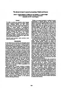
Figure 1. (A) Sample stimulus display of the main task of the fMRI study, consisting of two object images, each of which is overlaid with two texture features. Participants were asked to compare the presented objects and decide if they differed in terms of their texture. (B) Sample stimulus display with a texture-location difference between the two objects. On the left chair, the backrest area is covered with texture A and the seat area with texture B. On the right chair, the backrest area has the B texture and the seat area has the A texture. (C) Sample stimulus display with a texture-type difference between the two objects. On the left chair, the backrest area is covered with texture A and the seat area with texture B. On the right chair, the backrest area and seat are covered with textures C and D, which differ from A and B. D. All 22 objects used in Experiments 1 and 2.
2
Journal of Cognitive Neuroscience
Volume X, Number Y
surface texture to individual object parts. Using common, everyday objects composed of multiple parts as stimuli (Figure 1A), we contrasted the patterns of brain activation under two conditions: (1) texture-location, when participants detected differences in the assignment (binding) of the same surface features to different object parts (Figure 1B), and (2) texture-type, when participants detected differences in the texture features between two objects on the same within-object parts (Figure 1C). The texture-location and texture-type tasks were matched for difficulty in terms of accuracy (ACC) and RT and in the pattern and number of eye fixations. Consequently, the contributions of attentional load and/or difficulty of processing were minimized. In the subsequent psychophysical experiments, we investigated whether the assignment of surface texture to object parts interferes with visuospatial perception and whether whole-object or whole-shape level processing is required.
EXPERIMENT 1 Materials and Methods After independently localizing ROIs in dorsal and ventral cortex for each participant separately (described below), we explored the pattern of activity in response to texturelocation and texture-type differences within the localized ROIs as well as at the level of the whole brain. During this main task, participants viewed images of two objects presented simultaneously and indicated whether the objects were the same or different (Figure 1A). In separate blocks, the objects could be identical or could differ in either texture-location or texture-type. Participants Twenty-two healthy adults (15 women, 7 men, age range = 21–31) participated in the experiment. All were righthanded and had normal vision (corrected, if necessary). Informed consent was obtained from all participants under a protocol approved by the Institutional Review Board of the National Institute of Mental Health. Procedure The experiment, implemented using E-Prime 2.0, was run on a Windows 7-based PC. Stimuli were presented via an analog projector on a 240 × 180 mm screen (15° visual angle horizontally by 11° vertically at a distance of 92 cm away from the participants’ eyes), situated at the bore opening of the MRI scanner at a resolution of 1024 × 768 (0.02° per pixel), 1-msec RT. Participants viewed the screen through a mirror attached to the head coil of the scanner. Eye movement data were collected using an infrared technique (Avotec model RE-5701, Avotec, Inc., Stuart, FL). In a single 80-min scan, participants completed eight functional runs: The first four runs comprised the texture
and location localizer tasks (two runs per localizer), and the remaining four comprised the main texture-type and texture-location tasks. Before entering the scanner, the texture-type and texture-location tasks were titrated, separately for each participant, so as to be matched in terms of RT (msec) and ACC (% correct). Localizer Tasks Each run of the texture and location localizer tasks contained 14 blocks of trials (10 trials per block) in counterbalanced order. Participants were not required to maintain fixation during these trials. In both localizer tasks, participants viewed two images presented simultaneously on either side of the screen center for 1.7 sec. In half the trials of a block, the images appeared in a top left, bottom right configuration and in the remaining half, in a top right, bottom left configuration (in random order). The above presentation order was used to encourage participants to compare the objects instead of attending to particular locations of the display. In addition, visual information presented in the entire visual field yields stronger BOLD responses, which improves the signal to noise ratio. Each block of trials lasted 22 sec and was preceded and directly followed by 8 sec of fixation. Trials were separated by 300 msec of fixation. Texture Localizer The texture localizer, adapted from Cavina-Pratesi et al. (2010), compared activation evoked by a texture-matching task to that evoked by an object-matching task, thereby identifying brain regions more active in response to differences in texture compared to differences in the shape of objects. In separate blocks with counterbalanced order, the presented images depicted either gray-colored line drawings of object outlines (Figure 2A, D) or cubes overlaid with 1 of 22 types of surface texture (Figure 2B, C). Because the texture cubes are effectively object line drawings with surface texture applied on them, the line drawings of objects acted as a control to subtract out any activity that was not related to texture-processing per se, such as activity related to the shape and overall spatial position of the stimuli on the display. In half of the trials of each block, the two items differed (distinct object outlines or cubes overlaid with different textures), and participants indicated this difference by button press. Responses were withheld on matching trials. At the beginning of each block, a dummy trial consisting of two images with either a texture or an object difference between them (presented for 1.7 sec) informed participants of the task in the upcoming block. Location Localizer This task, adapted from Haxby et al. (1991), compared activation evoked by a distance-matching task (Figure 2E) with that evoked by a brightness-matching task (Figure 2F) Zachariou et al.
3
Figure 2. (A) Two sample object outline stimuli. (B) Two sample texture cube stimuli. (C) A series of three sample trials from a texture cube block of the texture localizer, with fixation interleaved. The last trial depicts different items, and a response is required. (D) A series of three sample trials from an object outline block of the texture localizer, with fixation interleaved. The last trial depicts different items and a response is required. (E) A series of three sample trials from a distancematching block of the location localizer, interleaved with fixation. The last trial depicts a distance mismatch between the ball and line across the two panels and a response is required. (F) A series of three sample trials from a brightnessmatching block of the location localizer, interleaved with fixation. The last trial depicts a brightness mismatch on the brightness of the ball across the two panels and a response is required.
to identify regions of the brain that process spatial relations between objects. The two tasks were presented in separate, counterbalanced blocks. In each trial, participants compared two panels (Figure 2E, F) and indicated via button press if the panels differed in distance or brightness. The distance- and brightness-matching tasks were visually identical: The display consisted of two panels, each containing a dot and a vertical black line. The panels always depicted the dot at opposite horizontal and vertical positions. In each trial, the distance between the dot and line was randomly drawn from a uniform distribution between 18 and 80 pixels. In addition, the brightness of the dot, on each trial, was randomly chosen to be one of eight brightness levels. Panels in identical trials in both distance- and brightness-matching tasks had the same dot-line distance and the same dot brightness.
4
Journal of Cognitive Neuroscience
In distance-matching blocks, participants compared the horizontal distance between the dot and the vertical line across the two panels. This horizontal distance differed in half of the trials of each block, and participants indicated detection of this difference by a button press (responses withheld on identical distance trials). A distance difference across panels was created by adding 18–30 pixels (drawn from a uniform distribution) to the original distance between the dot and line in one of the panels. In brightness-matching blocks, the horizontal distance between the dot and the vertical line was always identical across the two panels, and participants determined whether the brightness of the dot across the two panels was the same or different. In half of the trials in each block, the dots differed in brightness, and participants indicated this by a button press. Like the texture localizer
Volume X, Number Y
tasks, at the beginning of each block, a dummy trial with either a distance or brightness difference between the panels informed participants of the task in the upcoming block. The brightness-matching task, being visually identical to distance matching, was intended as a control task. More specifically, we used the brightness-matching task to subtract out any activity that was not related to spatial processing per se, such as activity related to the shape and overall spatial position of the stimuli on the display. Main Task On each trial, participants compared two objects, presented simultaneously on either side of the screen center and indicated with a button press if they differed in their surface (texture) features (Figure 1). Twenty-two familiar objects, some man-made, some natural, some manipulable, and some not (Figure 1D), acquired from wiki.cnbc. cmu.edu/TarrLab, were each rendered within a rectangle 5° high × 6° wide (average object size 5° visual angle). Across trials, the two object images appeared in one of two possible spatial configurations: In half the trials, objects appeared in a top left, bottom right configuration, and in the remaining half, in a top right, bottom left configuration, following the same procedure as the localizer tasks. Four different types of texture were used: brick, burlap, canvas, and sandstone (these textures were not used in the texture localizer task). For each of the four texturetypes, the scaling, intensity and direction of light were manipulated (using Photoshop) to generate random variants with equal contrast and luminosity. Two object parts were then chosen for each of the 22 objects and acted as placeholders for the four texture-types mentioned above. To create a texture-type difference, 1 of the 22 objects (randomly selected) was presented twice in the stimulus display. One instance of the object had two of the four texture-types assigned to its selected parts (one texturetype per object part), and the other instance of the object was assigned the remaining two texture-types. As a result, the two instances of the object in a texture-type difference trial had completely different texture-types between them. To create a texture-location difference, two instances of the same object (randomly selected) were also presented but both instances were assigned the exact same two texture-types (one texture-type per object part). However, the assignment of texture-type to object part on one of the two object instances was reversed in relation to the other instance of the object. Consequently, the two object instances in a texture-location difference trial had identical texture-types but bound to different within-object parts (the spatial position of the withinobject parts did not change, just the assignment of texturetype to object part). Before scanning, participants completed a training session that lasted 15–20 min. Over the course of training,
the running average RT for the two types of texture differences was compared, and the ratio of texture-type/ texture-location blocks was adjusted to allow more practice for the type of difference with the slower RT, until the absolute difference in RT was within 100 msec, indicating that performance on the two tasks was equated prior to the scan session. The RT of a training block was only included in the running average if ACC was above 90%; if ACC was below 90%, the training block repeated. In addition to RT and ACC, we also report inverse efficiency scores (IES, in msec), which equals the mean RT divided by the proportion of correct responses, calculated separately for each condition and each participant. Lower values on this measure indicate better performance (Christie & Klein, 1995; Akhtar & Enns, 1989; Townsend & Ashby, 1983). IES discount possible speed–ACC tradeoffs in performance and ensure that the two types of texture difference detection tasks are closely matched. Trials were blocked by task type (texture-type or texture-location). Critically, independent of condition, the same object images were presented at the same screen locations. Each block consisted of 10 trials (2.7 sec each), separated by 200 msec of fixation. A functional run of the main task consisted of twelve 32-sec blocks, each preceded and followed by 8 sec of fixation. At the beginning of each block, a dummy trial was presented consisting of a 2.8-sec display of two objects with the type of difference that would follow in the block (the objects presented in these dummy trials did not appear within the blocks). Participants were explicitly instructed to look for texture differences only. They were never told to determine the presence of each type of texture difference or how to determine texture-type or texture-location differences between objects. Participants were never given the labels “texture-type” and “texture-location” but were shown multiple examples of each of the two texture differences during the instruction section and during training. Participants were required to press a button if a difference was detected and to withhold a response otherwise. In each block, no difference was present in half the trials. fMRI Acquisition Participants were scanned in a General Electric MR750 3T scanner with a 32-channel head coil. Functional images were acquired with an EPI sequence (repetition time = 2 sec, echo time = 27 msec, flip angle 79°, 3.2 mm isotropic voxels, 72 × 72 matrix, field of view = 230 mm, 45 axial slices covering the whole brain). The 45 slices were acquired with in-plane acceleration, using the GE protocol ASSET (www.gehealthcare.com/usen/education/ tip_app/docs/fieldnotes_volume1-1_asset.pdf) with an acceleration factor of 2. An MPRAGE sequence (1-mm3 voxels; 176 slices, field of view 256 mm) was used for anatomical imaging and was acquired within the same scan session. Zachariou et al.
5
Functional scans were slice scan time-corrected, motioncorrected, coregistered to their constituent anatomical image, normalized to Talairach space (Talairach & Tournoux, 1988), smoothed with a Gaussian kernel of 6.0 mm FWHM, and mean-based intensity normalized (all volumes by the same factor) using AFNI (Cox, 1996). In addition, linear and nonlinear trends (where necessary) were removed during preprocessing of the data.
fMRI Statistical Analyses All imaging data were analyzed using AFNI (Cox, 1996). Data from the localizer and main task were analyzed using linear mixed-effects models (3dLME; Chen, Saad, Britton, Pine, & Cox, 2013). The resulting statistical maps were thresholded at a family-wise error-corrected α < 0.01 at p < .001, using the AFNI program 3dClustSim (afni.nimh. nih.gov/pub/dist/doc/program_help/3dClustSim.html). The motion parameters from the output of the volume registration step were regressed out in all AFNI analyses. Brain–behavior correlations were analyzed using linear mixed-effects models (3dLME; Chen et al., 2013), ANCOVAs using the AFNI program 3dttest++ (afni.nimh.nih.gov/pub/ dist/doc/program_help/3dttest++.html) and the SPSS software package. The resulting statistical maps were thresholded at a family-wise error-corrected α < 0.01 at p < .001. Behavioral data collected inside the scanner were analyzed using SPSS and a general linear mixed-effects model (with participants added as a random variable). Multiple comparisons used Sidak corrections where necessary. The eye-tracking data were analyzed using the Open Gaze And Mouse Analyzer package (OGAMA; Voßkühler, 2009). The following procedure was used to compare the pattern of eye fixations between texture-type and texturelocation tasks across trials. For each of the four on-screen locations where object images could appear (two locations per trial), nine areas of interest (AOIs) were constructed, covering the entire region occupied by an object image (36 AOIs in total). The width of all AOIs was 2° of visual angle and their height was 1.7°. An additional AOI with similar dimensions was placed at the center of the screen, at the same location where a fixation cross appeared between trials of the main task. The number of eye fixations, fixation duration and fixation duration divided by number of eye fixations (a measure of individual fixation duration) in each AOI, for each task type, were calculated for each participant. ANOVAs were then conducted separately for each dependent measure with task type and AOI (1–37) as factors.
Results Texture Localizer Statistical maps were created using the fMRI contrast of texture cubes > object outlines (Figure 3A). This contrast showed significant positive activations (stronger BOLD 6
Journal of Cognitive Neuroscience
response for texture cubes) bilaterally within the lingual gyrus, inferior and middle occipital gyrus, and posterior fusiform gyrus (bilateral Brodmann’s areas [BA] 17, 18, 19). A mask created from the activity of these regions was used as a ROI in the analysis of the main task data. The brain regions identified by the texture localizer are comparable with regions previously identified to be associated with texture processing (e.g., Cavina-Pratesi et al., 2010; Kastner, Weerd, & Ungerleider, 2000) with the exception that, in the left hemisphere, the pattern of activity in response to the texture cubes extended more laterally and farther into the lingual gyrus relative to these cited studies. To establish an additional, more inclusive ROI for texture processing that included more anterior regions of the ventral visual pathway, we ran the fMRI contrast of texture cubes > distance matching (Figure 3B), which revealed positive activations in bilateral lingual gyrus, inferior and middle occipital gyrus, and fusiform gyrus (bilateral BA 17–20 and 37). This contrast thus revealed a stronger BOLD response in those regions in response to differences in the type of texture compared to differences in location relative to a landmark, analogous to the functional segregation between the ventral and dorsal visual streams. We refer to this ROI as the extended ventral stream ROI. Location Localizer Statistical maps were created using the fMRI contrast of distance matching > brightness matching (Figure 3C). A single mask, which served as the dorsal ROI, was created from all positively activated regions: bilateral precuneus, superior and inferior parietal lobules (bilateral BA 7 and 40). Main Task: ROI-constrained Analyses fMRI activity evoked by each task type was explored by constraining linear mixed-effects model analyses (3dLME; Chen et al., 2013) within the localizer-identified ROIs (separate analyses performed for each localizer ROI). Statistical maps for task type were created using the contrast of texture-location versus texture-type, with positive BOLD activations corresponding to regions more active in response to texture-location and negative BOLD activations corresponding to regions more active in response to texture-type. Texture Localizer and Extended Ventral Stream ROIs We did not observe any significant BOLD responses in either the texture localizer or the extended ventral stream ROI (even at uncorrected p < .05), indicating that fMRI activation in regions within the ventral visual pathway (including the early visual areas associated with texture processing in previous studies and more anterior regions of the ventral visual pathway) did not differ between texture-type and texture-location trials (Figure 4A). Volume X, Number Y
Figure 3. (A) Cortical activation map (magnitude of activity; difference in beta-weight coefficients) revealed by the fMRI contrast of texture cubes > object outlines from the texture localizer. (B) Cortical activation map revealed by the fMRI contrast of texture cubes > distance-matching from the texture and location localizer tasks. (C) Cortical activation map revealed by the fMRI contrast of distance-matching > brightness-matching from the location localizer.
Zachariou et al.
7
To preclude the possibility that the absence of any significant BOLD activation was a result of overall poor brain signal within inferior temporal cortex, we ran additional analyses comparing texture-type and texture-location to baseline (the AFNI calculated baseline from the regression model), separately. There was substantial activation for each of the two texture detection tasks compared to baseline (α < 0.01), but both the magnitude and extent of the areas activated for each task were comparable. In addition, overall BOLD activity for the texture tasks in relation to baseline, within the ventral stream ROIs, was marginally stronger compared to the dorsal ROI (α < 0.06). This latter ROI effect could be explained by differences in signal-tonoise ratio, magnetic field inhomogeneity, physiological noise (pulsation), or simply because textured objects are slightly better at driving the inferior temporal cortex com-
pared to the PPC; the absence of a difference between texture-type and texture-location within the ventral ROIs, however, cannot be attributed to poor brain signal. Location Localizer ROI Within the location localizer ROI, texture-location difference detections produced stronger BOLD responses compared to texture-type difference detections within bilateral precuneus, bilateral superior parietal lobule, and right inferior parietal lobule (bilateral BA 7 and right BA 40; Figure 4A). The peak of activity in response to texturelocation difference detections, in relation to texture-type, occurred within the right precuneus (Talairach coordinates: X: 14, Y: −73, Z: 53, p = .0001, α < 0.001). The cluster of activity (size: 70 voxels) corresponding to the above peak
Figure 4. (A) Cortical activation maps (magnitude of activity; difference in beta-weight coefficients) revealed by the fMRI contrast of texturelocation > texture-type, constrained within the localizer ROIs. (B) Cortical activation maps revealed by the fMRI contrast of texture-location > texture-type, at the level of the whole brain. In A and B, positive activations (yellow– orange) correspond to regions more active for texture-location difference detections compared to texture-type difference detections. Negative activations (cyan–blue) correspond to regions more active for texturetype compared to texturelocation difference detections. The cyan outlines illustrate the brain regions that comprise the location localizer ROI, the yellow outlines illustrate the ROI identified by the fMRI contrast of texture cubes > distance matching (the extended ventral stream ROI), and the green outlines illustrate the texture localizer ROI.
8
Journal of Cognitive Neuroscience
Volume X, Number Y
has been associated with visuospatial perception (Sugio et al., 1999) and visuospatial attention (Silver, Ress, & Heeger, 2005). The findings of the dorsal stream ROI analysis indicate that spatial processing regions within PPC (as indicated by the distance matching task used in the location localizer) are also engaged when processing the assignment of texture to within-object parts. Main Task: Whole-brain Analysis An examination of the pattern of activity across the entire cortex was conducted using the same contrast of texturelocation > texture-type. Significant positive differences in BOLD activation were observed within bilateral precuneus, superior parietal lobule, right inferior parietal lobule, left middle temporal gyrus, and left inferior temporal gyrus (bilateral BA 7, right BA 40 and left BA 19 and 37; Figure 4B). The posterior parietal regions of activation overlapped substantially with the parietal regions observed in the location localizer ROI. The activations observed within the left middle and inferior temporal gyri overlapped partially with the activity observed in the texture localizer, namely BOLD activity stronger in response to the object outlines in relation to the texture cubes (shown in blue/negative activation on Figure 3A). To further explore this partial overlap, we used the texture localizer data to define an additional ROI, corresponding to activity stronger for object outlines in comparison to the texture cubes. We then conducted a linear mixed-effects model analysis of the main task (similar to the previous ROI analyses) constrained within this newly defined ROI. The above ROI analysis, however, did not yield any significant results, indicating that the texture-location-related activity within the middle and inferior temporal gyri is not robustly represented within the regions identified by our localizer tasks. At the level of the whole brain, we did not observe any brain regions more active in response to texture-type difference detections compared to texture-location difference detections. Brain–Behavior Correlation Analyses In both the ROI-constrained and whole-brain analyses, texture-location difference detections led to significant BOLD activation within the dorsal visual pathway. In addition, at the level of the whole brain, texture-location difference detections activated regions within the left middle temporal and inferior temporal gyri. To explore the functional contribution of these regions, we correlated the activity from the ROI-constrained and wholebrain analyses with behavioral performance using RT (msec) as the behavioral measure. We chose RT as it had the greatest variability among our behavioral measures and was therefore a good candidate for a correlation analysis. The linear mixed model used (see Methods) was constructed using one factor and one covariate: brain activity (beta-weight coefficients) for task type (texture-location
and texture-type) was the factor and RT for each task type was the covariate. The model was then run, testing for both main effects and any possible interactions between the factor and covariate. This analysis yielded no significant brain–behavior correlations at the level of the whole brain. When we repeated the analysis within each of the ROIs obtained from the localizer tasks, no significant brain activity–behavior correlations were found within the ventral stream ROIs (texture localizer ROI and extended ventral stream ROI). However, there was a significant fMRI–RT interaction within the right PPC (within the location localizer ROI; Figure 5A): The correlation between fMRI activation (betaweight coefficients) for texture-location and RT was stronger in the right precuneus and superior and inferior parietal lobules compared to the correlation between activity for texture-type (beta-weight coefficients) and RT (this interaction was significant in right BA 7 and 40). No other significant correlations were found. To unpack the significant interaction described above, we ran two additional ANCOVA analyses (using the AFNI program 3dttest++). The input to the first ANCOVA was magnitude of activation (expressed in difference between two sets of beta-weight coefficients per voxel) from the fMRI contrast of texture-location > texture-type (separately for each participant) and the covariate: RT for texture-location (Figure 5B). The second ANCOVA had the same brain activity input as the first but RT for texture-type was used as the covariate. Note that the fMRI contrast of texture-location > texture-type is bidirectional, including BOLD activations from both texture-location difference detections (positive if significant) and texture-type difference detections (negative if significant). Consequently, the covariate in each of the above two ANCOVAs had the potential to correlate with activity from either side of the contrast of texture-location > texture-type. As expected, the only significant correlation observed was within the right PPC (within the location localizer ROI) in the same anatomical regions we identified for the linear mixed effects model analysis above. The ANCOVA was only significant for the correlation between BOLD activation from the fMRI contrast of texture-location > texturetype and RT for texture-location detections (r of most significant voxel = .5, within the right precuneus; Figure 5B). Activity within the dorsal visual pathway for texture-location difference detections predicted behavioral performance, such that participants with greater activation within the location localizer ROI were slower at detecting texturelocation differences. The above finding is consistent with previous literature showing that longer RTs correlate with larger magnitude, task-related hemodynamic responses (Domagalik, Beldzik, Oginska, Marek, & Fafrowicz, 2014). Two additional correlation analyses were also performed using average activity per participant from the group level, location localizer ROI and are summarized in Figure 5C. The first correlation analysis is between average activity from the fMRI contrast texture-location > texture-type (difference Zachariou et al.
9
Figure 5. (A) Correlation maps revealed by the interaction term in the linear mixed model contrasting the brain activity– behavior correlations between the two texture-type tasks. Separate analyses were run within each of the localizer ROIs. Positive activations (yellow–orange) correspond to regions, within a localized ROI, where the correlation between brain activity (beta-weight coefficients) and RT (msec) was stronger for texture-location compared to texture-type difference detections (the unit is difference in r values: the difference in the correlation between texture-location and RT minus the correlation between texture-type and RT). Negative activations (cyan–blue) correspond to regions, within a localized ROI, where the correlation between brain activity (beta-weight coefficients) and RT (msec) was stronger for texture-type compared to texture-location difference detections. (B) Correlation maps of brain activity from the fMRI contrast of texture-location > texturetype (difference in beta-weight coefficients) correlated with RT (msec) for texture-location. No significant brain activity– behavior correlation was observed between the same activity and RT (msec) for texture-type. Warm colors indicate positive brain activity– behavior correlations, whereas cool colors indicate negative correlations. (C) Summary of B with average activity (from the fMRI contrast texture-location > texture-type) extracted separately for each participant using the group level distance estimation localizer ROIs and correlated with RT (msec). The left panel depicts the correlation with RT (msec) for texture-location change detections, and the right panel depicts the correlation with RT (msec) for texture-type change detections.
in beta-weight coefficients), extracted separately for each participant (using the group level distance estimation localizer ROI as a mask) and RT for texture-location change detections. The same brain activity was used for the second analysis but was correlated with RT for texture-type change detection. Parallel to the previous findings, the only significant correlation was between the average brain activity from the fMRI contrast texture-location > texture-type and RT for texture-location change detections (r 2 = .27, p < .01). Lastly, we correlated the brain activity (the beta-weight coefficients) for texture-location and constituent RT and, separately, the brain activity for texture-type and constituent RT within the group level, location localizer ROI. This analysis is similar to the previous correlation analy10
Journal of Cognitive Neuroscience
ses but explores the correlations separately for each texture task and RT instead of using the activity obtained from the fMRI contrast of texture-location > texture-type. Consistent with the previous analyses, the correlation between the activity for texture-location and constituent RT was significant ( p = .01, r2 = .26) with slower RTs predicting greater BOLD activity. In contrast, the correlation between texture-type and constituent RT was not significant ( p = .56, r2 = .01). Main Task: Behavioral Performance and Pattern of Eye Fixations To ensure that the performance and pattern of eye movements (which correlate with shifts of attention) were Volume X, Number Y
comparable across the two texture tasks, we conducted ANOVAs (separately for RT, ACC, and IES) with task type (texture-type vs. texture-location) as the sole factor. The results showed that task type was not significant in any analysis (RT: F(1, 42) = 0.12, p = .73; 1234 msec for texture-type vs. 1251 msec for texture-location; ACC: F(1, 42) = 2.45, p = .12; 95% ACC for texture-type vs. 93% ACC for texture-location; IES: F(1, 42) = 0.57, p = .45; 1306 msec for texture-type vs. 1352 msec for texturelocation), confirming that behavioral performance was matched across the two texture tasks. In the analyses of the pattern of eye fixations, all dependent measures yielded a significant main effect of AOI ( p < .001) but not of task type ( p = .30), and there were no significant interactions between the factors ( p = .64). The main effect of AOI showed that participants, irrespective of task type, mainly fixated mostly the center of each object image (vs. other parts of the object). Since task type and the interaction term between task type and AOI were not significant, the data as a whole indicate that participants fixated the same within-object locations for approximately the same amount of time, regardless of the task performed. Participants were interviewed immediately after their scan session and asked to describe how the textures differed between objects during the scan. All participants indicated that they observed two distinct types of texture differences during the scans. When asked to label the differences they most often referred to texture-type as “changes in texture” or “the objects had different textures” and texture-location as “in some trials the textures were at different parts of the objects.”
EXPERIMENT 2 Materials and Methods The results of the fMRI study showed that spatial processing and the mapping of surface (texture) features to within-object parts activate common dorsal stream brain regions, suggesting that these two processes might depend on the same neural mechanisms. To test this interpretation, we investigated whether spatial judgments about objects would be adversely affected by task-irrelevant distracters that differed in the binding of texture features to within-object parts (texture-location), but not by distracters that differed in the textures themselves (texture-type). A preferential decrement in object location performance due to texture-location differences is taken to indicate common processing mechanisms for localizing objects and textures within them.
Participants Twenty-four right-handed undergraduate Carnegie Mellon University students (13 women, 11 men, age range = 20– 24) with normal vision (or corrected to normal) received
course credit for participation. None of these participants completed Experiment 1. Informed consent was obtained from all participants, and the Institutional Review Board of Carnegie Mellon University approved all procedures. Procedure The experiment, implemented using E-Prime 2.0, was run on a Windows XP-based PC with a 22-in screen (33.4° horizontal × 22° vertical, at a distance of 80 cm from the participants’ eyes), 3-msec RT computer display at a resolution of 1680 × 1050 pixels (0.02° per pixel). On each trial, two side-by-side panels were shown, one to the left and the other to the right of the screen center. Each panel comprised two different objects separated by a horizontal reference line (see Figure 6A). The horizontal reference line in each panel was 4° wide and positioned so that its midpoint fell 6° from the horizontal center of the screen. The same 22 objects used in Experiment 1 were also used in Experiment 2. Each was rendered within a rectangle at a visual angle of 3° high × 3.7° wide (average object size about 2° visual angle). Objects in the same row (either top right, top left or bottom right, bottom left across panels) denoted corresponding objects, could be the same or differ with respect to their texture (either texture-type or texture-location) and/or their spatial distance from the reference line (closer or farther relative to the reference line, see examples in Figure 6B–E). If both a texture and a distance difference were present across panels, they occurred on distinct objects (i.e., corresponding objects could not differ with respect to both texture and distance). When a distance difference between the two panels was present, it could be either easy or difficult to detect. The ease/difficulty of distance judgments was controlled by varying the magnitude of the difference across the panels of a stimulus display, with respect to the distance between an object and the horizontal line.1 A vertical distance difference of 0.5° was used for the easy distance discriminations and a difference of 0.4° for the difficult distance discriminations. Participants pressed two different buttons (using different fingers for each) to indicate that a difference was present or that the panels were identical. The experiment was split into two separate sections. In Section 1, participants reported via button press whether the corresponding objects across panels were the same or different in distance from the reference line. In Section 2, they reported whether the objects differed in texture; such differences could be in texture-type or texture-location. Sections 1 and 2 were divided by a 2-min break. Participants were not informed that differences on the task-irrelevant dimension (texture in Section 1, distance in Section 2) could occur. Separation of the tasks in this way was intended to induce the use of processing mechanisms specific to the instruction, making differences on the alternative dimension clearly orthogonal to Zachariou et al.
11
Figure 6. (A) Sample stimulus display of Experiment 2. Each panel contained two objects separated by a black horizontal line. The rows of the two panels always matched with respect to the shape of the objects depicted, although the corresponding objects themselves could differ in terms of their texture features. (B) Stimulus display with one distance difference between the object pairs. A distance difference, if present, could be either easy or difficult to detect. (C) Stimulus display with a texture-location difference between the object pairs. (D) Stimulus display with a texture-type difference between the object pairs. (E) Stimulus display with two differences between the pairs, one in texture (texture-location depicted) and one in distance relative to the reference line. (F) Enlarged texture-location and texture-type differences. Gray brackets in the figure are used to highlight the corresponding objects with a difference between them.
the participant’s task. The fixed ordering of sections was used so that participants would be naive with respect to the texture difference distracters when processing spatial distance. Within each section, trials were blocked by type of texture difference (texture-type or texture-location); only one type of texture difference was present within a block and the order of the blocks was counterbalanced across participants. Within each block, texture difference trials occurred equally as often as spatial distance and nodifference trials. Before the experiment, participants completed a training session under instructions to detect distance differences, using all 22 objects without texture differences. If more than six errors occurred during the training, it was repeated. Psychophysical Experiment Statistical Analyses The design of Experiment 2 was within subjects with two independent variables: rule and task type. The rule variable comprised two levels, corresponding to the categories of difference detection, namely, distance and texture. The instruction to detect distance differences in Section 1 will be referred to as “Rdistance” (R stands for rule), and the instruction to detect texture differences used in Section 2 will be referred to as “Rtexture.” The task-type variable consisted of five levels, corresponding to the type of difference that could be present in each trial: easy dis12
Journal of Cognitive Neuroscience
tance difference, difficult distance difference, texture-type difference, texture-location difference and no difference. These five levels of the task-type variable were nested within the rule variable, occurring in both the Rdistance and Rtexture levels. The main analysis focused on data from Section 1, under Rdistance, when participants were detecting distance differences and texture differences acted as distracters. Data from Section 2, using Rtexture (detecting texture differences), are reported to confirm that the texture-type and texture-location tasks were equally demanding in terms of RT and ACC and to explore whether the distracter effects are bidirectional, that is, whether distance differences, as distracters, affect texture difference detection performance. Repeated-measures general linear mixed-effects models (with participants added as a random variable) were used to analyze all data, with p < .05, unless adjustments for multiple comparisons were needed. All multiple comparisons used Sidak corrections. The dependent variables of Experiment 2 were RT (msec), ACC (% correct), and IES (msec).
Results Effects of Texture-type and Texture-location Distracters under Rdistance This analysis evaluated the effects of texture-type and texture-location distracters in Section 1, when participants Volume X, Number Y
performed spatial distance judgments and texture differences acted as distracters. The analysis considered trials with distracter differences (texture differences) or no differences (identical trials). The data are shown in Figure 7. The analysis included task type as the sole factor with levels corresponding to no difference, a texturetype difference, or a texture-location difference. Note that correct responses for trials with distracter differences present required participants to press the button corresponding to no differences (identical trials), indicating that no distance differences existed across the panels of the stimulus display. Consequently, the comparison of the participants’ responses between identical trials (no difference trials) and trials with distracter differences present acted as a measure of distracter interference effectiveness. The effect of task type was not significant for ACC (F(2, 1896) = 2.26, p = .11; Figure 7A) but was significant for RT (F(2, 1646) = 4.18, p = .02; partial η 2 = 0.16; Figure 7B) and IES (F(2, 46) = 5.75, p = .01; partial η2 = 0.2). Pairwise comparisons indicated that participants were slower to respond on trials with a texture-location distracter difference (IES: 2683 msec) than on trials with either a texture-type distracter (IES: 2367 msec) difference (RT: p = .04; IES: p = .04) or no difference (RT: p = .03; IES: p < .01, IES: 2342 msec). The comparison of texturetype distracter trials with no difference trials was not significant (RT: p = .53; IES: p = .72; Figure 7B). The data thus indicate that texture-location distracter differences interfered with the distance judgments. This was not true
for texture-type distracter differences, which led to RTs and IES essentially equal to those on no difference trials, indicating that texture-type distracter differences did not affect distance judgments. Analysis of Trials with Texture Differences Only under Rtexture: Matching Texture-type and Texture-location Differences in Terms of RT, ACC and IES To test whether the RTs and accuracies for processing texture-type and texture-location were equally demanding, as in the fMRI experiment, an analysis with task type as a factor was conducted comparing performance on Section 2/Rtexture trials, when participants were looking for texture differences as targets. Trials with a single texture-location difference or a single texture-type difference were used for the analysis (trials with distracter location differences were not included). The effect of task type was not significant for ACC (F(1, 971) = 0.45, p = .50; Figure 7C), RT (F(1, 733) = 1.38, p = .24; Figure 7D), or IES (3174 msec for texture-type vs. 3025 msec for texture-location; IES F(1, 46) = 0.67, p = .52), confirming that performance for detecting texture-type and texture-location differences was matched. Effects of Distance Distracters under Rtexture (Section 2) This analysis evaluated the effect of distance distracters in Section 2, in which participants were required to look for
Figure 7. (A) ACC (% correct) and (B) RT (in msec) for Section 1 trials of Experiment 2 with a texture-type distracter difference, a texture-location distracter difference, or no difference (identical pairs) between the panels. In Section 1 of Experiment 2, participants performed spatial distance judgments (distance of objects relative to a reference line) and texture differences between objects acted as distracters. C and D depict ACC and RT, respectively, for Section 2 trials of Experiment 2 where differences in spatial distance (labeled “Location” on the graphs) acted as distracters and texture differences were the target. The error bars denote ±1 SE.
Zachariou et al.
13
texture differences between the objects. The effect of the distance distracters was evaluated on trials with no texture differences present (i.e., trials with a single distracter distance difference). The analysis included the factor task type with three levels: no difference, an easy distance difference, and a difficult distance difference. Similar to the texture distracter analysis above, correct responses required participants to press the button corresponding to no difference (identical trials), indicating that no texture difference was present on the target attribute across the panels of the stimulus. In the analyses, for ACC, RT (Figure 7C, D), and IES, the effect of task type was not significant (RT: F(2, 1936) = 1.56, p = .21; ACC: F(2, 1837) = 0.1, p = .91; IES: F(2, 46) = 0.74, p = .48), indicating that distance distracters at either level of difficulty had no effect on ACC, IES, or the latency to match targets in texture.
EXPERIMENT 3 Materials and Methods In the fMRI study, we showed that spatial processing regions within PPC are involved in the within-object assignment of surface texture to individual object parts. In the behavioral experiment, we demonstrated that task-irrelevant differences in the assignment of surface features (texture) to different parts (texture-location differences) within an object affect performance on object location judgments, as expressed in the form of a distance estimation task. Experiments 1 and 2, however, do not provide evidence of whether the effects observed are object-specific per se or can occur when the same differences are introduced across random shape parts at distinct spatial locations. We explored the above under two conditions: (i) when shape parts interlocked to form unified shapes (whole-shape condition, emulating whole, complex objects) or (ii) when the same shape parts appeared separated at distinct spatial locations (parts condition; Figure 8). In both the whole-shape and parts conditions, texture-type and texture-location differences occurred on the same shape parts at the same spatial positions (see details below). If the interference effect observed in Experiment 2 is specific to mapping surface texture within objects, then texture-location should interfere with location judgments only in the whole-shape condition and not in the parts condition.
Participants Twenty healthy adults (9 women, 11 men, age range = 22–37) who did not participate in the previous experiments participated in Experiment 3. All were right-handed and had normal vision (corrected, if necessary). Informed consent was obtained from all participants under a proto14
Journal of Cognitive Neuroscience
col approved by the Institutional Review Board of the National Institute of Mental Health. Procedure Experiment 3 was run on the same equipment as Experiment 2 and followed an almost identical procedure: On each trial, two side-by-side panels were shown on either side of the screen center. Each panel comprised two different shapes (in the whole-shape condition) or two different groups of shape parts (in the parts condition) separated by a horizontal reference line (see Figure 8A). The size of the stimuli was comparable to that in Experiment 2 (average shape size about 2° visual angle). Using randomly generated, interlocking polyominoes (Golomb, 1994), we created 18 shapes, consisting of a minimum of three to a maximum of five polyominoes (Figure 8E). The 18 shapes comprised the whole-shape condition of the experiment. The parts condition was created as follows: first, two noncontiguous polyominoes from each of the 18 shapes were chosen to remain fixed in terms of their spatial position. Then, the remaining polyominoes from each of the 18 shapes were moved slightly (either up/down or left/right) so they would not interlock, thus breaking up each whole shape into its constituent parts. As a result, two polyominoes always occupied the exact same spatial position in both the whole-shape and parts condition, but in the parts condition they appeared as spatially separated (Figure 8C, D). The same four different types of texture used in Experiments 1 and 2 were used here to create the two types of texture differences. The two fixed-position polyominoes from each of the 18 shapes acted as placeholders for the four texture-types above. The remaining polyominoes from each shape were all overlaid with the same surface texture, one that was different from the four textures above and never changed. Texture-type and texturelocation differences were created using the same procedure as Experiments 1 and 2 and always occupied the fixed-position polyominoes of each shape (Figure 8C, D). A distance difference between two panels was created in the same way as Experiment 2. A vertical distance difference of 0.4° across panels was used for the distance discriminations. Participants pressed two different buttons (using the same finger) to indicate that a difference was present or that the panels were identical. The experiment was split into the same two sections as Experiment 2. However, in Section 2 (when participants performed texture difference judgments), no distance differences were present because they were found to be ineffective as distracters in the previous experiment; participants were only presented with texture difference and no difference trials. Participants were not informed that differences on the task-irrelevant dimension (e.g., texture in Section 1) would occur. Before the experiment, participants completed a training session under instructions to detect distance differences, Volume X, Number Y
Figure 8. (A) Sample stimulus displays from Experiment 3. The left panel illustrates a sample whole-shape trial, and the right panel illustrates a sample parts trial. (B) Sample stimulus displays (left, whole shapes; right, parts condition) with one distance difference between the shape pairs. (C) A sample texture-type difference between two shapes, illustrated both in whole-shape and parts configuration. (D) A sample texture-location difference between two shapes, illustrated both in whole-shape and parts configuration. C and D, which appear within the stimulus displays shown in A and B, are depicted enlarged here for clarity. (E) A sample of six shapes (out of 18) together with their constituent parts configurations used in Experiment 3.
using all 18 whole shapes and groups of parts without texture differences. If more than six errors occurred during training, it was repeated.
Statistical Analyses Experiment 3 used the same statistical analyses as Experiment 2.
Results Effects of Texture-type and Texture-location Distracters under Rdistance This analysis evaluated the effects of texture-type and texture-location distracters in Section 1, when participants performed spatial distance judgments and texture differences acted as distracters. For this analysis, we considered the scenario where participants had to indicate that the panels were identical with respect to distance but single distracter texture differences were present as distracters. The results are shown in Figure 9. The analysis included task type and whole shapes versus parts as
factors. For task type, we only included the levels corresponding to no difference, a texture-type difference, or a texture-location difference. Note that correct responses for trials with distracter differences present required participants to press the button corresponding to no difference (identical trials), indicating that no distance differences existed across the panels of the stimulus display. Consequently, the comparison of the participants’ responses between identical trials (no difference trials) and trials with distracter differences present acted as a measure of distracter interference effectiveness. A postexperiment interview indicated that all participants were aware of texture difference distracters between the whole shapes and parts while they performed spatial judgments but could not differentiate between the two texture conditions (texture-location and texture-type). The effects of task type and whole shapes versus parts were not significant for ACC (task type: F(2, 5388) = 2.36, p = .10; whole shapes vs. parts: F(1, 5388) = 2.4, p = .13; Figure 9A), and there were no interactions between the factors (F(2, 5388) = 0.22, p = .80). In terms of RT and IES, the effects of task type and whole shapes versus parts were also not significant (for RT, task type: F(2, 4974) = 2.57, Zachariou et al.
15
p = .08; whole shapes vs. parts: F(1, 4974) = 0.16, p = .69; For IES, task type: F(2, 42) = 5.85, p = .85; whole shapes vs. parts: F(1, 21) = 3.62, p = .64), but there was a significant interaction between the factors (for RT, F(2, 4974) = 3.74, p = .03; partial η2 = 0.13; Figure 9B; For IES: F(2, 42) = 9.0, p < .01; partial η2 = 0.22). To explore the significant interaction, we considered the effect of task type separately for the whole-shape and parts conditions. For the wholeshape condition, task type was significant for both RT and IES (RT: F(2, 2479) = 9.9, p < .01; partial η2 = 0.17; IES: F(2, 42) = 7.18, p < .01; partial η2 = 0.26). Pairwise comparisons between the levels of task type indicated that participants were slower to respond on trials with a texturelocation distracter difference compared to trials with either a texture-type distracter difference ( p = .01) or no difference ( p = .03). The comparison of texture-type distracter trials with no difference trials was not significant ( p = .94; Figure 9B). In contrast to the findings in the whole-shape condition, the effect of task type was not significant in the parts condition for either RT or IES (RT: F(2, 2474) = 0.47, p = .63; IES: F(2, 42) = 1.26, p = .29). In summary, the interference effect of texture-location differences on distance estimation judgments was only present in the whole-shape condition and completely absent when the shape was split up into its constituent parts. As in Experiment 2, texture-type differences did not affect distance estimation judgments. Analysis of Trials with Location Differences Only: Matching the Whole-shape and Parts Conditions in Terms of RT, ACC, and IES To test whether distance estimation judgments were equally demanding between the whole-shape and parts
conditions (in terms of RT, ACC, and IES), an analysis with task type and whole shapes versus parts as factors was conducted, comparing trials in Section 1 with either distance differences or no differences between the panels. Task type was the only significant factor in the analysis of ACC and RT (ACC: F(1, 5394) = 54, p < .01; RT: F(1, 4780) = 36, p < .01). Whole shapes versus parts was not significant (ACC: F(1, 5394) = 1.0, p = .32; RT: F(1, 4780) = 2.5, p = .10), and there were no significant interactions between the two factors (ACC: F(1, 5394) = 0.1, p = .75; RT: F(1, 4780) = 0.2, p = .65). In the analysis of IES, both task type (F(1, 21) = 3.64, p = .07) and whole shapes versus parts (F(1, 21) = 1.51, p = .23) were not significant, and there were no significant interactions between the two factors (F(1, 21) = 1.62, p = .22). Irrespective of the whole-shape versus parts condition, ACC for no difference trials was significantly higher (93%) compared to trials with a distance difference (84%). In terms of RT, participants were slower to respond in trials with no differences between the panels (2420 msec) compared to trials with a distance difference present (1945 msec). Analysis of Trials with Texture Differences Only: Matching Texture-type and Texture-location Differences in Terms of RT and ACC To test whether the RTs and accuracies for processing texture-type and texture-location were equally demanding, an analysis with task type and whole shapes versus parts as factors was conducted, comparing performance on Section 2/Rtexture trials, when participants were looking for texture differences as targets (no distracters were
Figure 9. (A) ACC (% correct) and (B) RT (in msec) for Section 1 trials of Experiment 3 with a texture-type distracter difference, a texture-location distracter difference, or no differences (identical). In Section 1, participants performed spatial distance judgments and texture differences between shapes acted as distracters. C and D depict ACC (% correct) and RT (in msec), respectively, for Section 2 trials of Experiment 3. In Section 2, texture differences were always targets, and there were no distracter differences present. The error bars denote ±1 SE.
16
Journal of Cognitive Neuroscience
Volume X, Number Y
present). The effect of task type was significant for ACC (96% correct for texture-type vs. 90% correct for texturelocation; F(1, 2725) = 28, p < .01), and there was also a marginally significant interaction between the factors (F(1, 2725) = 3.9, p = .05; Figure 9C). The main effect of whole shapes versus parts was not significant (F(1, 2725) = 0.00, p = .99). To explore the marginally significant interaction, we considered the effect of task type, separately for the whole-shape and parts conditions. In the whole-shape condition, task type approached statistical significance (F(1, 1355) = 3.2, p = .08): texture-type 95% accurate compared to texturelocation 91% accurate. Task type was significant under the parts condition (F(1, 1349) = 42, p < .01) with texture-type being more accurate (97% accurate) compared to texture-location (88% accurate). The summary of the ACC analyses was as follows. In both the wholeshape and parts conditions, texture-type differences were easier to detect in terms of ACC compared to texturelocation differences. The significant interaction indicates that the magnitude of the difference in ACC between the two texture tasks was significantly larger in the parts condition. In the analysis of RT, the task type and whole-shape versus parts factors were not significant (F(1, 2519) = 0.86, p = .36; F(1, 2519) = 0.72, p = .4), and there were no significant interactions between the factors (F(1, 2519) = 0.16, p = .7; Figure 9D). Because of the significant main effects and interaction in the analysis of ACC, the effects of ACC on RT had to be accounted for and any possible speed ACC tradeoffs addressed. Here, to evaluate the effects of ACC on RT, we repeated the analysis using IES (msec) as the dependent measure. The above analysis yielded neither significant main effects: task type (F(1, 21) = 0.06, p = .80), whole shapes versus parts (F(1, 21) = 2.54, p = .16), nor significant interactions between the factors (F(1, 21) = 0.07, p = .79). Consequently, when we accounted for the effects of ACC on RT, texture-type and texture-location differences appear to have been matched as targets for both whole shapes and parts.
DISCUSSION Many categories of objects consist of multiple parts with unique surface features. Can spatial processing mechanisms, within the dorsal visual stream, mediate the mapping of discrete surface textures to different parts of an object? We explored the above question in fMRI and psychophysical experiments, within the context of novel difference detection tasks. In Experiment, 1 we demonstrated that regions within the dorsal visual pathway, specifically the localized regions active during distance estimation discriminations (namely, bilateral precuneus, bilateral superior parietal lobule, and right inferior parietal lobule), were also more active in response to reassignments of texture to within-
object parts (texture-location) in comparison to changes in the type of texture (texture-type). In addition, the magnitude of the activity related to texture-location detections, within the dorsal pathway, predicted the participants’ behavioral performance, such that greater activation predicted slower RTs. In contrast, the magnitude of activation in localized regions of the ventral visual pathway, regions identified here and previously to be involved in texture processing (e.g. Cavina-Pratesi et al., 2010; Kastner et al., 2000), was equivalent for differences detected in the type of texture features and in the assignment of these textures to within-object parts. In Experiment 1, the participants’ instructions were to look for any texture differences between stimuli, in essence forcing them to perform same–different judgments. Therefore, the detection of two texture difference types was not essential for the task. Hence, a possible easy strategy might have been to ignore the two types of differences altogether and treat all differences as texture-type (a qualitative difference in texture). However, the postexperiment interview clearly indicated that participants were sensitive to both types of texture differences during the scans and perceived texture-location as being qualitatively different from texture-type. Experiment 2 extended the findings of the fMRI study by showing that changes in the assignment of texture to within-object parts (texture-location) interfered with the detection of spatial distance relative to a reference line. In contrast, spatial processing was unaffected by differences in texture, per se. Finally, in Experiment 3, we demonstrated that this asymmetric interference pattern between texture-type and texture-location distracters during distance matching only occurred when whole shapes were presented and not when the same changes in texture assignment occurred on separated shape parts at distinct spatial locations. This result implicates object level processing as a basis for the interference. Given that detecting differences in the type and withinobject assignment of texture features were matched for difficulty and that the visual display and pattern of eye fixations were identical for both tasks, one cannot attribute the results to differential task demands or eye movements. The observed activation pattern is therefore task-specific, arising in the service of monitoring the within-object assignment of surface features to object parts. Interestingly, reassigning surface texture to different within-object parts is not considered a traditional dorsal stream function: Neither the spatial location of the object nor the spatial configuration of its edge-based constituent parts changed. More importantly, as observed in Experiment 3, texture-location differences did not interfere with location judgments when they occurred between disconnected shape parts. The underlying process thus appears to involve a within-shape or within-object Zachariou et al.
17
component used in monitoring separate surface texture features and their assignment to distinct edge-based parts of a single object. This within-shape spatial component appears to be mediated by spatial processing mechanisms within the dorsal stream, presumably providing the within-object spatial information necessary for this process. Another notable finding of Experiment 3 is that the asymmetric interference effects between texture-location and texture-type were observed without the participants’ awareness of two distinct types of texture differences, as reflected in the postexperiment interview. Consequently the findings are less likely to reflect different behavioral strategies used by the participants during the distracter conditions and more likely represent the underlying cognitive function of how participants processed the two types of texture differences: in the unified shape condition, participants had to attend the shapes, as unified wholes, in order to perform the spatial judgments. Hence, texture-location differences occurred in relation to whole objects or shapes. In the split shape condition, participants were unlikely to attend all constituent shape parts at once or to unify them perceptually into shapes. Instead, we think the participants treated the spilt parts as individual shapes. Consequently, because the spatial position of the individual shape components remained unchanged, texture-location was effectively transformed into texture-type: the shape parts only differed between them in the type of texture. Earlier studies investigating the assignment of surface features to individual simple shapes using illusory conjunction and conjunction search paradigms provide evidence that surface feature to shape binding is mediated by parietal lobe mechanisms (Baumgartner et al., 2013; Humphreys, Cinel, Wolfe, Olson, & Klempen, 2000; Friedman-Hill et al., 1995; Treisman & Gelade, 1980). Our experiments complement and extend these findings by showing that the feature binding effects observed with the simple stimuli used in illusory conjunction and conjunction search experiments extend to the binding of multiple surface features to individual parts within complex objects. Also, consistent with previous literature, our findings indicate that parietal lobe mechanisms mediate this mapping of surface texture to within-object parts. Additionally, our results demonstrate the dependence of this mapping on the same neural substrates that process the location of individual objects in space as changes in the mapping of surface texture to object parts interfered specifically with location judgments whereas changes in the type of texture did not. Role of Attention Certain regions within the PPC that were more active in response to texture-location relative to texture-type have been associated with visuospatial attention (Silver et al., 18
Journal of Cognitive Neuroscience
2005). Could attention-related mechanisms, instead of spatial processing mechanisms, account for our results? An attention-based hypothesis is appealing, but there are certain aspects of our findings that are difficult to account for using an attention-based approach. Such an approach would imply that our finding of stronger BOLD responses within PPC evoked by texture-location relative to texture-type difference detections is due to a greater attentional load for the former task. But this runs counter to our data indicating that the two tasks were matched in difficulty and the attention maps obtained from the eye-tracking data were identical between the two tasks. In addition, an attention-based hypothesis would not explain why, under matched conditions, texture-location detection interferes with spatial judgments but texturetype detection does not. The visuospatial hypothesis we propose suggests that the remapping of texture requires spatial processing, which leads to activity in PPC, whereas changes in the type of texture, per se, do not. Both processes are equally demanding but depend on different substrates: primarily the ventral visual pathway processes changes in texture, whereas both the ventral and dorsal visual pathways process changes in the assignment of texture. ACC Differences between Texture-type and Texture-location in Section 2 Trials of Experiment 3 (Rtexture) In the analyses of the Section 2 trials of Experiment 3, when participants were looking for texture differences as targets, we found significant differences in ACC between the two texture tasks and a significant interaction between texture task (task type) and the whole-shape versus parts condition. Given this result, there could be concern that ACC differences in Section 2 explained the findings of Section 1, namely, the interference effects of texture-location on distance estimation judgments, which were found only in the whole-shape condition. The findings of Experiment 3, however, do not support the above concern. For example, the significant difference in ACC observed in Section 2 indicates that differences in texture-location were more difficult to detect compared to texture-type. As such, the easier to detect texture-type differences are expected to be more effective as distracters in Section 1. Yet, the more difficult to detect texture-location differences caused the distracter effects we observed. Furthermore, the significant interaction between the texture tasks and the whole/parts conditions shows that the greatest difference in ACC between the texture-type and texture-location differences occurred in the parts condition. Consequently, in Section 1, differences between the two texture tasks as distracters on distance estimation judgments are expected predominantly in the parts condition and less so in the whole-shape condition. In contrast to the above, it was in the whole-shape condition that we observed the Volume X, Number Y
biggest difference in distracter effectiveness between texture-type and texture-location. Spatial Distance Distracters Did Not Affect Texture Judgments On the basis of the findings of Experiments 1 and 2, we concluded that common neural processes mediate the representation of an object’s location in space and the mapping of surface features to within-object parts. As a result, the finding of Experiment 2 that texture matching (both texture-type and texture-location) was unaffected by distance distracters seems counterintuitive, especially with respect to texture-location difference detections. We believe that this result may be explained by the nature of the tasks: To perform distance judgments, participants required attention to be allocated on both the reference line and the objects. Consequently, differences on either the objects or the reference lines held the potential to interfere with the distance estimation task. Conversely, for the texture tasks in Section 2, attention to the reference line was unimportant for correct performance. Without attending to the reference line, distance differences are impossible to detect. For the above reason, distance differences between the reference lines and the objects may have been far less effective as distracters during texture matching in Section 2 compared to the distracting effect of differences in texture during distance matching in Section 1.
some support for this idea by showing that dorsal stream regions process spatial information within the boundary of an object and not just the spatial location information related to whole objects. Consequently, a remaining question is whether detecting differences in the configural arrangement of the parts that constitute an object also reflects spatial computations mediated by dorsal stream mechanisms. This possibility will also be explored in future experiments.
UNCITED REFERENCES Biederman, 1987 Biederman & Ju, 1988 Hayworth & Biederman, 2006 Robertson & Treisman, 2006 Shalev & Humphreys, 2002 Ungerleider & Mishkin, 1982 Acknowledgments The research reported here was supported by the NIMH Intramural Research Program (NIH Clinical Study Protocol, 93-M0170, NCT00001360), a fellowship to V. Z. (NIH/ NIDA R90 DA023420: MNTP) at the Center for the Neural Basis of Cognition, University of Pittsburgh and Carnegie Mellon University, and a grant from the National Science Foundation to M. B. (BCS 1354350). Reprint requests should be sent to Valentinos Zachariou, Laboratory of Brain and Cognition, National Institutes of Health, Bethesda, MD 20814, or via e-mail:
[email protected].
Future Directions The magnitude of the brain activity within the ventral visual pathway did not differ for texture-type and texturelocation difference detections, as demonstrated in Experiment 1. Consequently, we concluded that, in terms of the magnitude of activity, ventral stream mechanisms could not differentiate between the two texture tasks. The above result was anticipated because both texture-type and texture-location differences both require texture processing for correct identification. As such, there is no clear reason to assume that one type of texture difference should be preferred more than another, in terms of magnitude of activity, within regions a priori identified to be texture selective. In terms of the pattern of activity, however, the two texture tasks could well be differentiable, a possibility we intend to explore using multivoxel pattern analysis in future studies. The results of the current experiments extend the findings of our earlier work (Zachariou, Klatzky, & Behrmann, 2013), in which we found comparable fMRI activation for shape-difference detections in both ventral and dorsal visual streams. We proposed that the dorsal shape-related activity might be in response to a spatial component, instead of shape features per se, associated with the configural arrangement of the parts that constitute an object, that is, the object’s structure. Our current results provide
Notes 1. The actual distance difference between the easy and difficult conditions was defined in terms of number of pixels away from the reference line at a resolution of 1680 × 1050 that were later converted into degrees of visual angle. At the above resolution, an easy difference was defined as 12 pixels away from the line and the difficult condition as 8 pixels away from the line. The difference is small in absolute distance but was enough to lead to RT and ACC differences between the easy/difficult conditions.
REFERENCES Akhtar, N., & Enns, J. T. (1989). Relations between convert orienting and filtering in the development of visual attention. Journal of Experimental Child Psychology, 48, 315–334. Baumgartner, F., Hanke, M., Geringswald, F., Zinke, W., Speck, O., & Pollmann, S. (2013). Evidence for feature binding in the superior parietal lobule. Neuroimage, 68, 173–180. Biederman, I. (1987). Recognition-by-components: A theory of human image understanding. Psychological Review, 94, 115–147. Biederman, I., & Ju, G. (1988). Surface versus edge-based determinants of visual recognition. Cognitive Psychology, 20, 38–64. Cavina-Pratesi, C., Kentridge, R. W., Heywood, C. A., & Milner, A. D. (2010). Separate channels for processing form, texture, and color: Evidence from fMRI adaptation and visual object agnosia. Cerebral Cortex, 20, 2319–2332.
Zachariou et al.
19
Chen, G., Saad, Z. S., Britton, J. C., Pine, D. S., & Cox, R. W. (2013). Linear mixed-effects modeling approach to fMRI group analysis. Neuroimage, 73, 176–190. Christie, J., & Klein, R. (1995). Familiarity and attention: Does what we know affect what we notice? Memory & Cognition, 23, 547–550. Cohen, A., & Rafal, R. D. (1991). Attention and feature integration: Illusory conjunctions in a patient with a parietal lobe lesion. Psychological Science, 2, 106–110. Cox, R. W. (1996). AFNI: Software for analysis and visualization of functional magnetic resonance neuroimages. Computers in Biomedical Research, 29, 162–173. Di Lollo, V. (2012). The feature-binding problem is an ill-posed problem. Trends in Cognitive Sciences, 16, 317–321. Domagalik, A., Beldzik, E., Oginska, H., Marek, T., & Fafrowicz, M. (2014). Inconvenient correlation–RT–BOLD relationship for homogeneous and fast reactions. Neuroscience, 278, 211–221. Erez, J., Cusack, R., Kendall, W., & Barense, M. D. (2015). Conjunctive coding of complex object features. Cerebral Cortex, bhv081. Esterman, M., Verstynen, T., & Robertson, L. C. (2007). Attenuating illusory binding with TMS of the right parietal cortex. Neuroimage, 35, 1247–1255. Friedman-Hill, S. R., Robertson, L. C., & Treisman, A. (1995). Parietal contributions to visual feature binding: Evidence from a patient with bilateral lesions. Science, 269, 853–855. Golomb, S. W. (1994). Polyominoes (2nd ed.). Princeton, NJ: Princeton University Press. Haxby, J. V., Grady, C. L., Horwitz, B., Ungerleider, L. G., Mishkin, M., Carson, R. E., et al. (1991). Dissociation of object and spatial visual processing pathways in human extrastriate cortex. Proceedings of the National Academy of Sciences, U.S.A., 88, 1621–1625. Hayworth, K. J., & Biederman, I. (2006). Neural evidence for intermediate representations in object recognition. Vision Research, 46, 4024–4031. Humphreys, G. W. (2003). Conscious visual representations built from multiple binding processes: Evidence from neuropsychology. Progress in Brain Research, 142, 243–255. Humphreys, G. W., Cinel, C., Wolfe, J., Olson, A., & Klempen, N. (2000). Fractionating the binding process: Neuropsychological evidence distinguishing binding of form from binding of surface features. Vision Research, 40, 1569–1596. Humphreys, G. W., Hodsoll, J., & Riddoch, M. J. (2009). Fractionating the binding process: Neuropsychological evidence from reversed search efficiencies. Journal of Experimental Psychology: Human Perception and Performance, 35, 627. Kastner, S., Weerd, P. D., & Ungerleider, L. G. (2000). Texture segregation in the human visual cortex: A functional MRI study. Journal of Neurophysiology, 83, 2453–2457.
20
Journal of Cognitive Neuroscience
Riesenhuber, M., & Poggio, T. (1999). Hierarchical models of object recognition in cortex. Nature Neuroscience, 2, 1019–1025. Robertson, L. C., & Treisman, A. (1995). Parietal contributions to visual feature binding: Evidence from a patient with bilateral lesions. Science, 269, 853–855. Robertson, L. C., & Treisman, A. (2006). Attending to space within and between objects: Implications from a patient with Balint’s syndrome. Cognitive Neuropsychology, 23, 448–462. Shafritz, K. M., Gore, J. C., & Marois, R. (2002). The role of the parietal cortex in visual feature binding. Proceedings of the National Academy of Sciences, 99, 10917–10922. Shalev, L., & Humphreys, G. W. (2002). Implicit location encoding via stored representations of familiar objects: Neuropsychological evidence. Cognitive Neuropsychology, 19, 721–744. Silver, M. A., Ress, D., & Heeger, D. J. (2005). Topographic maps of visual spatial attention in human parietal cortex. Journal of Neurophysiology, 94, 1358–1371. Sugio, T., Inui, T., Matsuo, K., Matsuzawa, M., Glover, G. H., & Nakai, T. (1999). The role of the posterior parietal cortex in human object recognition: A functional magnetic resonance imaging study. Neuroscience Letters, 276, 45–48. Talairach, J., & Tournoux, P. (1988). Co-planar stereotaxic atlas of the human brain. 3-Dimensional proportional system: An approach to cerebral imaging. Thieme. Tarr, M. J. (1999). News on views: Pandemonium revisited. Nature Neuroscience, 2, 932–934. Townsend, J. T., & Ashby, F. G. (1983). The stochastic modeling of elementary psychological processes. New York: Cambridge University Press. Treisman, A., & Schmidt, H. (1982). Illusory conjunctions in the perception of objects. Cognitive Psychology, 14, 107–141. Treisman, A. M., & Gelade, G. (1980). A feature-integration theory of attention. Cognitive Psychology, 12, 97–136. Ungerleider, L. G., & Haxby, J. V. (1994). “What” and “where” in the human brain. Current Opinion in Neurobiology, 4, 157–165. Ungerleider, L. G., & Mishkin, M. (1982). Two cortical visual systems. In Analysis of visual behavior (pp. 549–586). Cambridge, MA: MIT Press. Voßkühler, A. (2009). OGAMA description (for Version 2.5). Berlin, Germany: Freie Universität Berlin, Fachbereich Physik. Yue, X., Pourladian, I. S., Tootell, R. B., & Ungerleider, L. G. (2014). Curvature-processing network in macaque visual cortex. Proceedings of the National Academy of Sciences, 111, E3467–E3475. Zachariou, V., Klatzky, R., & Behrmann, M. (2013). Ventral and dorsal visual stream contributions to the perception of object shape and object location. Journal of Cognitive Neuroscience, 26, 189–209.
Volume X, Number Y