Jul 26, 1976 - umn of ECNSS-M (ethylene succinate-cyanoethyl silicone .... ELLSWORTH ET AL. c.;_. 25P. U. = L1 l'. 20. CS. cK.. C; . r. 5 i. ~~0. 9 t. ~-j. - -'1 .... 1113. 1129. 1155. 1166. 1239. 1911. 2196. 177. 180. 198. 351. 399. 623. 839.
JOURNAL OF CLINICAL MICROBIOLOGY, Jan. 1977, p. 91-99 Copyright ©D 1977 American Society for Microbiology
Vol. 5, No. 1
Printed in U.S.A.
Comparative Serological and Cutaneous Reactivity of Candidal Cytoplasmic Proteins and Mannan Separated by Affinity for Concanavalin A J. H. ELLSWORTH,' E. REISS,* R. L. BRADLEY,2 H. CHMEL,3 AND D. ARMSTRONG Mycology Division, Center for Disease Control, Atlanta, Georgia 30333,* and Department of Medicine, Infectious Disease Service, Memorial Sloan-Kettering Cancer Center, New York, New York 10021
Received for publication 26 July 1976
Yeast-form Candida albicans cells were disrupted for 1.5 min in a Braun homogenizer and centrifuged at 100,000 x g. The supernatant was concentrated by ammonium sulfate precipitation and then dialyzed. The resulting material (650 mg), containing 81.2% protein and 11.5% carbohydrate, was subjected to affinity chromatography on concanavalin A (Con A) linked to agarose. A protein fraction was eluted from the column with buffer, and a fraction containing mannan was eluted with 0.2 M a-methyl mannoside. The candidal soluble proteins had 19 components which were resolvable by polyacrylamide gel electrophoresis. The material with affinity for Con A contained mannan and 17% complexed protein. Antigenic differences between the soluble proteins and the mannan-protein complex were shown by lines of intersection in immunodiffusion. The soluble proteins devoid of mannan reacted in immunoelectrophoresis with sera from infected rabbits and patients with chronic candidiasis. These same sera also reacted with a mannan-protein complex eluted from the Con A column with amethyl mannoside. The comparative ability of candidal proteins and cell wallderived mannan to elicit skin test reactions in guinea pigs sensitized by infection or with formaldehyde-killed yeast was studied. Candidal proteins at a 10-,lg dose elicited positive reactions at 6 and 21 days after sensitization. The reactions persisted for 48 h and showed minimal tendency to an arthus response, which was marked when mannan-containing antigens were used. The antigenicity of cell wall-derived mannans and candidal soluble proteins devoid of mannan was compared in immunodiffusion tests of sera from 39 patients with neoplastic disease. Of these patients with documented candidiasis, 13 of 20 reacted to one or more mannan antigens, and 3 of 20 reacted to candidal soluble proteins. In contrast, of those patients who were uninfected or had superficial Candida spp. infections, 5 of 19 reacted to candidal soluble proteins, and 16 of 19 reacted to one or more mannan antigens.
The mannan of the Candida albicans cell ise of this work is that in disease of great severwall is recognized as the dominant antigen to ity or long duration, the proliferation and phagwhich 20% of the normal population gives posi- ocytosis of Candida spp. cells should allow the tive reactions in agglutinin tests (7, 18, 21). internal cytoplasmic proteins of the fungus to This level of background agglutination, result- provide an antigenic stimulus. Therefore, in ing from the presence of C. albicans as a com- immunological assays for measuring the antimensal in the oral, gynecological, and intes- body response to somatic, internal antigens, an tinal tracts of 20 to 50% of the population (22), array of soluble proteins obtained from a wholeobscures the serodiagnosis of disseminated in- cell homogenate should be used. A pitfall in fection in the immunologically compromised this approach is that, during the process of cell host and has led to a search for antigens capa- disruption, mannan becomes associated with ble of discriminating between superficial and the supernatant; moreover, a mannan-containinvasive candidiasis (1-3, 10, 15, 20). The prem- ing glycoprotein with antigenic properties has been shown to occur naturally in the cytoplasm Present addresses: Electro Nucleonics Laboratories, (13). The approach taken in the present work Bethesda, MD 20014. 2 Laboratory of Streptococcal Dis- was to quantify the polysaccharide present in eases, National Institutes of Health, Bethesda, MD 20014. 3Infectious Disease Section, East Orange Veterans Ad- an homogenate and separate it from protein by affinity chromatography. The serological propministration Hospital, East Orange, NJ 07019. I
91
92
ELLSWORTH ET AL.
erties of mannan and the candidal proteins were then compared with hyperimmune rabbit sera and sera from patients having either chronic mucocutaneous candidiasis or neoplastic disease, with and without invasive Candida spp. infections. MATERIALS AND METHODS Source and culture of organisms. An isolate of C. albicans (group A) was obtained in 1973 from the nasal sinus of a patient at the Clinical Center, National Institutes of Health (NIH), Bethesda, Md. The yeast was grown in 27 1-liter flasks, each containing 250 ml of medium consisting of: yeast extract, 0.05%; neopeptone (Difco), 1%; glucose, 2%; potassium phosphate buffer, 5 x 10⁻³ M, pH 6.5; and an initial yeast concentration of 105 cells/ml. Liquid cultures were incubated on a gyratory shaker for 48 h at 25°C. The yeast-form cells were harvested by centrifugation and washed twice with distilled water. Cell disruption and antigen extraction. The washed cells were suspended in water at a concentration corresponding to a 550-nm optical density of 0.325 at a 1:1,000 dilution, were disrupted by shaking with 0.5-mm glass beads for 1.5 min in a Braun MSK cell homogenizer (Bronwill Scientific, Inc., Rochester, N.Y.), and were cooled to 4°C with liquid CO2. The homogenate was centrifuged in three stages at 4°C: 10,000 x g for 20 min to remove intact cells and cell walls, 25,000 x g for 30 min to sediment cell organelles, and 100,000 x g for 1 h to obtain a clear supernatant fluid. The 100,000 x g supernatant (125 ml) was brought to 70% saturation with (NH4)2S04 and allowed to stand overnight at 4°C. The resulting precipitate was sedimented at 29,000 x g for 1 h, dissolved in 30 ml of 0.05 M potassium phosphatebuffered saline (PBS, pH 7.4), and dialyzed against 6 liters of PBS diluted 1:1, with one change of dialysate. Lyophilization of the dialyzed solution yielded 1.11 g, of which 400 mg was calculated to be buffer salts. This material was designated "candidal extract I." Mannan was prepared by the method of Peat et al. (14). Other polysaccharide antigens were extracted from isolated C. albicans cell walls of antigenic groups A and B with cold 0.5 N NaOH (15). C. albicans cultures 20A and 526B used for this purpose were obtained from H. F. Hasenclever. The cell wall-derived antigens were designated peptidoglucomannan (PGM, glucose-mannose [2:3]) and soluble mannoglucan (sMG, glucose-mannose [6:1]). Affinity chromatography. The candidal extract I was subjected to affinity chromatography on concanavalin A (Con A) bound to agarose. A column (0.9 by 10 cm) of Con A-Sepharose (Pharmacia Fine Chemicals, Piscataway, N.J.) was equilibrated with 0.01 M tris(hydroxymethyl)aminomethane-buffered saline, pH 7.2, containing 10-3 M CaCl2 and 10-3 M MnCl2 (12). A 200-mg sample of the candidal extract I was dissolved in buffer, placed onto the column, and eluted overnight at 4°C at a flow rate of 15 ml/h. Fractions of 2 ml were collected and screened for protein content by the Folin phenol method (11);
J. CLIN. MICROBIOL. those containing protein were pooled (22 ml), dialyzed against PBS, and lyophilized. Most of the material bound to Con A was eluted with a single buffer containing 0.2 M a-methyl mannoside (Nutritional Biochemicals Corp., Cleveland, Ohio), the competitive inhibitor of mannan-Con A complex formation. A volume of 166 ml was collected, concentrated 2 times in vacuo, dialyzed against distilled water, and lyophilized. Characterization of antigens. The total protein of candidal extract I and the two components separated by affinity chromatography was estimated with Folin phenol (11), total N was estimated by the micro-Kjeldahl method, and total carbohydrate was estimated with phenol and sulfuric acid (8). A 1-mg sample of the material recovered after binding to Con A was hydrolyzed (2 N trifluoroacetic acid, 3 h, 100°C, under N2), and the acid was removed in vacuo. The alditol-acetate derivative was prepared (19) and separated by gas chromatography on a column of ECNSS-M (ethylene succinate-cyanoethyl silicone polymer) in a Glowall model 330 instrument (Glowall Corp., Willow Grove, Pa.). The candidal extract I and the two components were also analyzed by polyacrylamide disc gel electrophoresis (4). Preparation of antisera. Antisera were obtained from immunized rabbits and from two patients with chronic mucocutaneous candidiasis being treated by Charles H. Kirkpatrick at the NIH Clinical Center. Sera from 39 patients with neoplastic diseases from the Memorial Sloan-Kettering Cancer Center, New York, N. Y., were also analyzed. Male New Zealand albino rabbits were intravenously infected with 106 live C. albicans yeast cells and reinfected 2 weeks later. Agglutinin titers of 1:160 were observed after 4 weeks. Titers were elevated to 1:640 after three intravenous doses of 1 ml of candidal extract I given over 10 days. In addition, when rabbits were immunized with 1-mg doses of formaldehyde-killed yeast cells (0.25 M formaldehyde, 10 h, 4°C) given 3 times weekly for 1 month, agglutinin titers were 1:1,280. Clinical specimens. Sera were obtained from selected patients with neoplastic disease who were studied well enough to document (i) the absence of infection, (ii) superficial infection, (iii) superficial infection with candidemia, or (iv) disseminated candidiasis. Patients with positive cultures for Candida spp. from skin, oropharynx, gastrointestinal tract, urinary tract, and blood, as well as patients on broad-spectrum antibiotic therapy and/or chemotherapy who developed the clinical picture of fever, hypotension, or abnormal behavior in absence of positive blood cultures for bacteria, were then placed into one of the four groups defined below (H. Chmel and D. Armstrong, manuscript in preparation). (i) No candidiasis. This category included patients who were carefully studied at the time the serum was obtained and had no evidence ofCandida spp. infection on physical examination or culture. (ii) Superficial candidiasis. These were patients in the proper clinical setting with evidence of Candida spp. infection confined to the skin, pharynx, or gastrointestinal tract, as demonstrated on physical examination and culture. These patients were moni-
VOL. 5, 1977
93
CANDIDAL PROTEINS AND MANNAN ANTIGENS
tored, but were not treated with a systemic antifungal agent, did not have positive blood cultures, and, if an autopsy was performed, showed no candidiasis. (iii) Candidemia with or without superficial candidiasis. Patients in this group had positive blood cultures for Candida spp., and both physical examination and culture yielded either no evidence or only superficial candidiasis. These patients were treated either with catheter removal, amphotericin B, or a combination of both. Since blood cultures were positive, they were considered as having invasive candidiasis (see Table 3). (iv) Disseminated candidiasis. The patients in this category had the proper clinical setting and were culture positive for Candida spp. (including blood culture). Some were treated with amphotericin B, and some were not. Two patients in this group received prolonged amphotericin B therapy for disseminated candidiasis and recovered from the infection. In the others, disseminated candidiasis was demonstrated at autopsy. Cutaneous hypersensitivity tests of guinea pigs. Guinea pigs, NIH strain 13, were sensitized either by infection or by injection of antigens in adjuvant. The infective dose of 106 live yeast cells was injected intraperitoneally. The candidal extract I (2 mg/animal) was given in the nuchal area with either incomplete Freund adjuvant or complete adjuvant containing Mycobacterium tuberculosis var. hominis. Formaldehyde-killed yeast cells were supplied in doses of 2.5 mg/animal in incomplete adjuvant. Groups of three animals, each group sensitized by one of the above routes, were skin tested in areas other than the injection sites 6 days after sensitization with the homologous yeast antigen and at 21 days with various mannan and glucomannan antigens, as well as with candidal soluble proteins devoid of mannan (15). (Note: different test sites were used for each challenge.) Results were expressed as the mean of the squared radii of induration from the three animals per group. The serological activity of the candidal extract I and the afflnity column-derived components were measured by immunodiffusion. In addition, immunoelectrophoresis was carried out in a Gelman apparatus with barbital buffer, ionic strength 0.03, pH 8.9, at a voltage of 10 V/cm for 1.5 h. (6). RESULTS
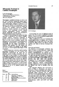
Chemical analysis. A total of 650 mg of (NH4)2S04 precipitable, nondialyzable material was recovered from the 100,000 x g supernatant (see "Cell disruption and antigen extraction"). This candidal extract I contained 11.5% carbohydrate, 13.9% N, and 81.2% protein (Table 1). Of the 200 mg placed on the Con ASepharose column, 77.4 mg was eluted with buffer and 21 mg was eluted with a-methyl mannoside (Fig. 1). The material eluted with buffer contained no detectable carbohydrate and was composed of 19 proteins resolved by disc gel electrophoresis (Fig. 2). This column fraction was referred to as candidal soluble pro-
teins because it had no detectable carbohydrate (Table 1). The material binding to Con A consisted of 80.2% carbohydrate and 17% protein. Since this carbohydrate fraction was found by gas chromatography (compared to mannoseglucose standard) to contain only mannose, it was designated as the mannan-protein complex. Part of this material migrated in disc gel electrophoresis and is shown in comparison with candidal extract I and the soluble proteins in Fig. 2. Serological activity. Immunoelectrophoresis and immunodiffusion. The soluble proteins reacted in immunoelectrophoresis with the sera of infected rabbits, with sera from two patients with chronic mucocutaneous candidiasis (Fig. 3), and with 9 of 39 sera from patients having neoplastic disease (Tables 2, 3). The sera from rabbits immunized with formaldehyde-killed cells were not reactive with the soluble proteins. The mannan-protein complex reacted equally in immunodiffusion tests with sera from rabbits immunized both by infection and by formaldehyde-killed cells, as well as with the human sera. The specificity of the sera was demonstrated in immunodiffusion by three lines of intersection between the soluble proteins and the mannan-protein complex and the PGM extracted from C. albicans cell walls by the method of Reiss et al. (15) (Fig. 4). A 10-,ug dose of the candidal soluble proteins elicited positive skin reactions in infected guinea pigs as well as in those sensitized with formaldehyde-killed yeast cells. At 6 days after sensitization with candidal extract I in complete Freund adjuvant, a strong cutaneous response to candidal soluble proteins devoid of mannan was observed, an indication of early development of cell-mediated immunity. The comparative ability of various cell wall and cytoplasmic antigens to elicit cutaneous hypersensitivity in guinea pigs was estimated at 21 days postinfection. The strongest reaction was obtained with the candidal soluble proteins at a 100-,ug dose; there was little evidence of TABLE 1. Quantitative analysis of whole and fractionated cytoplasmic extract of C. albicans Percentage of dry wt
Component
Carbohydratea
Extract
Slbeipo
11.5 81.2
0 91.0 15.9
Proteinb Total nitrogenc 13.9 a Phenol-sulfuric. b Folin phenol. c
Micro-Kjeldahl.
Mannan-
protein complex 80.2 17.5 3.8
94
J. CLIN. MICROBIOL.
ELLSWORTH ET AL.
c.;_
25P
U
I
L1 l'=
20
~-j
t
9
CS
- -'1
cK..
C;
r~ ~ ~ ~ ~ ~.
.
a ~~MM -
t
5 i
~~0 20
40 30 VOLUME, ML
50
216
FIG. 1. Affinity chromatography of the candidal extract I on a Con A-Sepharose column. aMM refers to amethyl mannoside. Immunoelectrophoresis of material eluted in 9 to 30 ml is shown in (a). The trough contained serum of a patient with chronic candidiasis. Immunodiffusion of the material recovered in 50 to 216 ml of effluent volume is shown in (b). Antigen (1 mg/ml) was in the center well, and the outer wells contained rabbit antisera (1, 2, 3) or sera of human patients with chronic candidiasis (4, 5).
early 5-h response, and the reaction persisted at 48 h. The candidal soluble proteins were not toxic as judged by skin tests of nonimmunized guinea pigs. When doses of 10, 50, 100, 500 ,ug, and 1 mg of protein were injected intradermally into nonimmunized animals, only the 1-mg dose caused a nonspecific skin reaction. The PGM from the cell wall resulted in an early 3-h reaction of the arthus type as well as a delayed response. Heat-stable mannan elicited the smallest amount of induration at 24 h in infected animals. DISCUSSION To investigate the specificity of the immune response to Candida spp. infection, it was esan
sential in the present work to separate mannan from the soluble cytoplasmic proteins. Affinity chromatography on Con A insolubilized on agarose was found to be a facile method of separating the mannan component of the candidal extract I under conditions that would not denature proteins, unlike the Sevag procedure (16) previously applied to the separation of protein and carbohydrate components of fungal antigens. When rabbits were immunized by infection and then were given three doses of candidal cytoplasmic extract, they responded with a multiplicity of antibodies specific for the cytoplasmic proteins which were separated in immunoelectrophoresis, in addition to antibody
CANDIDAL PROTEINS AND MANNAN ANTIGENS
VOL. 5, 1977
95
directed against mannan, the dominant cell wall antigen (15, 18, 21). Furthermore, patients with chronic mucocutaneous candidiasis were also sources of antibodies to candidal proteins as well as to polysaccharide antigens. Previously, Axelsen (1) found that 78 proteins from the cytoplasm of C. albicans were capable of reacting with antibody from rabbits immunized with a yeast supernatant in adjuvant. Buckley et al. (5) added Con A dropwise to a homogenate supernatant fraction of C. albicans as a means of removing mannan. Preliminary concentration by salt precipitation and dialysis before affinity chromatography on insolubilized Con A allowed more accurate quantitation of the protein content of candidal extract. Separation of mannan and protein components made possible the use of soluble proteins in immunodiffusion to demonstrate lines of intersection between polysaccharide and candidal FIG. 2. Disc gel electrophoretic patterns of the protein-containing immune precipitates. Data candidal extract before and after affinity chromatog- obtained from immunodiffusion tests performed raphy. (1) Candidal extract I; (2) candidal soluble on sera of cancer patients with known or susproteins devoid of mannan; (3) mannan-protein com- pected candidal disease were useful in a complex. parison of the relative antigenicity of candidal
1
2
3
FIG. 3. Immunoelectrophoresis of candidal proteins devoid of mannan. Antigens in wells (20 mg/ml) were electrophoresed and diffused against serum obtained from (A) a patient with chronic candidiasis, and (B) an infected rabbit.
96
J. CLIN. MICROBIOL.
ELLSWORTH ET AL.
TABLE 2. Immunodiffusion patterns of human cancer patients' sera with various C. albicans antigens Patient serum no.
Immunodiffusion reactivity profilea
Agglutinin titer-'
1 106 167 168 169 171 172 175 176 201 208 234 236 532 542 560 754 852 899 905 934 1042 1113 1129 1155 1166 1239 1911 2196 177 180 198 351 399 623 839 942 2284 6172
1, 3, 4, 5 1, 2, 3, 4, 6 2, 4, 6 1, 3, 4, 5 1, 6 1, 2, 3, 4, 6 1, 2, 4, 6 1, 2, 3, 4, 6 1, 3, 4 1, 2, 3, 4 1, 2, 3, 4, 6 2, 3, 4, 5, 6 3 1, 3, 5 1, 2, 3, 4, 5, 6 1, 2, 3, 4, 6 1, 3, 4, 6 1, 2, 3, 4, 6 1, 3, 4, 6 1, 2, 3, 4, 6 1, 2, 3, 4, 5, 6 4 1, 2, 3, 4, 5, 6 1, 3, 6 5 1, 2, 3, 4, 6 1, 3, 4, 6 1, 2, 3, 4, 6 1, 4 0 0 0 0 0 0 0 0 0 0
160 640 80 80 20 80 40 320 160 640 160 80 160 160 0 160 80 320 320 80 80 80 640 160 80 160 20 640 20 0 0 0 80 0 0 0 0 0 0
Primary diseaseb
Type of Candida infectionc
Esophageal Ca Acute leukemia Ca liver and lung AML AML AML RCS AML Ca lung AMML Bladder Ca RCS CML Liver Ca ALL Bladder Ca Breast Ca AMML AML AML AMML
Dissem., clinical Superficial, candidemia None
MM
AML EL AML ALL AML Bladder Ca AML ALL HD ALL Diverticulosis AML CLL RCS HD AMML Esophageal Ca
Superficial Superficial
Superficial Superficial Superficial
None Superficial
Dissem.-PM Superficial
Dissem.-PM Superficial, candidemia None Superficial, candidemia None Candidemia None Dissem.-PM None Superficial, candidemia Dissem., PM Superficial
Superficial
Dissem., PM Superficial
Dissem., clinical Dissem., PM Dissem., PM None Dissem., clinical Superficial, candidemia None Superficial, candidemia Superficial
Dissem., PM Dissem., PM Candidemia
a Code of antigen identification: 1, Peptidoglucomannan group A; 2, mannan group B; 3, soluble mannoglucan group A; 4, soluble mannoglucan group B; 5, C. albicans cytoplasmic proteins devoid of mannan group A; 6, peptidoglucomannan group B. bCode of primary disease abbreviations: AML, acute myelogenous leukemia; AMML, acute myelo monocytic; CLL, chronic lymphocytic; CML, chronic myelogenous; EL, erythro; HD, Hodgkin's disease; MM, multiple myeloma; RCS, reticular cell sarcoma; CA, cancer. e Dissem., Disseminated, see Materials and Methods; PM, postmortem.
soluble proteins devoid of mannan and mannan-containing, cell wall-derived antigens. With serum samples obtained from banked sera of carefully evaluated patients, a clear relationship could not be obtained between clinical diagnosis of candidal disease and a positive serological response. The relative sensitivity of Candida antigens in detecting precipitins in the sera of cancer patients is shown in Tables 2 and 3; in order of descending sensitivity, they were PGM group A > sMG group B > PGM group B > mannan
group B > cytoplasmic soluble proteins group A. Of 20 patients with invasive (positive blood culture or disseminated) candidiasis, 16 reacted positively with one or more antigens and 3 reacted with the candidal soluble proteins. Thus a 15% correlation could be made between response to candidal proteins and disseminated disease. On the other hand, of those patients who were uninfected or had only superficial Candida spp. infections, 5 of 19 reacted with candidal proteins, a correlation of more than 25%.
CANDIDAL PROTEINS AND MANNAN ANTIGENS
VOL. 5, 1977
97
TABLE 3. Relation of invasive candidiasis to immunodiffusion responses of sera from cancer patients to mannan-containing cell wall antigens and cytoplasmic soluble proteins of C. albicans Antigen
Serologically tive patients posi(%)
88% (24/29)b Peptidoglucomannan, group A (1)a 55% (16/29) Mannan, group B (2) 79% (23/29) Soluble mannoglucan, group A (3) 79% (23/29) Soluble mannoglucan, group B (4)' 55% (16/29) Cytoplasmic soluble proteins, group A (5) 68% (20/29) Peptidoglucomannan, group B (6? a Antigen code from Table 2. b Number of positive reactors of total patients in category. rCategories 3 and 4 defined in Materials and Methods.
Patients with invasive candidiasis (%)
55% 40% 55% 50% 15% 40%
(11/20) (8/20) (11/20) (10/20) (3/20) (8/20)
Pauienisiat infectionCandida (%) 68% 42% 63% 68% 26% 63%
(13/19) (8/19) (12/19) (13/19) (5/19) (12/19)
Of the eight patients who manifested a positive precipitin response to candidal soluble proteins, three were found to have invasive candidiasis. Therefore, the hypothesis that precipitins directed against protein determinants reflect invasiveness in contrast to those directed against determinants containing mannan was not supported. In further investigation of the diagnostic value of antigens for predicting the onset and severity of candidiasis, it would be useful to coat latex particles with PGM, group A, and a separate latex preparation coated with candidal cytoplasmic proteins devoid of mannan. In this way, a rapid-screening test would permit the enumeration of a titer that could be followed prospectively. Taschdjian et al. (22) have emphasized the importance, in human infections, of the presence of antibody directed against candidal proteins as a specific indication of disseminated disease, whereas Filice et al. have suggested that superficial infections may stimulate the production of antibody to cytoplasmic protein antigens (G. Filice, B. Yu, and D. Armstrong, J. Infect. Dis., in press). We support the latter view and expansion of this type of study is still warranted. Guinea pigs sensitized by infection gave positive skin reactions to candidal proteins devoid of carbohydrate as well as to heat-stable mannan (0.9% N) and to the PGM of the cell wall (7.5% amino acid) (15). Both the candidal soluble proteins and the cell wall mannan antigens elicited delayed-type hypersensitivity in infected guinea pigs. However, the animals gave an arthus response to the polysaccharide antigens at 4 h after intradermal change, which ter well contained rabbit antiserum, and antigens (1
FIG. 4. Serological specificity of the candidal soluible proteins and mannan-protein complex. (A) Cen-
mg/ml) were in outer wells: (1) and (4) candidal soluble proteins; (2) and (5) cell wall PGM, group A; (3) and (6) cytoplasmic mannan-protein complex. (B) Rabbit antiserum versus: (1) PGM, group A; (2) mannan, group B; (3) sMG, group A; (4) SMG, group B; (5) candidal soluble proteins; (6) PGM, group B.
98
J. CLIN. MICROBIOL.
ELLSWORTH ET AL.
TABLE 4. Skin test responses elicited by various components of C. albicans in sensitized guinea pigs Sensitization Method
Formaldehyde-killed yeast
Skin test antigen
Formaldehyde-killed
Days post
6
yeast
21
Candidal extract I CFAa
Soluble proteins
21
Soluble proteins
6 21
Candidal extract I IFAb
Soluble proteins
6 21
Infection
a b c
Soluble proteins
21
Soluble mannoglucan
21
Peptidoglucomannan
21
Mannan
21
Antigen dose (.Lg)
10 100 10 100 10 100 10 100 10 100 10 100 10 100 10 100 10 100 10 100 10 100
Induration (r2, mm 2)c 5h 0
24h 11
48 h 10
8 0
32 8
o
12 36 100 36 146 28 146 6 17 32 112 26 44 0 30 36 36 0 20
28 9 25 25 62 6 31 12 81 0 0 12 36 9 25 0 0
0 31 11
28 21 39 0 0 20 37 9 13 25 21 27 30 0 27
0 25 0 25
CFA, Complete Freund adjuvant. IFA, Incomplete Freund adjuvant. r2, Mean square radius of induration.
was not evident in the challenge with the soluble proteins. It was noteworthy that animals who were sensitized with adjuvant and who were skin tested 6 days later with candidal soluble proteins showed a strong reaction indicative of an early development of delayed-type hypersensitivity, possibly a Jones-Mote reaction (17). Therefore, it is possible that, as mentioned earlier, the response to the candidal proteins is strictly one of hypersensitivity and not a good estimator of invasive candidiasis.
LITERATURE CITED 1. Axelsen, N. H. 1973. Quantitative immunoelectrophoretic methods as tools for a polyvalent approach to standardization in the immunochemistry of Candida albicans. Infect. Immun. 7:949-960. 2. Axelsen, N. H., and C. H. Kirkpatrick. 1973. Simultaneous characterization of free Candida antigens and Candida precipitins in a patient's serum by means of crossed immunoelectrophoresis with intermediate gel. J. Immunol. Methods 2:245-250. 3. Biguet, J., P. TranVanKy, S. Andrieu, and R. Degaey. 1965. Etude immun6electrophoretique de la nature et de l'ordre d'apparition des anticorps precipitants du serum de lapins en fonction de leur mode d'immunisation contre Candida albicans. Sabouraudia 4:148-157. 4. Brewer, J. M., and R. B. Ashworth. 1969. Disc electrophoresis. J. Chem. Educ. 46:41-45. 5. Buckley, H. R., E. W. Lapa, and S. S. Hipp. 1970. Immunological significance of some antigens of Candida albicans, p. 21-24. Proceedings of the Fifth International Congress of Infectious Diseases, Vienna, Austria.
6. Gelman Instrument Co. 1970. Clinical electrophoresis. Gelman Instrument Co., Ann Arbor, Mich. 7. Hasenclever, H. F., and W. 0. Mitchell. 1964. Immunochemical studies on polysaccharides of yeasts. J. Immunol. 93:763-771. 8. Hodge, J. E., and B. T. Hofreiter. 1962. Determination of reducing sugars and carbohydrates, p. 380-394. In R. L. Whistler and M. L. Wolfrom (ed.), Methods in carbohydrate chemistry, vol. 1. Academic Press Inc., New York. 9. Kirkpatrick, C. H., R. R. Rich, and J. E. Bennett. 1971. Chronic mucocutaneous candidiasis: model-building in cellular immunity. Ann. Intern. Med. 74:955-978. 10. Lehner, T., H. R. Buckley, and I. G. Murray. 1972. The relationship between fluorescent, agglutinating, and precipitating antibodies to Candida albicans and their immunoglobulin classes. J. Clin. Pathol. 25:344-348. 11. Lowry, 0. H., N. J. Rosebrough, A. L. Farr, and R. J. Randall. 1951. Protein measurement with the Folin phenol reagent. J. Biol. Chem. 193:265-275. 12. Neurath, A. R., A. M. Prince, and A. Lippen. 1973. Affinity chromatography of hepatitis B antigen on concanavalin A linked to sepharose. J. Gen. Virol. 19:391-395. 13. Odds, F. C., and J. C. Hierholzer. 1972. Purification and properties of a glycoprotein acid phophatase from Candida albicans. J. Bacteriol. 114:257-266. 14. Peat, S., W. J. Whelan, and T. E. Edwards. 1961. Polysaccharides of baker's yeast. Part IV. Mannan. J. Chem. Soc., p. 29-34. 15. Reiss, E., S. H. Stone, and H. F. Hasenclever. 1974. Serological and cellular immune activity of peptidoglucomannan fractions of Candida albicans cell walls. Infect. Immun. 9:881-890. 16. Restrepo-Moreno, A., and J. D. Schneidau. 1967. Nature of skin reactive principle in culture filtrates prepared from Paracoccidioides brasiliensis. J. Bacte-
VOL. 5, 1977
CANDIDAL PROTEINS AND MANNAN ANTIGENS
riol. 93:1741-1748. 17. Richerson, H. B., H. F. Dvorak, and S. Leskowitz. 1970. Cutaneous basophil hypersensitivity I. A new look at the Jones-Mote reaction, general characteristics. J. Exp. Med. 132:546-557. 18. Salvin, S. B. 1959. Current concepts of diagnostic serology and skin hypersensitivity in the mycoses. Am. J. Med. 27:97-114. 19. Shaw, D. H., and G. W. Moss. 1969. Quantitative estimation of neutral sugars by gas-liquid chromatography. J. Chromatogr. 41:350-357.
99
20. Stickle, D., L. Kaufman, S. 0. Blumer, and D. W. McLaughlin. 1972. Comparison of a newly developed latex agglutination test and an immunodiffusion test in the diagnosis of systemic candidiasis. Appl. Microbiol. 23:490-499. 21. Summers, D. F., A. P. Grollman, and H. F. Hasenclever. 1964. Polysaccharide antigens of the Candida cell wall. J. Immunol. 92:491-499. 22. Taschdjian, C. L., M. S. Seelig, and P. J. Kozinn. 1973. Serological diagnosis of candidal infections. C.R.C. Crit. Rev. Clin. Lab. Sci. 4:19-59.