310
Akshatha C et al / International Journal of Biomedical and Advance Research 2016; 7(7): 310-314.
International Journal of Biomedical and Advance Research ISSN: 2229-3809 (Online); 2455-0558 (Print) Journal DOI: 10.7439/ijbar CODEN: IJBABN
Original Research Article
Correlation of p53 over expression with the clinicopathological prognostic factors in gastric adenocarcinoma Akshatha C*1, Vijaya Mysorekar2, Arundhathi S3, Arul P1 and Smitha1 1Assistant
Professor of Pathology, Dhanalakshmi Srinivasan Medical College, Perambalur, India of Pathology, M. S. Ramaiah Medical College, Bangalore, India 3Lecturer in Pathology, Sanjay Gandhi Institute of Trauma and Orthopaedics, Bangalore, India 2Professor
*Correspondence Info: Dr. Akshatha C Assistant Professor of Pathology, Dhanalakshmi Srinivasan Medical College, Perambalur, India E-mail:
[email protected]
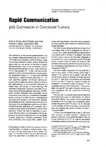
Abstract Background: Gastric cancer is one of the leading causes of cancer deaths. The etiology of gastric cancer is complex multistep process characterised by histopathologic precursor lesions and molecular genetic alterations involving APC, K-ras and p53 genes. Overexpression of p53 in gastric carcinoma is associated with bad prognosis. Aim: To establish correlation between p53 overexpression and clinicopathological features of gastric carcinoma. Materials and methods: A cross-sectional, descriptive hospital based study of clinical, histopathological and immunohistochemistry (IHC) features of gastric carcinoma was conducted on gastrectomy specimens received to the Department of Pathology from January 2008 to June 2013. The clinical features, tumour morphology and p53 status by immunohistochemistry, were evaluated in each case. Results: The most common histologic type of gastric carcinoma was of intestinal type and predominantly were grade III tumours (60%). Most (75%) of them presented with lymph node metastasis. Majority were in stage IIIA (27.5%) or stage IV (20.0%). Majority of gastric carcinomas (35%) had 1+ intensity of p53 expression but and it was not statistically significant. Majority of gastric adenocarcinomas showed p53 positivity in 4cm in size. Intestinal type of adenocarcionma (55%) were predominant according to Lauren’s classification (Table 2) and according to WHO classification diffuse type was frequently seen (11/40, 27.5%) (Table 3). Majority of patients presented with grade III gastric carcinomas (24/40, 60%) (Table 4). Gastric carcinomas more commonly (30/40, 75%) presented in advanced stage with lymph node metastasis. Majority of patients presented in stage IIIA (27.5%) or stage IV (20.0%) (Table 5). There was no statistical significance between TNM stage and histological type of gastric carcinoma (Table 6). Majority of gastric carcinomas (14/40, 35%) had 1+ intensity of p53 positivity, followed by 2+ intensity (12/40, 30%), 4+ intensity (7/40, 17.5%); however this difference was not statistically significant. Majority of gastric adenocarcinomas showed p53 positivity in 75% (intense) in a stomach adenocarcinoma, intestinal type (anti-p53-polyhorse radish peroxidase-DAB chromogen, x400)
4. Discussion The p53 gene codes a protein that is important in cell cycle and thus functions as a tumor suppressor. It has been attributed as the “guardian of genome” as it plays a pivotal role by preventing genome mutation and conserving stability. It is involved in cell cycle regulation, DNA repair and in apoptosis. Since it is involved in many biological processes it plays a pivotal role in carcinogenesis. Thus, loss of p53 function plays a crucial role in tumor development. [6] In nonmalignant stomach, p53 immunopositive cells are found in neck, gastric proliferative zone and in IJBAR (2016) 07 (07)
Stage III B 0 .0% 2 18 % 0 .0% 2 22.2% 0 .0% 0 0% 0 .0% 4 10.%
Stage III C 0 .0% 0 .0% 0 .0% 1 11.1% 0 .0% 1 14.% 0 .0% 2 5.0%
Stage IV 1 14% 2 18.% 0 .0% 1 11.1% 1 33.3% 1 14.% 1 100% 7 17.%
Total
2 value
p’ value
7 100.% 11 100.% 2 100.% 9 100.0% 3 100.0% 7 100.% 1 100.% 40 100.%
44.0
0.67
Helicobacter pylori infection. In H. pylori gastritis, free radicals produced by activated leucocytes cause mucosal DNA damage and thus nuclear p53 expression can be expected to be seen in proliferative zone.[7] p53 overexpression has been reported in 17-90.7% of invasive tumors. The nuclear staining of p53 can be seen in both intestinal and diffuse type of gastric tumors even though common in intestinal type. The degree of p53 expression correlates with proliferative rate of tumors which can be explained by higher incidence of p53 positivity in intestinal type of tumors.[8] p53 expression is common in poorly differentiated tumors than well differentiated carcinomas.[9] Its expression is more common in tumors from proximal stomach compared to distal tumors.[10] Evaluation of p53 as a prongostic marker has yielded conflicting results. Thus, this study was conducted to establish correlation between p53 overexpression in gastric cancer by IHC and various prognostic factors including the clinical features, tumour histopathology, tumour grade, lymph node status, and stage of the tumour. In the present study, 40 gastrectomy specimens were studied and correlation of p53 status with clinical features and tumor histopathology was analysed. The peak age group who presented with gastric carcinoma was between 60 and 69 years and thus elderly age is a risk factor for development of gastric carcinoma. Lymph node metastasis at time of diagnosis was seen in 75% of patients whereas in studies by Ghaffarzadegan et al[11] and Aurello et al[12] it was 57.6% and 64% respectively. Intestinal type of gastric adenocarcinoma was the common type of gastric adenocarcinoma observed in our study which was similar to that noted in studies by Nabi et al[13] and Omran et al[14]. In our study most of the tumors were of grade III (60%) whereas it was grade II in studies by Chiaravalli et al[15] and Choi sang-wook et al[16]. In the present study p53 positivity was seen in 25 out of 40 cases (62.5%) of gastric adenocarcinoma. 59.3% of tumours in the pyloric antrum (14/24 cases) and 100% of gastroesophageal junction tumours (3/3 cases) showed p53 www.ssjournals.com
Akshatha C et al / Correlation of p53 over expression with the clinicopathological prognostic factors in gastric adenocarcinoma
positivity. Studies by Fenoglio-Preiser et al[17] and Brito et al[18] showed p53 immunoreactivity in 17-19% and 35% of gastric carcinomas respectively. Ghaffarzadegan et al[11] noted p53 positivity in 75% of gastric carcinomas and found that p53 alterations occur much more commonly in proximal lesions than in distal ones, suggesting that the molecular events leading to the development of gastric carcinoma may be very different in proximal versus distal tumors. Overall, however, it appears that p53 alterations occur early in the development of gastric carcinoma, being present even in the non neoplastic mucosa and they increase in frequency as one progress along the pathway of gastric carcinoma development. In our study, p53 positivity was seen in 4 out of 7 cases (57.1%) of signet ring cell adenocarcinomas and 5 out of 9 (55.6%) of tubular adenocarcinomas. P53 positivity was seen in 18 out of 30 (60%) cases with lymph node metastasis and 7 out of 10 cases (70%) without lymph node metastases, but this was not statistically significant (p=0.646). A study by Brito et al[18] showed that the frequency of p53 positivity in tumours of tubular histological type (46%) was significantly higher than that in signet ring tumours (10%) (p = 0.006), and neoplasms that invaded deeply into the submucosa were more frequently positive (45%) than others (30%). In their study, five of eight (62%) T1 tumours with lymph node metastases were p53 immunoreactive. Their findings showed that immunocytochemically demonstrable overexpression of p53 correlates with other morphological markers of aggressiveness in T1 gastric adenocarcinoma. Azarhoush et al[3] showed that p53 alterations correlate well with gastric location, and are frequent in adenocarcinomas of cardia than that of antrum. There was no difference in clinicopathologic characteristics between p53 positive and p53 negative gastric carcinomas. In our present study, p53 alterations did not correlate with clinicopathological parameters. Filiz et al[19] found no relation between p53 staining and parameters such as nuclear grade, invasion depth, lymph node involvement. The present study showed p53 positivity to be equal in intestinal and diffuse type of gastric adenocarcinoma. Filiz et al[19] found p53 positivity in 17 out of 20 (85%) intestinal type and 13 out of 22 (59%) diffuse carcinoma. Ghaffarzadegan et al found p53 positivity in 59 out of 100 cases and they noted a significant correlation between rate of p53 overexpression and histologic type of tumor.[11] There was no significant association between protein accumulation and lymph node status in our study which was supported by study by Lazar et al[20].
5. Conclusion Mutation in p53 plays a vital role in development of gastric carcinoma. However, a significant correlation between gastric carcinoma and clinicopathological parameters could not be established. A prolonged follow up study on a large IJBAR (2016) 07 (07)
313
number of cases can help to find out whether p53 overexpression by standard IHC could be a useful marker for identification of gastric carcinoma.
References [1] Karim S, Ali A. Correlation of p53 over-expression and alteration in p53 gene detected by polymerase chain reaction-single strand conformation polymorphism in adenocarcinoma of gastric cancer patients from India. World J Gastroenterol 2009; 15:1381-1387. [2] Ghavam-Nasiri MR, Razaei E, Ghafarzadegan K, Seilanian-Toosi M, Malekifard H.Expression of p53 in colorectal carcinoma: correlation with clinicopathologic features. Arch Iran Med 2007; 10:38-42. [3] Azarhoush R, Keshtkar A A, Amiriani T, Kazemi-Nejad V. Relationship between p53 expression and gastric cancers in cardia and antrum. Arch Iran Med 2008; 11:502-506. [4] Georgescu CV, Saftoiu A, Georgescu CC, Ciurea R, Ciurea T. Correlations of proliferation markers, p53 expression and histological findings in colorectal carcinoma. J Gastrointestin Liver Dis 2007; 16:133-9. [5] Huh JW, Lee JH, Kim HR. Expression of p16, p53, and Ki-67 in colorectal adenocarcinoma: a study of 356 surgically resected cases. Hepatogastroenterology 2010; 57:734-40. [6] Dan-ping Z, Xiao-wen D, Jia-ping P, Yi-xiong Z, Suzhan Z. Prognostic significance of bcl-2 and p53 expression in colorectal carcinoma. Journal of Zhejiang Univ Science B 2005; 6(12):1163-1169. [7] Correa P, Shiao Y. Phenotypic and genotypic events in gastric carcinogenesis. Cancer Res 1994; 54:1914s-1943. [8] Ioachim E, Stefanou GD, Agnantis NJ. Expression of p53 protein in gastric cancer: an immunohistochemical study with correlation to proliferative activity. Anticancer Res 1997; 17:513–517. [9] Sasaki I, Yao T, Nawata H, Tsuneyoshi M. Minute gastric carcinoma of differentiated type with special reference to the significance of intestinal metaplasia, proliferative zone and p53 protein during tumor development. Cancer 1999; 85: 1719–1729. [10] Fenoglio-Preiser CM, Wang J, Stemmermann GN, and Noffsinger A. TP53 and Gastric Carcinoma: A Review. Human Mutation 2003; 21:258-270. [11] Ghaffarzadegan K, Zali MR, Ahmadi Pharm KJ, Asadzadeh H. Correlation of nuclear p53 immunoreactions with the histopathologic features in gastric carcinoma. Arch Iranian Med 2004; 7 (4): 279 – 283. [12] Aurello P, D'Angelo F, Rossi S, Bellagamba R, Cicchini C, Nigri G, Ercolani G, De Angelis R, Ramacciato G. Classification of lymph node metastases from gastric cancer: comparison between N-site and N-number www.ssjournals.com
Akshatha C et al / Correlation of p53 over expression with the clinicopathological prognostic factors in gastric adenocarcinoma
systems. Our experience and review of the literature. Am Surg. 2007; 73(4):359-366. [13] Nabi U, Hannan Nagi A, Riaz S, Sami W. Morphological Evaluation of Colorectal Carcinoma with Grading Staging and Histological types. JPMA 2010; 60:996-999. [14] Omran OM, Thabet M. Gelatinases A and B expression in human colorectal cancer in upper Egypt: a clinicopathological study. Ultrastruct Pathol. 2012; 36(2):108-116. [15] Chiaravalli AM, Klersy C, Vanoli A, Ferretti A, Capella C, Solcia E. Histotype-based prognostic classification of gastric cancer. World J Gastroenterol. 2012; 18(9):896904. [16] Sang-wook C, Won-sang P, Jin-mo Y, Chan-su S.P53 protein overexpression and allele loss of p53 gene in gastric adenocarcinoma. Journal of Korean Medical Science 1994; 9: 299-303.
IJBAR (2016) 07 (07)
314
[17] Fenoglio-preiser C M, Wang N J, Stemmermann G N, and Noffsinger. A p53 review article TP53 and gastric carcinoma: A review, Human Mutation 2003; 21:58-270. [18] Brito MJ, Williams GT, Thompson H, Filipe MI. Expression of p53 in early (T1) gastric carcinoma and precancerous adjacent mucosa. Gut 1994; 35(12):16971700. [19] Filiz O, Semsi A, Kutlu A. Kemal. Detection of protein p53 in gastric carcinomas: An immunohistochemical study of 50 cases. The Turkish Journal of Gastroenterology 2000; 11 (4):62-71. [20] Lazar D, Sorina Taban S, Sporea I, Dema A, Mario M, Lazar E, Goldiş A, The immunohistochemical expression of the p53-protein in gastric carcinomas. Correlation with clinicopathological factors and survival of patients. Romanian Journal of Morphology and Embryology 2010; 51(2):249-257.
www.ssjournals.com