Decay-accelerating factor (DAF) and intercellular adhesion molecule-1 (ICAM-1), despite being ... viruses, to DAF is insufficient to mediate cell infection, unless.
Journal of General Virology (2000), 81, 889–894. Printed in Great Britain .........................................................................................................................................................................................................
SHORT COMMUNICATION
Cytoplasmic interactions between decay-accelerating factor and intercellular adhesion molecule-1 are not required for coxsackievirus A21 cell infection Darren R. Shafren,1 Douglas J. Dorahy,2 Rick F. Thorne2 and Richard D. Barry1 1 Picornaviral Research Unit, Discipline of Immunology and Microbiology and 2 Cancer Research Unit, Faculty of Medicine and Health Sciences, The University of Newcastle, Level 3, David Maddison Clinical Sciences Building, Royal Newcastle Hospital, Newcastle, 2300 New South Wales, Australia
Coxsackievirus A21 (CAV-21) employs a cell receptor complex of decay-accelerating factor (DAF) and intercellular adhesion molecule-1 (ICAM-1) for cell infectivity. In this study, the nature of potential extra- and/or intracellular interactions between DAF and ICAM-1 involved in picornaviral cell entry was investigated. Firstly, it was shown that intracellular interplay between DAF and ICAM-1 is not required for CAV-21 infection, as CAV-21 lytic infection mediated via the DAF/ICAM-1 receptor complex is not inhibited by replacement of the transmembrane and cytoplasmic domains of ICAM1 with those from an unrelated cell surface molecule, CD36. By immunoprecipitation, chemical cross-linking and picornaviral binding assays, the existence of a close spatial association between DAF and ICAM-1 on the surface of ICAM-1transfected RD cells was confirmed. Furthermore, it was shown that potential extracellular DAF/ICAM-1 interactions are likely to occur in an area on or proximal to DAF SCR3 and may influence the route of CAV-21 cell entry.
Decay-accelerating factor (DAF) and intercellular adhesion molecule-1 (ICAM-1), despite being structurally dissimilar, mediate signal transduction following antibody cross-linking (Shenoy-Scaria et al., 1992 ; Chirathaworn et al., 1995 ; Rothlein et al., 1994) and are both used as cellular attachment receptors for many human picornaviruses (Bergelson et al., 1994, 1995 ; Casasnovas & Springer, 1994 ; Clarkson et al., 1995 ; Shafren et al., 1995, 1997 a ; Staunton et al., 1992 ; Ward et al., 1994). In particular, the human enterovirus coxsackievirus A21 (CAV-21) can bind to the first short consensus repeat (SCR) of DAF (Shafren et al., 1997 b ; unpublished data) or the Author for correspondence : Darren Shafren. Fax j61 2 4923 6814. e-mail dshafren!mail.newcastle.edu.au
0001-6614 # 2000 SGM
N-terminal domain (d1) of ICAM-1 (Shafren et al., 1997 a). Binding of CAV-21, as well as many other human enteroviruses, to DAF is insufficient to mediate cell infection, unless DAF is cross-linked by specific anti-DAF MAbs (Shafren, 1998 ; Shafren et al., 1998). Receptor-mediated capsid conformational changes are considered by many to be a prerequisite for picornaviral cell entry (Fricks & Hogle, 1990 ; Casasnovas & Springer, 1994 ; Arita et al., 1998). Recent findings that surface-expressed ICAM-1 is able to induce relatively rapid conformational changes in CAV-21 virion structure, whereas surface DAF appears to maintain sequestered CAV-21 in a conformationally unaltered state may explain why ICAM-1 alone, in contrast to DAF, is able to mediate CAV-21 cell entry (Shafren, 1998). CAV-21 lytic infection of cells expressing both ICAM-1 and DAF can only be completely inhibited by the combined action of anti-ICAM-1 (d1) and anti-DAF SCR1 MAb blockade (Shafren et al., 1997 b). Although, human rhinovirus 14 (HRV14) and CAV-21 compete for the same binding epitope on ICAM-1 (Lonberg-Holm et al., 1976), replication of HRV-14 in HeLa cells, in contrast to that of CAV-21, is completely inhibited by anti-ICAM-1 (d1) MAb blockade but is not affected by anti-DAF MAb pretreatment (Shafren et al., 1997 b). Interestingly, anti-DAF SCR3 MAb blockade partially blocks CAV-21 infection of HeLa cells without interfering with CAV-21 attachment to either DAF or ICAM-1 (Shafren et al., 1997 b).Taken together, these findings raise the interesting possibility that CAV-21 cell entry, unlike that of HRV-14, may be influenced by either extra- and\or intracellular interplay between DAF and ICAM-1. In this study, using chimeric ICAM-1 receptors, we have shown that CAV-21 lytic cell infection mediated via the DAF\ICAM-1 receptor complex is not dependent on the presence of the transmembrane and cytoplasmic domains of ICAM-1. Secondly, employing chemical cross-linking, antibody and picornavirus binding techniques, we have confirmed that a close extracellular spatial association exists between DAF and ICAM-1 on the surface of ICAM-1-expressing RD cells and that this interaction is most likely to occur in an area on or proximal to DAF SCR3. IIJ
D. R. Shafren and others
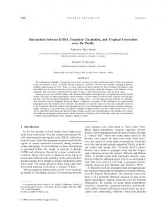
(a)
(c)
(b)
(d)
Fig. 1. CAV-21 infection of cells expressing ICAM-1, CD36 and chimeric ICAM-1/CD36. (a) A schematic representation of surface-expressed ICAM-1, CD36 and chimeric ICAM-1/CD36. (b) Immunoprecipitation of biotinylated surface-expressed wild-type ICAM-1, CD36 and chimeric ICAM-1/CD36. RD cells transiently expressing ICAM-1, CD36 and chimeric ICAM-1/CD36 cells were surface-biotinylated and immunoprecipitated with either an anti-ICAM-1 MAb or anti-CD36 MAb. (c) Flow cytometric analysis of binding of anti-ICAM-1 and anti-CD36 MAbs to RD cells transiently expressing ICAM-1, CD36 and chimeric ICAM-1/CD36. The open histogram represents the binding of the FITC–anti-mouse conjugate, whereas the closed histogram represents the binding of the anti-ICAM-1 or CD36 MAbs where appropriate. (d) CAV-21-induced cell lysis of ICAM-1-expressing RD cells. Monolayers of transfected RD cells (Fig. 1 c) were inoculated with CAV-21 (105 TCID50) and, following incubation for 24 h at 37 mC, were inspected for signs of cell lysis.
Firstly, we investigated the potential role of intracellular DAF\ICAM-1 interactions in the CAV-21 cell infection cycle. To address this question, a chimeric ICAM-1\CD36 molecule was constructed in which the fifth extracellular domain, the transmembrane and cytoplasmic domains of ICAM-1 were removed and replaced with the transmembrane and cytoplasmic domains from CD36. CD36 is an 88 kDa polypeptide and a cellular receptor for thrombospondin (Greenwalt et al., 1992). A schematic representation of wild-type ICAM-1, CD36 and chimeric ICAM-1\CD36 is shown in Fig. 1 (a). To confirm the fidelity of surface-expressed chimeric ICAM1\CD36, transfected RD cells were surface-labelled with biotin, immunoprecipitated with anti-ICAM-1 (Boyd et al., 1988) or anti-CD36 (Mazurov et al., 1992) MAbs where appropriate and analysed by SDS–PAGE ; specific proteins were visualized IJA
using enhanced chemiluminescence (Shafren et al., 1997 b). The results shown in Fig. 1 (b) reveal that ICAM-1 is expressed in RD cells as a 90 kDa polypeptide, and that CD36 and chimeric ICAM-1\CD36 are expressed as 85 and 70 kDa proteins, respectively. The identity of a second minor polypeptide in the ICAM-1\CD36 immunoprecipitate which possesses a similar molecular mass to native ICAM-1 has been shown to be a chimeric ICAM-1\CD36 molecule with a different glycosylation pattern compared to the major species (data not shown). The capacity of the anti-ICAM-1 MAb (d1) to efficiently immunoprecipitate the chimeric ICAM-1\CD36 molecule further confirms that the conformation of the d1 was retained despite the deletion of the fifth extracellular and the transmembrane\cytoplasmic domains. Next, we investigated the capacity of the chimeric ICAM-
DAF/ICAM-1 interactions in CAV-21 infection
1\CD36 to mediate CAV-21 lytic infection of RD cells. RD cells were transiently transfected with ICAM-1, CD36, chimeric ICAM-1\CD36 cDNAs or vector alone (pEF-BOS ; Mizushima & Nagata, 1990) and, following incubation for 48 h, cells were challenged with 10& TCID of CAV-21. At &! 24 h post-infection, cells were monitored for signs of virusinduced cell lysis and photographed at i20. The fluorescence histograms shown in Fig. 1 (c) indicate a comparable level of surface expression of ICAM-1 and chimeric ICAM-1\CD36. CD36 was expressed only on the surface of cells transfected with CD36 cDNA (Fig. 1 c). RD cells expressing ICAM-1 or chimeric ICAM-1\CD36 were highly susceptible to CAV-21-mediated cell lysis, whereas cells transfected with either vector alone (BOS) or CD36 were refractile to infection (Fig. 1 d). Next, we investigated whether DAF and ICAM-1 shared an extracellular relationship on the surface of ICAM-1expressing RD cells. Previously, we have shown a spatial association between ubiquitously surface-expressed DAF and ICAM-1 on HeLa cells (Shafren et al., 1997 b) and to address this question we transiently transfected DAF-expressing RD cells with either ICAM-1 or CD36 cDNA (Shafren et al., 1997 b). Cell transfectants were iodinated, chemically crosslinked and immunoprecipitated with anti-DAF (Kinoshita et al., 1985), anti-ICAM-1 (Boyd et al., 1988) or anti-CD36 MAbs (Mazurov et al., 1992). In the ICAM-1-transfected RD cells, in the absence of cross-linking, the anti-DAF MAb and the antiICAM-1 MAb immunoprecipitated polypeptides of approximately 75 kDa and 90 kDa, respectively, whereas the antiCD36 MAb did not recognize any proteins (Fig. 2 a). Following chemical cross-linking of these cells, immunoprecipitation with the anti-DAF MAb identified the DAF band at 75 kDa as previously seen in non-cross-linked samples, but also revealed an additional polypeptide of 90 kDa, similar to that observed in both ICAM-1 MAb immunoprecipitates. Immunoprecipitation of cross-linked cells with the anti-ICAM-1 MAb identified the ICAM-1 band at 90 kDa and a slight band at 75 kDa, similar to that observed in both anti-DAF immunoprecipitates. The finding that little detectable DAF was observed in the cross-linked anti-ICAM-1 immunoprecipitate may reflect the fact that DAF was expressed at a lower level than ICAM-1 on the surface of transfected RD cells (Fig. 2 a). Thus, a large proportion of cell surface ICAM-1 may not be associated with DAF. The DAF and ICAM-1 spatial association was shown to be specific and not an artefact of cell transfection, as CD36 MAb and DAF MAb immunoprecipitates of CD36transfected RD cell lysates (cross-linked and non-cross-linked) revealed only the presence of polypeptides specific to the appropriate MAbs (Fig. 2 b). In an attempt to map the regions of DAF involved in the DAF\ICAM-1 extracellular relationship, we generated a stable ICAM-1-expressing RD cell line. The RD cell surfaceexpressed ICAM-1 was shown to be functional by facilitating the lytic infection of these cells by both HRV-14 and CAV-21
(a)
(b)
Fig. 2. Immunoprecipitation of surface-expressed DAF and ICAM-1 before and after chemical cross-linking. (a) ICAM-1- and (b) CD36-transfected RD cells were surface-iodinated and immunoprecipitated with either an anti-DAF (SCR1) MAb, anti-ICAM-1 MAb or anti-CD36 MAb prior to [noncross-linked (NCL)] and following chemical cross-linking (DSP). The immunoprecipitates were separated by SDS–PAGE and labelled polypeptide(s) were visualized by autoradiography.
(Fig. 3 a). However, as previously observed in HeLa B cells (Shafren et al., 1997 b), MAb blockade of ICAM-1 domain 1 (MAb IH4 ; Boyd et al., 1988) completely inhibited HRV-14 cell infection over a range of input multiplicities, while only partially blocking CAV-21 cell infectivity at relatively low levels of virus challenge (Fig. 3 a). Flow cytometry was employed to assess the relative binding level of MAbs directed against individual SCR of DAF on the surface of RD cells in the presence and absence of coexpressed ICAM-1. Anti-DAF SCR1 and SCR4 MAbs bound to DAF at a comparable level on the surface of RD and ICAM1-transfected RD cells, whereas a MAb directed at DAF SCR2\3 and more striking a MAb to DAF SCR3 bound at levels significantly higher on RD cells compared to ICAM-1transfected RD cells (Fig. 3 c). The apparent reduction in spatial accessibility to DAF SCR3 mediated by ICAM-1 co-expression was confirmed by the reduced capacity of a known DAF SCR3 IJB
D. R. Shafren and others
(a)
(b)
(c)
(d)
Fig. 3. Antibody and virus binding to individual SCR of DAF. (a) ICAM-1 MAb blockade of HRV-14 and CAV-21 infection. Serial dilutions of stock preparations of HRV-14 and CAV-21 were used to inoculate ICAM-1-transfected RD cell monolayers in 96well plates that had been preincubated in the presence or absence of an anti-ICAM-1 (d1) MAb (20 µg/ml) for 1 h at 37 mC. Following incubation for 48 h, cell lysis was assessed as previously described (Shafren et al., 1997 b). Results are expressed as the mean percentage of cell lysis of duplicate wells relative to the uninfected control cell monolayers. (b) A schematic representation of the putative extracellular spatial relationship between surface-expressed DAF and ICAM-1. The black region represents the cell membrane, whereas the grey region represents the putative site for the DAF/ICAM-1 interaction. SCR, short consensus repeat ; D1–D5, extracellular domains. (c) Antibody binding to DAF. Suspensions of RD or ICAM-1-transfected RD (RD-ICAM-1) cells were incubated with anti-DAF MAbs (Kinoshita et al., 1985), IA10 (SCR1), VIIIA7 (SCR2), IH4 (SCR3), IIH6 (SCR4) and phycoerythrin-conjugated anti-mouse F(ab)2. The results [triplicate tubesj(3iSD)] are expressed as the percentage geometric mean of fluorescence intensities of MAb binding to RD-ICAM-1 relative to RD cells. (d ) Picornavirus binding. Suspensions of RD or RD-ICAM-1 cells were incubated with 35S-labelled preparations of purified poliovirus type 1 (PV1 ; 10 000 c.p.m.) or echovirus type 7 (E7 ; 40 000 c.p.m.) and processed as previously described (Shafren et al., 1997 b). Results represent the means of triplicate wellspSD.
binding virus, echovirus 7 (Clarkson et al., 1995), to bind to DAF on ICAM-1-expressing RD cells compared to nontransfected RD cells (Fig. 3 d). Surface expression of ICAM1 on RD cells did not interfere with the attachment of poliovirus type 1, as a comparable level of binding to both cell lines was observed (Fig. 3 d ). Preincubation of the ICAM-1transfected RD cells with the above anti-DAF MAbs did not IJC
reduce binding of the anti-ICAM-1 (d1) MAb. Furthermore, pretreatment of ICAM-1-transfected RD cells with a combination of anti-DAF SCR1 and SCR3 MAbs did not inhibit HRV-14 replication in these cells (unpublished data). In this study, we have shown that CAV-21 lytically infects RD cells expressing either wild-type ICAM-1 or a chimeric form, in which the transmembrane and cytoplasmic domains
DAF/ICAM-1 interactions in CAV-21 infection
are replaced with those from an unrelated molecule, CD36 (Fig. 1). This result is consistent with earlier findings in which HRV14 and group B coxsackieviruses were shown to infect cells expressing a chimeric form of ICAM-1 and CAR, respectively, where the transmembrane and cytoplasmic domains were replaced with a glycosyl phosphatidylinositol anchor (Staunton et al., 1992 ; Wang & Bergelson, 1999). Overall, the data herein suggest that the postulated interplay between DAF and ICAM-1 in regulating CAV-21 cell entry probably occurs in an extracellular fashion rather than in an intracellular environment. A close spatial association between DAF and ICAM-1 on the surface of ICAM-1-transfected RD cells was confirmed (Fig. 2). Antibody and virus binding patterns to surfaceexpressed DAF in the presence or absence of co-expressed ICAM-1 suggest that the interactions between DAF and ICAM-1 occur at an epitope located in or proximal to DAF SCR3 (Fig. 3 c). Molecular modelling studies have predicted that DAF is expressed on the cell surface in an ‘ S-shaped ’ form (Kuttner-Kondo et al., 1996) and, based on this structure, a schematic representation of the putative DAF\ICAM-1 spatial relationship is shown in Fig. 3 (b). Previously, we have shown that anti-DAF SCR3 pretreatment of HeLa B cells does not inhibit CAV-21 cell binding but partially blocks CAV-21 replication (Shafren et al., 1997 b). The findings from this study that the DAF\ICAM-1 spatial relationship occurs at a region located near SCR3 of DAF suggest that DAF SCR3 MAb pretreatment may inhibit CAV-21 infection by impeding extracellular DAF\ICAM-1 interactions at this location. HRV-14 and CAV-21 compete for the same epitope on ICAM-1 d1 (Lonberg-Holm et al., 1976), but while MAb blockade of this domain affords total protection against HRV14 infection it does not do so for CAV-21 (Fig. 3 a). This finding raises the interesting possibility that when the ICAM1-mediated route of cell entry is blocked, CAV-21, unlike HRV-14, may employ an alternate route of cell entry, possibility mediated via cross-linked-DAF (Shafren, 1998). Studies employing HeLa cells expressing dominant-negative dynamin mutants lacking normal GTPase activity have confirmed that HRV-14 enters cells via a clathrin-coated pit endocytotic mechanism (De Tulleo & Kirchhausen, 1998 ; Grunert et al., 1997). However, the structurally similar poliovirus, using a cellular receptor from the same family as ICAM-1, employs a different cell entry mechanism (De Tulleo & Kirchhausen, 1998). Recently, simian virus 40 (SV-40) has been shown to enter cells via endocytotic structures known as caveolae (Anderson et al., 1996). Caveolae are specialized cell surface invaginations with several known functions, including endocytosis of macromolecules using mechanisms independent of clathrincoated pits (Rothberg et al., 1990 ; Sargiacomo et al., 1993). This SV-40 route of cell internalization is mediated by the viral cross-linking of MHC class I molecules (Anderson et al., 1996). Interestingly, DAF cross-linked by specific anti-DAF MAbs is
sequestered to caveolae (Mayor et al., 1994 ; Rothberg et al., 1992). In contrast to HRV-14, CAV-21 binds to DAF SCR1 (Shafren et al., 1997 b ; unpublished data) and can lytically infect RD cells in the absence of ICAM-1 when cell surface DAF is cross-linked with MAbs directed against DAF SCR2\3 (Shafren, 1998). Therefore, as the putative DAF\ICAM-1 extracellular interaction is likely to occur on or proximal to DAF SCR3, one may postulate that ICAM-1 may in fact crosslink DAF, allowing translocation of DAF-bound CAV-21 to caveolae for subsequent cell internalization ; this area is currently under investigation. Recent functional studies indicate that dynamin mediates both clathrin-dependent endocytosis and the internalization of caveolae in mammalian cells (Henley et al., 1998). Therefore, it would be of interest to investigate whether expression of a dominant-negative dynamin mutant in ICAM-1-transfected RD cells would inhibit CAV-21 cell infection. The author thanks Andrew Boyd, Bruce Loveland and Taroh Kinoshita for their generous gift of the MAbs used in this study ; and Rebecca Ingham for excellent technical assistance. This research was supported by a project grant from the National Health and Medical Research Council of Australia.
References Anderson, H. A., Chen, Y. & Norkin, L. C. (1996). Bound simian virus 40
translocates to caveolin-enriched membrane domains, and its entry is inhibited by drugs that selectively disrupt caveolae. Molecular Biology of the Cell 7, 1825–1834. Arita, M., Koike, S., Aoki, J., Horie, H. & Nomoto, A. (1998). Interaction of poliovirus with its purified receptor and conformational alteration in the virion. Journal of Virology 72, 3578–3586. Bergelson, J. M., Chan, B. M., Solomon, K. R., St John, J. N. & Finberg, R. W. (1994). Decay-accelerating factor (CD55), a glycosylphosphatidyl-
inositol-anchored complement regulatory protein, is a receptor for several echoviruses. Proceedings of the National Academy of Sciences, USA 91, 6245–6249. Bergelson, J. M., Mohanty, J. G., Crowell, R. L., St John, N. F., Lublin, D. M. & Finberg, R. W. (1995). Coxsackievirus B3 adapted to growth in
RD cells binds to decay-accelerating factor (CD55). Journal of Virology 69, 1903–1906. Boyd, A. W., Wawryk, S. O., Burns, G. F. & Fecondo, J. V. (1988).
Intercellular adhesion molecule-1 (ICAM-1) has a central role in cell-cell contact-mediated immune mechanisms. Proceedings of the National Academy of Sciences, USA 85, 3095–3099. Casasnovas, J. M. & Springer, T. M. (1994). Pathway of rhinovirus disruption by soluble intercellular adhesion molecule 1 (ICAM-1) : an intermediator in which ICAM-1 is bound and RNA is released. Journal of Virology 68, 5882–5889. Chirathaworn, C., Tibbetts, S. A., Chan, M. A. & Benedict, S. H. (1995).
Cross-linking of ICAM-1 on T cells induces transient tyrosine phosphorylation and inactivation of cdc2 kinase. Journal of Immunology 155, 5479–5482. Clarkson, N. A., Kaufman, R., Lublin, D. M., Ward, T., Pipkin, P. A., Minor, P. D., Evans, D. J. & Almond, J. W. (1995). Characterisation of
the echovirus 7 receptor : domains of CD55 critical for virus binding. Journal of Virology 69, 5497–5501. IJD
D. R. Shafren and others De Tulleo, L. & Kirchhausen, T. (1998). The clathrin endocytic pathway in viral infection. EMBO Journal 17, 4585–4593. Fricks, C. E. & Hogle, J. M. (1990). Cell-induced conformational change in poliovirus : externalization of the amino terminus of VP1 is responsible for liposome binding. Journal of Virology 64, 1934–1945. Greenwalt, D. E., Lipsky, R. H., Ockenhouse, C. F., Ikeda, H., Tandon, N. N. & Jamieson, G. A. (1992). Membrane glycoprotein CD36 : a
review of its roles in adherence, signal transduction, and transfusion medicine. Blood 5, 1105–1115. Grunert, H. P., Wolf, K. U., Langner, K. D., Sawitzky, D., Habermehl, K. O. & Zeichhardt, H. (1997). Internalization of human rhinovirus 14
into HeLa and ICAM-1-transfected BHK cells. Medical Microbiology and Immunology 186, 1–9. Henley, J. R., Krueger, E. W., Oswald, B. J. & McNiven, M. A. (1998).
Dynamin-mediated internalization of caveolae. Journal of Cell Biology 141, 85–99. Kinoshita, T., Medof, M. E., Silber, R. & Nussenzweig, V. (1985).
Distribution of decay accelerating factor in the peripheral blood of normal individuals and patients with paroxysmal nocturnal hemoglobinuria. Journal of Experimental Medicine 162, 75. Kuttner-Kondo, L., Medof, M. E., Brodbeck, W. & Shoham, M. (1996).
Molecular modelling and mechanism of action of human decayaccelerating factor. Protein Engineering 12, 1143–1149. Lonberg-Holm, K., Crowell, R. L. & Philipson, L. (1976). Unrelated animal viruses share receptors. Nature 259, 679–681. Mayor, S., Rothberg, K. G. & Maxfield, F. R. (1994). Sequestration of GPI-anchored proteins in caveolae triggered by cross-linking. Science 264, 1948–1951. Mazurov, A. V., Vinogradov, D. V., Vlasik, T. N., Burns, G. F. & Berndt, M. C. (1992). Heterogeneity of platelet Fc-receptor-dependent response
to activating monoclonal antibodies. Platelets 3, 181–188. Mizushima, S. & Nagata, S. (1990). pEF-BOS, a powerful mammalian expression vector. Nucleic Acids Research 18, 5322. Rothberg, K. G., Ying, Y. S., Kolhouse, J. F., Kamen, B. A. & Anderson, R. G. (1990). The glycophospholipid-linked folate receptor internalizes
folate without entering the clathrin-coated pit endocytic pathway. Journal of Cell Biology 110, 637–649. Rothberg, K. G., Heuser, J. E., Donzell, W. C., Ying, Y. S., Glenney, J. R. & Anderson, R. G. (1992). Caveolin, a protein component of
caveolae membrane coats. Cell 68, 673–682.
Sargiacomo, M., Sudol, M., Tang, Z. & Lisanti, M. P. (1993). Signal
transducing molecules and glycosyl-phosphatidylinositol-linked proteins form a caveolin-rich insoluble complex in MDCK cells. Journal of Cell Biology 122, 789–807. Shafren, D. R. (1998). Viral cell entry induced by crosslinked decayaccelerating factor. Journal of Virology 72, 9407–9412. Shafren, D. R., Bates, R. C., Agrez, M. V., Herd, R. L., Burns, G. F. & Barry, R. D. (1995). Coxsackieviruses B1, B3 and B5 use decay-
accelerating factor as a receptor for cell attachment. Journal of Virology 69, 3873–3877. Shafren, D. R., Dorahy, D. J., Greive, S. J., Burns, G. F. & Barry, R. D. (1997 a). Mouse cells expressing human intercellular adhesion
molecule-1 are susceptible to infection by coxsackievirus A21. Journal of Virology 71, 785–789. Shafren, D. R., Dorahy, D. J., Ingham, R. A., Burns, G. F. & Barry, R. D. (1997 b). Coxsackievirus A21 binds to decay accelerating factor but
requires intercellular adhesion molecule-1 for cell entry. Journal of Virology 71, 4736–4743. Shafren, D. R., Dorahy, D. J., Thorne, R. F., Kinoshita, T., Barry, R. D. & Burns, G. F. (1998). Antibody binding to individual short consensus
repeats of decay-accelerating factor enhance enterovirus binding and cell infection. Journal of Immunology 160, 2318–2323. Shenoy-Scaria, A. M., Kwong, J., Fujita, T., Olszowy, M. W., Shaw, A. S. & Lublin, D. M. (1992). Signal transduction through decay-
accelerating factor. Interaction of glycosyl-phosphatidylinositol anchor and protein tyrosine kinases p56lck and p59fyn 1. Journal of Immunology 149, 3535–3541. Staunton, D. E., Gaur, A., Chan, P. Y. & Springer, T. A. (1992).
Internalization of a major group human rhinovirus does not require cytoplasmic or transmembrane domains of ICAM-1. Journal of Immunology 148, 3271–3274. Wang, X. & Bergelson, J. M. (1999). Coxsackievirus and adenovirus receptor cytoplasmic and transmembrane domains are not essential for coxsackievirus and adenovirus infection. Journal of Virology 73, 2559–2562. Ward, T., Pipkin, P. A., Clarkson, N. A., Stone, D. M., Minor, P. D. & Almond, J. W. (1994). Decay accelerating factor CD55 is identified as
the receptor for echovirus 7 using CELICS, a rapid immuno-focal cloning method. EMBO Journal 13, 5070–5074.
Rothlein, R., Kishimoto, T. K. & Mainolfi, E. (1994). Cross-linking of
ICAM-1 induces co-signaling of an oxidative burst from mononuclear leukocytes. Journal of Immunology 152, 2488–2495.
IJE
Received 2 August 1999 ; Accepted 6 December 1999