Journal of
Functional Biomaterials Article
Dental Composite Formulation Design with Bioactivity on Protein Adsorption Combined with Crack-Healing Capability Chen Chen 1,2,† , Junling Wu 2,3,† , Michael D. Weir 2 , Lin Wang 2,4 , Xuedong Zhou 1 , Hockin H. K. Xu 2,5,6 and Mary Anne S. Melo 2, * 1 2
3 4 5 6
* †
State Key Laboratory of Oral Diseases, West China Hospital of Stomatology, Sichuan University, Chengdu 610041, China;
[email protected] (C.C.);
[email protected] (X.Z.) Department of Prosthodontics, Endodontics and Operative Dentistry, University of Maryland School of Dentistry, Baltimore, MD 21201, USA;
[email protected] (J.W.);
[email protected] (M.D.W.);
[email protected] (L.W.);
[email protected] (H.H.K.X.) Shandong Provincial Key Laboratory of Oral Biomedicine, Jinan 250012, China VIP Integrated Department, Stomatological Hospital of Jilin University, Changchun 130011, China Center for Stem Cell Biology & Regenerative Medicine, University of Maryland School of Medicine, Baltimore, MD 21201, USA Department of Mechanical Engineering, University of Maryland, Baltimore County, MD 21250, USA Correspondence:
[email protected] These authors contributed equally to this work.
Received: 9 August 2017; Accepted: 30 August 2017; Published: 7 September 2017
Abstract: Fracture and secondary caries are the primary reasons for the failure of dental restorations. To face this omnipresent problem, we report the formulation design and synthesis of a protein-resistant dental composite composed of 2-methacryloyloxyethyl phosphorylcholine (MPC) that also can self-repair damage and recover the load-bearing capability via microencapsulated triethylene glycol dimethacrylate (TEGDMA) and N,N-dihydroxy ethyl-p-toluidine (DHEPT). The bioactivity of the resulting MPC-microencapsulated TEGDMA-DHEPT was evaluated on protein adsorption through early bacterial attachment. Its mechanical properties were also investigated, including self-healing assessment. Microcapsules of poly (urea-formaldehyde) (PUF) were synthesized by incorporating a TEGDMA-DHEPT healing liquid. A set of composites that contained 7.5% of MPC, 10% of microcapsules, and without MPC/microcapsules were also prepared as controls. The two distinct characteristics of strong protein repellency and load-bearing recovery were achieved by the combined strategies. The novel dual composite with a combination of protein-repellent MPC and PUF microcapsules for restoring microcracks is a promising strategy for dental restorations to address the two main challenges of fracture and secondary caries. The new dual composite formulation design has the potential to improve the longevity of dental restorations significantly. Keywords: self-healing; microcapsules; mechanical property; protein repellent; dental composite
1. Introduction In a range of materials available for the restoration of the tooth cavity, composites are the predominantly selected choice as they offer advantages in their aesthetics and less invasive preparation techniques [1,2]. Prospective randomized controlled trials (RCTs) and systematic reviews have highlighted that even though the overall survival rates were satisfactory, there are high annual failure rates associated with composites [3,4]. The replacement of a failed composite restoration leads to an increase in cavity size and destruction of the remaining tooth structure [5,6]. Replacement costs
J. Funct. Biomater. 2017, 8, 40; doi:10.3390/jfb8030040
www.mdpi.com/journal/jfb
J. Funct. Biomater. 2017, 8, 40
2 of 12
represent an enormous annual expense in the United States, considering that the annual cost for tooth cavity restorations in the United States was $46 billion in 2005 [7]. The predominant reasons for composite restoration failures are secondary caries and restoration fractures, which represent more than 90% of recorded failures [8]. Short-term survival rates report secondary caries often occurred after three years or later, which contribute to the high annual failure rates related to this material [9]. Besides the lack of suitable mechanical properties reflected by fracture, the incidence rate for biological complications represented by secondary caries, with or without fracture of the restoration, has been reported to be close to twice as high as technical complications [9]. New rational materials design based on prior knowledge have been developed for finding advanced designs to address these ongoing problems of dental composites [10]. One step on this path addresses the inhibition of nonspecific adsorption of proteins at the surface of dental restorative materials [11]. Since protein adsorption is believed to be the first step in the salivary pellicle formation, which also leads to bacterial attachment and biofilm formation, the inhibition of this process is a potential target for antibacterial approaches. Among the many strategies for imparting high resistance to protein adsorption so far investigated, the polymer 2-Methacryloylothelxyethyl-phosphorylcholine (MPC), in particular, has been one of the most promising approaches [12,13]. Since proteins are hydrophobic, MPC shows a high resistance toward protein adsorption due to the low polymer–water interfacial energy and high hydrophilicity [14,15]. This feature plays a critical role in reducing protein adsorption and preventing the formation of any conditioning layer that might otherwise enable the bacteria to gain anchorage to the surface [15,16]. The excellent biocompatibility of MPC-containing polymers has also been confirmed [17]. To date, recently resin-based direct restorative materials modified with MPC have shown lower protein adsorption, which has been associated with oral bacterial reduction [18–23]. Given the major requirements of clinical services and materials after fracture, an autonomous crack-healing ability has recently captured a lot of attention due to the recovery strength of biomaterials after being forced to break [24]. This approach employs a liquid healing agent encapsulated in a polymeric shell to form microcapsules, which are then incorporated into a matrix material [25]. When cracking occurs, the propagating crack will rupture the microcapsules, release the healing liquid that has its polymerization trigger when in contact with capsules, and lead to the healing of the composite [26,27]. More recently, novel self-healing poly (urea-formaldehyde) (PUF) microcapsules containing polymerizable TEGDMA and N,N-dihydroxy ethyl-p-toluidine (DHEPT) were synthesized and incorporated into dental resin good self-healing efficacy [28,29]. A combination of adequately selected strategies can bring synergistic effect that can help to overcome the two most predominant reasons for failures in composite restorations. The design and development of a protein-repellent dental composite with autonomous crack-healing ability would have a significant impact on the longevity of the composite, which would be reflected in their service lives. However, while these two approaches are successful individually, they have never been evaluated together. In the present study, we report for the first time the design formulation of a dual-loaded dental composite that combines and provides both high protein repellency and self-healing simultaneously in their core and surface. 2. Materials and Methods 2.1. Chemicals and Reagents Bisphenol A glycidyl dimethacrylate (Bis-GMA) and triethylene glycol dimethacrylate (TEGDMA) were obtained from Esstech (Essington, PA, USA) and used as received. N,N-dihydroxy ethyl-p-toluidine (DHEPT), ethylene-maleic anhydride (EMA), ammonium chloride, resorcinol, formaldehyde, bovine serum albumin (BSA), camphorquinone (CQ), ethyl 4-(diamethylamino) benzoate (4E), 2-Methacryloylothelxyethyl-phosphorylcholine (MPC), 3 methacryloxypropyltrimethoxysilane, and N-propylamine purchased from Sigma-Aldrich (Saint Louis, MO, USA) were used without further
J. Funct. Biomater. 2017, 8, 40 J. Funct. Biomater. 2017, 8, 40
3 of 12 3 of 12
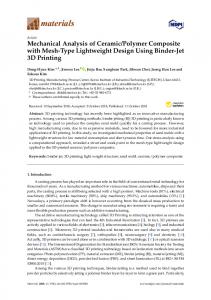
Louis, MO, USA) were used without further purification. Barium boroaluminosilicate glass particles purification. Barium boroaluminosilicate glass particles were obtained from Caulk/ Dentsply (Milford, were obtained from Caulk/ Dentsply (Milford, DE, USA). DE, USA). 2.2. Synthesis of Self-Healing Microcapsules 2.2. Synthesis of Self‐Healing Microcapsules Microcapsules (MCS) were prepared using an in-situ polymerization of formaldehyde via the Microcapsules (MCS) were prepared using an in‐situ polymerization of formaldehyde via the urea method, as described previously [28]. First, DHEPT was added to the TEGDMA monomer at urea method, as described previously [28]. First, DHEPT was added to the TEGDMA monomer at 1 1wt %. A mixture of 50 mL of distilled water and 13 mL of a 2.5% aqueous solution of EMA copolymer wt %. A mixture of 50 mL of distilled water and 13 mL of a 2.5% aqueous solution of EMA copolymer was prepared in a 250 mL-Erlenmeyer flask. The flask was suspended in a water bath on a hot was prepared in a 250 mL‐Erlenmeyer flask. The flask was suspended in a water bath on a hot plate. plate. Second, the shell-forming material urea (1.25 g), ammonium chloride (0.125 g), and resorcinol Second, the shell‐forming material urea (1.25 g), ammonium chloride (0.125 g), and resorcinol (0.125 (0.125 g) were added into the solution under 300 rpm agitation by a magnetic stir bar (Ø = 7.8 mm, g) were added into the solution under 300 rpm agitation by a magnetic stir bar (Ø = 7.8 mm, length = length = Fisher 50 mm,Scientific). Fisher Scientific). Resorcinol was added the reaction of shell formation to enhance 50 mm, Resorcinol was added in the in reaction of shell formation to enhance the the rigidity of the shell [30]. The pH was adjusted to 3.5 by the drop-wise addition of 1 M NaOH. rigidity of the shell [30]. The pH was adjusted to 3.5 by the drop‐wise addition of 1 M NaOH. Then, Then, the agitation rate was increased to 400 rpm, and 30 mL of the TEGDMA-DHEPT liquid was the agitation rate was increased to 400 rpm, and 30 mL of the TEGDMA‐DHEPT liquid was added added into the flask [31]. After 10 min of agitation, a stabilized emulsion of fine TEGDMA-DHEPT into the flask [31]. After 10 min of agitation, a stabilized emulsion of fine TEGDMA‐DHEPT droplets droplets were formed. Then, 3.15 g of a 37% aqueous solution of formaldehyde was added. The stirring were formed. Then, 3.15 g of a 37% aqueous solution of formaldehyde was added. The stirring was was continued heating to °C 55 ◦for C for 4h [28].In Inthis thisprocess, process,ammonium ammonium chloride chloride catalyzed catalyzed the continued with with heating to 55 4 h [28]. the reaction of urea with formaldehyde to form PUF at the oil–water interface to develop the shell [29]. reaction of urea with formaldehyde to form PUF at the oil–water interface to develop the shell [29]. The resulting microcapsules were rinsed with water and acetone, vacuum filtered, and air dried for The resulting microcapsules were rinsed with water and acetone, vacuum filtered, and air dried for 24 h. The microcapsules’ structure was confirmed by optical microscope 4× (TE2000-S, Nikon, Japan) 24 h. The microcapsules’ structure was confirmed by optical microscope 4× (TE2000‐S, Nikon, Japan) and SEM (Quanta 200, FEI, Hillsboro, OR, USA), as shown in Figure 1. and SEM (Quanta 200, FEI, Hillsboro, OR, USA), as shown in Figure 1.
Figure 1. 1. Transmitting image of of resulting resulting poly poly (urea-formaldehyde) (urea‐formaldehyde) (PUF) (PUF) microcapsules microcapsules Figure Transmitting optical optical image loaded with polymerizable TEGDMA and N,N‐dihydroxy ethyl‐p‐toluidine (DHEPT) (an average average loaded with polymerizable TEGDMA and N,N-dihydroxy ethyl-p-toluidine (DHEPT) (an diameter of 73 ± 31 μm). diameter of 73 ± 31 µm).
2.3. Concepting MPC and Self‐Healing Microcapsules into Composite 2.3. Concepting MPC and Self-Healing Microcapsules into Composite A parental composite formulation was made by mixing the following components: Bis‐GMA A parental composite formulation was made by mixing the following components: Bis-GMA and TEGDMA at a mass ratio of 1:1, 0.2% camphorquinone, 0.8% ethyl 4‐N,N‐dimethylamino benzo, and TEGDMA at a mass ratio of 1:1, 0.2% camphorquinone, 0.8% ethyl 4-N,N-dimethylamino and barium boroaluminosilicate glass (mean particle size of 1.4 mm) silanized with 4% 3‐ benzo, and barium boroaluminosilicate glass (mean particle size of 1.4 mm) silanized with methacryloxypropyltrimethoxy silane and 2% n‐propylamine. MPC, a methacrylate with a 4% 3-methacryloxypropyltrimethoxy silane and 2% n-propylamine. MPC, a methacrylate with phospholipid polar group in the side chain, was used as the protein‐repellent agent. MPC was a phospholipid polar group in the side chain, was used as the protein-repellent agent. MPC was synthesized according to a reported method [15]. synthesized according to a reported method [15]. The MPC powder was mixed with photo‐activated BisGMA‐TEGDMA resin at mass fractions The MPC powder was mixed with photo-activated BisGMA-TEGDMA resin at mass fractions of of 7.5%. Then, the composite was then mixed with microcapsules at microcapsule mass fractions of 7.5%. Then, the composite was then mixed with microcapsules at microcapsule mass fractions of 10%. 10%. The chosen mass fractions were selected considering previous investigations of the relationship between the MPC and microcapsules mass fraction and the mechanical properties of the composite [21,28].
J. Funct. Biomater. 2017, 8, 40
4 of 12
The chosen mass fractions were selected considering previous investigations of the relationship between the MPC and microcapsules mass fraction and the mechanical properties of the composite [21,28]. 2.4. Protein Repellence Essay For the protein adsorption and live/dead essays, each composite paste was placed into disc molds (Ø = 9 mm; 2 mm in thickness). They were light-cured, stored in distilled water at 98.6 ◦ F for 24 h, and sterilized by ethylene oxide, following a previous study [20]. The amount of protein adsorbed on the composite discs was determined by the micro bicinchoninic acid method [32]. Six disks were evaluated for each group. Each disk was immersed in phosphate buffered saline (PBS) for 2 h before immersing in 4.5 g/L bovine serum albumin (BSA) solutions at 37 ◦ C for 2 h. The disks then were rinsed with fresh PBS by stirring method (300 rpm for 5 min). The adsorbed protein was detached in sodium dodecyl sulfate (SDS) 1 wt % in PBS by sonication for 20 min. A protein analysis kit (Micro BCA protein assay kit, Fisher Scientific, Pittsburgh, PA, USA) was used to determine the BSA concentration in the SDS solution. The amount of protein adsorbed on the resin disk surface was calculated from the concentration of protein [22]. 2.5. Live/Dead Staining of Biofilms Fluorescence microscopy via live/dead assay was used to directly visualize the early bacterial attachment (4 h after inoculum) over the studied materials. A dental plaque microcosm biofilm model using human saliva was used to promote the biofilm grown over the composites, according to a previous report [33]. The biofilms on the disks were gently washed three times with phosphate-buffered saline (PBS), and then stained using a live/dead bacterial viability kit (Molecular Probes, Eugene, OR, USA). Live bacteria were stained with Syto 9 to produce a green fluorescence, and bacteria with compromised membranes were stained with propidium iodide to produce a red fluorescence [19]. The corresponding images were acquired using appropriate selective filters in the epifluorescence microscope (TE2000-S, Nikon, Melville, NY, USA). The area of green staining (live bacteria) was computed with NIS-Elements imaging software (Nikon, Melville, NY, USA). The area fraction of live bacteria was calculated based on green staining area/total area of the image. 2.6. Assessment of Mechanical Properties 2.6.1. Flexural Strength and Elastic Modulus Testing Following previous studies, composites specimens were placed in metal molds, photo-cured (Triad 2000, Dentsply, York, PA, USA) for 1 min on each side and then smoothed, which produced bar specimens with dimensions of 2 mm× 2 mm× 25 mm (n = 6) [29,34]. Twenty-four hours after manufacturing and water storage, specimens were subjected to three-point flexural testing using a computer-controlled Universal Testing Machine—UTM (5500R, MTS, Cary, NC, USA). All tests were performed using a span of 10 mm and a crosshead speed of 1 mm/min. Flexural strength (FS) was measured as FS = 3PmaxL/(2bh2 ), where P max is the load-at-failure, L is a span, b is specimen width, and h is thickness. Elastic modulus (E) was measured as E = (P/d)(L3/[4bh3]), where load P divided by displacement d is the slope in the linear elastic region of the load-displacement curve [34,35]. 2.6.2. Fracture Toughness and Self-Healing Assessment Fracture toughness (KIC ) was measured using a single edge V-notched beam (SEVNB) method [24]. The SEVNB composite bars specimens for fracture toughness measurement were prepared following a previous protocol [28,29]. After notching the bending bars with a razor blade and 3 µm-diamond suspensions (average notch: depth 700–800 µm; tip radius = 20 µm), the fracture toughness was determined by the same three-point bending test. The photo-cured composites containing the MPC and microcapsules as well the related controls were tested. This yielded the original virgin KIC of the specimens (KIC-virgin ).
J. Funct. Biomater. 2017, 8, 40
5 of 12
Before testing the self-healing assessment, the two halves of the specimen were attached to the metal mold and placed in a humidor at 37 ◦ C for 24 h. After the fracture, the healing process was trigged [30]. In this period, the disrupted microcapsules released the healing agent TEGDMA-DHEPT, J. Funct. Biomater. 2017, 8, 40 5 of 12 which reacts with the BPO in the resin matrix. The mix of these two components would cause the cause the polymerization of the released liquid to heal and bond the two cracked planes into one polymerization of the released liquid to heal and bond the two cracked planes into one cohesive cohesive specimen. The sample was fractured again using the same method, and the new fracture specimen. The sample was fractured again using the same method, and the new fracture toughness toughness (K IC healed ) was calculated.[31 ]The self‐healing efficiency (ή) was assessed as the percentage (K calculated [31]. The self-healing efficiency (ή) was assessed as the percentage of fracture IC healed ) was of fracture toughness after the healing in comparison with the virgin (ή = K IC‐healed K toughness after the healing in comparison with the virgin (ή = KIC-healed KIC-virgin × IC‐virgin 100). × 100).
2.7. Statistical Analysis 2.7. Statistical Analysis Statistical 3.5 3.5 (Systat, San San Jose, Jose, CA, CA, USA). The Statistical evaluations evaluations were wereperformed performedwith withSigmaStat SigmaStat (Systat, USA). normality distribution of the data and equality of variances were checked using the Kolmogorov– The normality distribution of the data and equality of variances were checked using the Smirnov test and Levene’s test, Levene’s respectively. As the data As were distributed, analysis of Kolmogorov–Smirnov test and test, respectively. thenormally data were normally distributed, variance (ANOVA) and the Tukey test were applied at a significance level of p