126
Int. J. Advanced Media and Communication, Vol. 3, Nos. 1/2, 2009
Designing a wireless sensor system for continuous monitoring of the cervical dilation of a pregnant woman Pramode Verma*, Anjan K. Ghosh, Robert C. Huck, Samuel Cheng and Shanshan Chen Telecommunication Engineering Program, School of Electrical and Computer Eng., University of Oklahoma Tulsa, Tulsa, OK 74135, USA E-mail:
[email protected] E-mail:
[email protected] E-mail:
[email protected] E-mail:
[email protected] E-mail:
[email protected] *Corresponding author
Mark Martens Planned Parenthood of Arkansas and Eastern Oklahoma, Inc., 5780 S. Peoria Ave., Tulsa, OK 74105, USA E-mail:
[email protected]
Anil Kaul Houston Center Ob. Gyn., Oklahoma State University – Center for Health Sciences, Tulsa, OK 74107, USA E-mail:
[email protected] Abstract: A novel electronic sensor system is designed to continuously monitor the dilation of the cervix of a pregnant female approaching labour and delivery. The system is capable of wirelessly transmitting the progress of dilation to a central monitoring agency that can get in touch with the attending physician and/or the hospital of the pregnant female. Using wireless telephony, the monitoring agency and/or the physician can advise the female about the medical care she needs at that moment, even though the female might be at her home or away from home, and away from the medical facility. This is an expanded version of a paper presented at the 3rd IEEE International Workshop on Medical Measurements and Applications, 9–10 May 2008, Ottawa, ON, Canada.
Copyright © 2009 Inderscience Enterprises Ltd.
Designing a wireless sensor system Keywords: wireless networking; electronic sensors; birth monitoring; RF design; ZigBee protocol. Reference to this paper should be made as follows: Verma, P., Ghosh, A.K., Huck, R.C., Cheng, S., Chen, S., Martens, M. and Kaul, A. (2009) ‘Designing a wireless sensor system for continuous monitoring of the cervical dilation of a pregnant woman’, Int. J. Advanced Media and Communication, Vol. 3, Nos. 1/2, pp.126–138. Biographical notes: Pramode Verma obtained his Doctorate in Electrical Engineering from Concordia University in Montreal, Canada in 1970 and an MBA from the Wharton School of the University of Pennsylvania in 1984. He is the author/co-author of over 75 publications and several books in telecommunications, computer communications and related fields. In his last position with Lucent Technologies as Managing Director – Business Development, Global Service Providers Business and Business Communications System, his responsibilities included creating strategic alliances and partnerships with leading organisations. He also held professional and management positions with Lucent Technologies – Bell Laboratories for 15 years. Anjan K. Ghosh has more than 25 years of research and teaching experience in the areas of optical information processing, optical communications and photonic sensors and instrumentation. He obtained his Doctorate in Electrical Engineering from Carnegie-Mellon University, Pittsburgh. He was the Head of the Department of Electrical Engineering, Adv. Center for Electronic Sciences, Laser Technology Program and the Center for Laser Technology, all at NT Kanpur. He has published over 140 papers in journals and conference proceedings. Robert C. Huck retired from the US Air Force in 1997 after 20 years of service. Upon retirement, he received his BS in Computer Engineering and an MS in Electrical and Computer Engineering at the University of Oklahoma. He is currently pursuing his PhD in Engineering. He is currently the manager of the Telecommunications Interoperability Lab at OU-Tulsa. He has been awarded two US patents and has authored and co-authored several conference publications in the areas of FSO and FSO tracking. He is a member of Tau Beta Pi, Eta Kappa Nu, and the IEEE since 1998. Samuel Cheng received the PhD Degree in Electrical Engineering from Texas A&M University in 2004. He worked in Microsoft Asia, China, and Panasonic Technologies Company, New Jersey, in the areas of texture compression and digital watermarking during the summers of 2000 and 2001. He joined the School of Electrical and Computer Engineering at the University of Oklahoma as an Assistant Professor since 2006. He has been awarded four US patents in various signal processing areas. He is a member of IEEE, Sigma Xi, and AAAS. His research interests include image/signal processing, information theory, and pattern recognition. Shanshan Chen received her Bachelor’s Degree in Electrical Engineering from Nanjing University of Science and Technology, China in 2006. Later she continued to pursue her Master’s Degree at the University of Oklahoma Tulsa, and received her MS in Telecommunication Engineering in 2008. She is currently working as a research associate in the Interoperability Lab at OU-Tulsa. Her general research interests include wireless sensor network, network security and network design.
127
128
P. Verma et al. Mark Martens graduated from Kenyon College in Ohio and attended graduate school in microbiology at Northwestern University in Chicago. He then attended medical school at George Washington University School of Medicine and Health Sciences in Washington, DC. He completed his residency in Obstetrics and Gynecology at Hartford Hospital in Connecticut, followed by a Fellowship in Obstetrics and Gynecology Infectious Diseases at Baylor College of Medicine, Houston, Texas. He is currently Chief Medical Officer for the Heartland Health Institute in Tulsa, Oklahoma and Medical Director and Vice President for Research for Planned Parenthood of Arkansas and Eastern Oklahoma, Inc., and Clinical Professor at the Oklahoma State University, Department of Obstetrics and Gynecology. Anil Kaul is a Graduate in Medicine and Dentistry with a Master’s in Public Health Administration from the University of Minnesota. After his fellowship in the Department of Obstetrics and Gynecology at the University of Texas Medical Branch in Galveston, he joined University of Minnesota as a faculty member and director of Women’s health research. He is currently a visiting faculty at Oklahoma State University. He has published more than 30 scientific papers with more than 100 scientific presentations at national and international scientific meetings; holds multiple patents, particularly in the area of early detection of preterm births. He also serves as a member of the Tulsa Community College biotechnology advisory board.
1
Introduction
Preterm labour and birth has always been a serious issue that challenges obstetricians. In the developed countries, 60–80% of deaths of infants are caused by preterm births without any congenital anomalies (Goldenberg, 2002). As a main cause of neonatal mortality and a substantial portion of all birth-related short- and long-term morbidity, many obstetrical studies have been done in preventing preterm birth before the preterm delivery begins. According to the World Health Organization, a preterm labour is a labour that happens before the 37th week but after the 20th week. Even though a preterm labour does not necessarily lead to a premature delivery, it is responsible for more than half of the preterm births (Goldenberg, 2002). With existing medical techniques, preterm delivery can be prevented if a preterm labour is detected after the 23rd week in gestation. Therefore, it is important to predict the beginning of preterm labour and to inform obstetricians that their patients are under preterm labour. Today, advances in obstetrics have considerably eased the process of assessment of the stage of labour. However, it is still difficult to predict the beginning of labour, even for the experienced mothers. For those who may experience preterm labour, the misjudging of the beginning of labour can lead to premature birth because of the lack of professional medical care. Moreover, infections caused by traditional examination may also lead to preterm births. Thus an accurate and sanitary means of predicting the beginning of labour for patients under the risk of preterm labour is urgently needed. To indicate the beginning of labour, obstetricians use the results of accumulated experience to recognise the symptoms of labour. Most symptoms are related to cervical changes. The cervix is the lower part of the uterus projecting into the vagina. Generally, it is a canal with 2.5–3 cm in diameter and 3–5 cm in length. It is the channel that a baby
Designing a wireless sensor system
129
has to pass through from the uterus into the vagina during delivery. Therefore, symptoms in the cervix during pregnancy can be used to indicate the beginning of labour. The symptoms that are used as indicators of the beginning of labour nowadays are: the degree of softening of the cervix, frequency of contractions of the pregnant woman, and the degree of dilation of the cervix. The softening of the cervix can be tested by experienced examiners with fingers inserted into the vagina. This is rather a late symptom which takes place after the cervix starts to dilate, and it cannot be tested by the pregnant woman herself. As for the frequency of contractions, the pregnant woman has to pass through the whole contraction phase and count the frequency of all the contractions; which makes it impractical to indicate when labour begins. Fortunately, the cervical dilation is a good indicator of the beginning of labour since it is an important precursor to delivery. For most women in labour, their cervices dilate from 0 cm (no dilation at all) to 10 cm (full dilation), so that a full-term baby’s head (around 10 cm) can come across (Rodriguez and Greenfield, 2004). After the cervix dilates to 10 cm, the pushing begins and the baby is delivered. Specifically, from no dilation to full dilation, there are three phases. The first phase is called the latent phase, during which the cervix dilates from 0 cm to 4 cm. The other two phases are Active Labour and Transition phase, respectively, with the indicator of cervical diameter as 4–8 cm, and 8–10 cm (Rodriguez and Greenfield, 2004). Cervical diameter can be defined as the distance across the cervical opening. When the cervix dilates, this diameter increases (van Dessel et al., 1991). However, for monitoring preterm difficulties, we only need to monitor the first stage of labour because after the first phase, a premature delivery cannot be prevented. Therefore, we need to measure cervical diameter continuously over the range of 0–4 cm. However, current monitoring devices for cervical dilation are all limited to hospital usage and performed by trained examiners. This means that the pregnant woman cannot measure the cervical dilation outside the hospital. Because of the disadvantages of the current cervical dilation monitoring devices, there is motivation to explore novel solutions to monitoring cervical dilation. The new solution should be able to give accurate information that indicates the progress of labour. Then, when the device detects a certain degree of cervical dilation, this information should trigger an alarm and notify both the pregnant woman and her medical services provider simultaneously. During this process, the pregnant woman should be able to move freely outside the hospital. Furthermore, the final solution should be affordable. By monitoring the process of cervical dilation, we expect that once the preterm labour begins, the pregnant woman has access to professional medical guidance. We also expect that the accuracy of monitoring can assure the pregnant woman so that she does not need to panic if she notices false indicators of labour not corroborated by dilation of the cervix. The devices for monitoring cervical dilation should give continuous information and should be reliable. It has been proven that the traditional digital examination of cervical opening is inaccurate since the result is not reproducible even when the examination is carried out by the same clinician. This traditional method only allows the examination intermittently thus the variation of cervical change cannot really be measured (Lucidi et al., 2000). Therefore, it is desirable to develop a new sensing technology in order to produce and record accurate and continuous results. Further, the final encased system should fit within the acceptable limits of space around the cervix. The accuracy of measurement of cervical dilation is very important. The information on the progress
130
P. Verma et al.
of cervical dilation actually affects the obstetrician’s decision. Also accurate clinical data can help research in this area. Thus the estimate and test of accuracy of the system should be important for both clinical practice and research (Chen, 2008).
2
An overview of currently available devices for measuring cervical dilation
Several researchers have strived to develop a small, efficient and inexpensive device for measuring cervical dilation. ‘Google Patent’ was used to search existing patents dealing with measuring cervical dilation, with the key word ‘cervical dilation’ and ‘cervix’. The results are listed below in Table 1. Table 1
Chronological list of a few patents on cervical dilation measurement
Patent name
Patent number Inventors
1
Cervical dilation measuring device
US 3768459
Emerson T. Cannon, Utah Research Richard M. Hebertson and Development Co., Inc
28 June 1971
2
Cervical dilation US 4141345 measurement instrument
David W. Allen, John. National Research A. Richardson, Ian A. Development Sutherland Corporation
27 February 1979
3
Cervical dilation meter
US 4682609
Natan Parsons
28 July 1987
4
Cervical dilation meter
US 4719925
Natan Parsons
19 January 1988
5
Method for the diagnosis US 5450857 of cervical changes
R.E. Garfield, W.S. Glassman
6
Cervical ring to detect labour
Robert A. Welch
7
Uterine cervix dilation, US 5876357 effacement, and consistency monitoring system
David Tomer
Labour Control 2 March System (L.C.S.) Ltd. 1999
8
Method and apparatus US 6039701 for monitoring cervical diameter
Jack Sliwa, Lee A. Blumenfeld
OB Innovations, Inc.
9
Device for cervical and US 6066104 pelvic measurement in medical obstetrics
Leland H. Dao, Christopher S. Miura, Michael James Bradley
10 Cervix dilation and labour progression monitor
US 5807281
Assignees
Board of Regents, The University of Texas System
Issue date
19 September 1995 15 September 1998
21 March 2000 23 May 2000
US 6270458
Ofer Barnea
Barnev Inc.
7 August 2001
11 Devices and methods for US 6419646 cervix measurement
Rosalyn P. Baxter-Jones
Cervilenz, Inc.
16 July 2002
12 Devices and methods for US 6450977 cervix measurement
Rosalyn P. Baxter-Jones
Cervilenz, Inc.
17 September 2002
Designing a wireless sensor system Table 1
131
Chronological list of a few patents on cervical dilation measurement (continued)
Patent name
Patent number Inventors
Assignees
Issue date
13 Devices and methods US 6524259 for cervix measurement
Rosalyn P. Cervilenz, Inc. Baxter-Jones, Joseph Stemler, Lindy Yow
25 February 2003
14 Devices and methods US 6802817 for cervix measurement
Rosalyn P. Cervilenz, Inc. Baxter-Jones, Joseph Stemler, Lindy Yow
12 October 2004
15 Cervical dilation monitor
Miki Ben-Cnaan, Ilan Halevi, Barak Halevi
22 November 2005
Rosalyn P. Cervilenz, Inc. Baxter-Jones, Joseph Stemler, Lindy Yow
7 February 2006
US 6966881
16 Devices and methods US 6994678 for cervix measurement
By an examination of all the patents, we classify them into four categories: digital examination, mechanical measurement, electromagnetic measurement and ultrasonic measurement. The advantages and disadvantages of each category are also summarised below. Most of the devices listed above are not automated and require bed-rest and hospitalisation (Chen, 2008).
2.1 Digital examination Digital examination is probably the oldest way of cervical examination. It is based on palpation of the cervix by inserting fingers into the vagina to “detect changes in cervical texture such as softening” (American College of Radiology, 2005). There are three disadvantages of digital examination. First, symptoms such as ripening and softening in cervix actually happen very late in the process of cervical dilation or even too late in some cases to function as a useful indicator. Using these symptoms to indicate cervical dilation might delay the necessary obstetrical intervention during delivery. Second, as a subjective measurement, digital examination cannot guarantee accuracy since the results vary from one examiner to another. In such examination, the same examiner can neither repeat the judgment on the degree of dilation, nor can he or she reliably communicate that degree of dilation to another medical practitioner. Further, this method increases the risk of infection to the uterine cavity, which may cause preterm delivery, as well as causing discomfort to the patient.
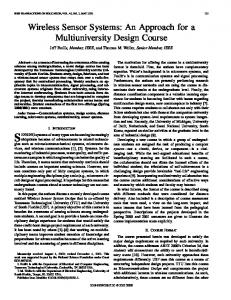
2.2 Mechanical measurement The mechanical calipers with graduation at one end and another end extending into cervix testing cervical dilation are developed to replace the digital measurement. Among all the patents listed in Table 1, patents 2, 3, 4, 6, 7, 8, 10, 11, 12, 13 and 14, are mechanical measurement devices. Among the first attempts to measure cervical dilation, Fireman’s cervimeter (Figure 1) is very typical. With two clips attached to the cervix, the caliper extend when the cervix is dilating, and the distance of the extension
132
P. Verma et al.
is measured on a ruler attached to the device. It is possible to improve this device by introducing a potentiometer to the distal ends of the lever arms and eliminating the ruler (Lucidi et al., 2000). Figure 1
Friedman’s mechanical cervimeter
Source: Lucidi et al. (2000)
When the arms separated of the calipers are separated because of the cervical dilation, the voltage applied on the potentiometer changes. The potentiometric device gives the readings directly and the ruler is not needed. Also the lever arms could be shorter, which reduces the weight of the device (Lucidi et al., 2000). The disadvantage of these devices is the difficulty of placing it in the cervix without causing any cervical distortion.
2.3 Electromagnetic measurement This class of devices is based on the principle of electromagnetic induction. Patent 1 falls into this category. In this patent, a pair of coils forms a transceiver. The coil connected to the signal generator (coil A) will generate a magnetic field. This magnetic field will then induce electric current through the receiving coil (coil B) that is connected to the signal processing system, an amplifier and a visual display. Once the distance between coil A and coil B starts to change, the magnitude of the current received at coil B will be changed. By processing the signal at the receiving end, the distance can be calculated by converting the voltage and displaying it on the visual display. The continuous monitoring feature of electromagnetic measurement makes it distinguished from other measurements, though its accuracy is low. Apart from the distance change, the planes of the induction coils can also cause change in the induced current. It is difficult to keep the planes of the two coils parallel (Lucidi et al., 2000). Due to this reason, the accuracy of this system is reduced.
2.4 Ultrasonic measurement Ultrasonic measurements are based on estimating the travelling time of an ultrasound signal and thus calculating the distance between two objects based on the velocity of the ultrasound. The first ultrasonic cervimeter only consisted of two transducers as small as 1 mm × 1 mm × 5 mm. The two traducers are attached to the opposite sides of the cervix, one generating the ultrasonic signal and the other receiving it. By computing the
Designing a wireless sensor system
133
transmission time, the distance between the two transducers is calculated. The advantages of ultrasonic devices are: •
the device does not bring discomfort or trauma to the pregnant women
•
it has a much higher accuracy compared with the mechanical alternatives.
Ultrasonic measurements can give a maximum error of 3 mm over the range from 2 cm to 10 cm (Lucidi et al., 2000; Cook and Ellwood, 1996). In measuring cervical dilation, ultrasonic devices, seem perfect at first sight. However, the expensive terminals associated with ultrasonic measurements, limit its usefulness as a clinical device. It is largely used as a research tool. This kind of device also limits the freedom of movement of the pregnant woman. Thus, new solutions still need exploring (Yost et al., 1999).
3
The sensor developed for dilation measurement
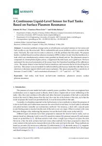
After examining various techniques for monitoring the cervical dilation we decided on an electronic device because such devices can be made in a small size, can be inexpensive, can be packaged to make them implantable inside the body, and such devices can transmit the data wirelessly to the outside world (Kriewall and Work, 1977; Zador et al., 1976; Sharf et al., 2006). In our project, we designed and developed a wireless sensor system for the continuous monitoring of cervical dilation of a female approaching labour and delivery. We designed a sensor that can continuously measure the dilation and transmit this information wirelessly to a transponder worn like a mobile phone or PDA. This transponder can automatically transmit the data on dilation to a remote data-processing agency in contact with the pregnant woman and her doctor and hospital. A schematic diagram of our system is shown in Figure 2 (Chen, 2008). Figure 2
A schematic diagram of the system for monitoring cervical dilation. The arrows represent wireless RF links
134
P. Verma et al.
This sensor will be attached to the cervix of the pregnant female by her doctor. This sensor consists of an electro-mechanical device encased in a non-reactive plastic tube. The sensor stretches as the cervix dilates. A doctor can pull out the whole unit easily when the monitoring is over and the delivery starts. A small ‘active’ circuit or tag with its own battery is attached to the sensor. This circuit determines the diameter of the dilated cervix and transmits the value of the diameter in centimeters through RF to a small wireless unit carried by the pregnant female like a ‘mobile phone’ as shown in Figure 2. This wireless unit is typically worn on a belt or attached to the clothing of the female. The main component of the electromechanical sensor is a solenoid coil. The self-inductance L of an air-core coil is given by: L = 10πµ0 N 2 a 2 (9a + 10l ) −1
(1)
where l is the length of the coil, µ0 is the permeability of air, a is the radius of the coil and N is its number of turns (Thompson, 1999). Equation (1) is an empirical formula for calculating the inductance of a single-layer coil. This formula achieves an accuracy of 1% when the length l > 0.8a. Equation (1) clearly shows that if the length l of the coil is changed the self-inductance also changes inversely. Thus, we envision an especially prepared coil attached to the two diametrically opposite sides of the cervical opening for measuring the diameter of the dilating cervix. A simple circuit is designed to convert the change in the inductance L to a voltage V(L). Since the frequency of the cervical dilation is less than 10 Hz, the voltage V(L) is a reliable measure of the length l and hence of the diameter of the cervix. One of the considerations in our design is the fact that the sensor will be implanted around the cervix along with other miniaturised devices. Before dilation, the cervix, on average, is 3 cm long and 2.5 cm wide. This greatly limits the size of the final device. In order to accommodate the device, the space at the end of the vagina with a diameter around 6 cm can be utilised. Meanwhile, the sensing core of our system will still be attached to the cervix in order to sense the cervical dilation. But with the extra room in the vagina, we can actually manufacture a device with a size limited of 6 cm. Another consideration in the design concerns the mechanical properties of the device. Since the cervix should actually stretch the coil using very little force, the coil should be easily expandable. According to Hooke’s law (Hibbeler, 2007), we have:
χ = Fl /(π Ea 2 ).
(2)
where χ is the distance that the coil is elongated, F is the force exerted on the coil and E is the young’s modulus of the wire. Therefore, given l and E a coil with a larger area will need less force to be elongated. After various trials and calculations, we fabricated solenoids of outer diameter of 6.35 mm with about 60 turns. We chose a silver based alloy as our material. Silver gives a good ductility to the wire. The material chosen provides excellent fatigue and strength properties and is nontoxic. The solenoids fabricated were ‘soft’ and required about a little force to stretch. Since the electromechanical sensor is attached to the cervix in the vagina and no wire should be attached from the sensor to the outside the data must be sent wirelessly from the sensor. This data delivery has to be wireless to allow mobility of the patient. Among the many wireless technologies prevailing today, it was decided to use ZigBee-based wireless transceivers.
Designing a wireless sensor system
135
ZigBee is a wireless transmission protocol implemented on top of the IEEE 802.15.4 radio communication standard. As it is designed for Wireless Personal Area Networks (WPANs), IEEE 802.15.4 differentiates itself by focusing on lower data rate transmission and lower power consumption compared to other wireless data transmission standards. Inheriting these features from IEEE 802.15.4, ZigBee technology is “targeted towards automation and remote control applications” (Ergen, 2004). ZigBee has the advantage of being a low-power and low-cost device, though allowable transmission distance is limited to 10–75 m, depending on the environment. The data rate ranges from 20 kbits/s to 250 kbits/s, which is not a high-data rate compared to other peer technologies such as Bluetooth or Ultra Wideband (UWB). ZigBee is used widely in Radio-Frequency (RF) applications that require a low data rate, a long battery life, and a secure network. In our case, only a short-distance wireless transmission is needed. This transmission does not require a high data rate for two reasons. First, although real-time update of the cervical dilation data is required, a delay of up to 1 min is acceptable. Second, the update interval can be up to 5 min or longer. This is based on the average interval of the cervical dilation during labour, which is counted in minutes (Turon, 2005). A data rate of 250 kbps can satisfy our requirements of data delivery. Meanwhile, since the final device is sealed and will only be powered once, the power supply has to last until the device is disposed. This requires that the device consume very little power. The features of ZigBee technology such as low-data rate and low-power consumption meet these requirements. The wireless unit has the ability of automatically dialing the telephone number of a medical data processing agency. The wireless unit does the dialing at a predetermined interval, for example once every several minutes. Once the contact with the data processing agency is established, the unit downloads the cervical dilation data and other information on the female to the data processing agency. The agency checks the progress of the cervical dilation and provides some basic instructions to the female over her mobile phone. If necessary, the agency also gets in touch with the physician and the clinic of the pregnant female. The doctor and the clinic may advise the female as appropriate. The basic sensor and the wireless transceiver system were assembled in our communications laboratory. The result of an experiment on the behaviour of the sensor is shown in Figure 3. The horizontal axis of the graph represents the length of the sensor in centimeters. Figure 3 clearly shows that the dc voltage generated by the sensor is inversely proportional to the length of the sensor as it is stretched from its original length of 3–10 cm. In Figure 3, the straight line without small circles represents the best-fit model obtained through linear regression analysis of the experimental data. Since passive and non-magnetic electromechanical device is used for monitoring the length, there is almost no hysteresis in the observed response of the sensor system. The information on the length of the sensor was transmitted wirelessly using a commercially available wireless sensor network component called MICA(TM) motes numbered MDA320, MIB510 and MPR2400 made by Crossbow, Inc. to the sensor circuit (Turon, 2005; Crossbow, 2005). The motes converted the voltage to digital data that was transmitted wirelessly at the frequency of 2.54 GHz to a RF receiver-mote kept several meters away from the sensor. In Figure 4, we show the value of the data received in the remote receiver. The horizontal axis in Figure 4 shows time in seconds and the vertical axis represents the voltage output of the sensor. The length measured by the sensor was varied manually and randomly during the observation period.
136
P. Verma et al.
Figure 3
Experimental data on linear variation of output voltage vs. length of the sensor (in cm) as it is stretched by pulling at its ends
Figure 4
An oscilloscope trace showing the output of the RF receiver corresponding to the voltage data transmitted wirelessly
The wireless unit should be designed with the capability of automatically dialing the telephone number of a medical data processing agency, such as Matria(TM) in Atlanta, GA (Verma et al., 2008). The dialing is done at a predetermined interval, say, every 20 min. Once the contact with the data processing agency is established the unit downloads the cervical dilation data and other information on the female to the data processing agency. The dialing interval can be remotely adjusted as required for medical reasons. The agency checks the progress of the cervical dilation and provides some basic instructions to the female over her mobile phone. If necessary, the agency also gets in touch with the doctor and the clinic of the pregnant female. The doctor and the clinic may then advise the female as appropriate. The authors note that some studies have related wireless transmission, such as the use of a mobile phone close to the brain, to a potentially higher incidence of brain tumour. Although within the context of this research no specific theoretical or practical studies/measurements have been carried out, we believe that there will be no adverse
Designing a wireless sensor system
137
impact due to the use of the wireless technology on the health of the fetus. The reasons are based on the following two factors. First, the wireless signal in our case will have an en extremely low level of power because it has to travel a distance of only one or two feet, as opposed to several miles in the case of mobile telephony. Second, the frequency of transmission in our case can be controlled so that the wireless signal is transmitted every few minutes as opposed to on a continuous basis in the case of mobile telephony. Taken together, both these factors should mitigate the adverse impact of the device on the fetus.
4
Conclusions
In this paper, we have reviewed the need for measuring cervical dilation as a warning for preterm labour. Various inventions for measuring the cervical dilations have been reviewed. We have presented the design of a novel measuring device that transfers the actual dilation information to hand-held device worn on the body of a pregnant female. This information can also be transferred to a remote point, enabling intervention by the medical personnel as necessary when indicated by the instrument.
References American College of Radiology (2005) Premature Cervical Dilation, ACR Appropriateness Criteria®. Chen, S. (2008) Continuous Wireless Monitoring of Cervical Dilation, Master’s Thesis, The University of Oklahoma, August. Cook, C.M. and Ellwood, D.A. (1996) ‘A longitudinal study of the cervix in pregnancy using transvaginal ultrasound’, British Journal of Obstetrics and Gynecology, Vol. 103, January, pp.16–18. Crossbow (2005) Crossbow Technology, Inc., XServe User’s Manual, Xbow.com, 2005–2007. Ergen, S.C. (2004) ZigBee/IEEE 802.15.4 Summary, Technical Document, University of Berkeley, September. Goldenberg, R.L. (2002) ‘The management of preterm labor’, The American College of Obstetricians and Gynecologists, Vol. 100, November, pp.1020–1037. Hibbeler, R.C. (2007) Mechanics of Materials, 7th ed., Prentice-Hall, Englewood Cliffs, NJ. Kriewall, T.J. and Work, B.A. (1977) ‘Measuring cervical dilation in human parturition using the Hall effect’, Medical Instrumentation, Vol. 11, February, pp.26–31. Lucidi, R.S., Blumenfeld, L.A. and Chez, R.A. (2000) ‘Cervimetry: a review of methods for measuring cervical dilation during labor’, Obstetrical and Gynecological Survey, Vol. 55, May, pp.312–320. Rodriguez, L. and Greenfield, M. (2004) Examination During Labor, DrSpock.com, 14 August. Sharf, Y., Farine, D., Batzalel, M., Megel, Y., Shenhav, M., Jaffa, A. and Barnea, O. (2006) ‘Continuous monitoring of cervical dilatation and fetal head station during labor’, Medical Engineering and Physics, Vol. 29, January, pp.61–67. Thompson, M.T. (1999) ‘Inductance calculation techniques’, Power Control and Intelligent Motion, Vol. 25, December, pp.40–45. Turon, M. (2005) ‘MOTE-VIEW: a sensor network monitoring and management tool’, IEEE Embedded Networked Sensors, EmNetS-II, Nos. 30–31, May, pp.11–18.
138
P. Verma et al.
van Dessel, T., Frijns, J.H.M., Kok, F.Th.J.G.Th. and Wallenburg, H.C.S. (1991) ‘Assessment of cervical dilation during labor: a review’, European Journal of Obstetrics and Gynecology and Reproductive Biology, Vol. 41, March, pp.165–171. Verma, P., Ghosh, A.K., Huck, R.C. et al. (2008) ‘Continuous wireless monitoring of the cervical dilation of a pregnant woman’, MeMeA 2008 – IEEE International Workshop on Medical Measurements and Applications Proceedings, Ottawa, Canada, pp.93–96. Yost, N.P., Bloom, S.L., Twickler, D.M. and Leveno, K.J. (1999) ‘Pitfalls in ultrasonic cervical length measurement for predicting preterm birth’, Obstetrics and Gynecology, Vol. 93, April, pp.511–516. Zador, I., Neuman, M.R. and Wolfson, R.N. (1976) ‘Continuous monitoring of cervical dilatation during labor by ultrasonic transit-time measurement’, Medical and Biological Engineering, Vol. 14, pp.299–305.