REVIEW published: 31 January 2017 doi: 10.3389/fmicb.2017.00109
Detection and Enumeration of Spore-Forming Bacteria in Powdered Dairy Products Aoife J. McHugh 1,2 , Conor Feehily 1,3 , Colin Hill 2,3 and Paul D. Cotter 1,3* 1
Food Bioscience Department, Teagasc Food Research Centre, Cork, Ireland, 2 School of Microbiology, University College Cork, Cork, Ireland, 3 APC Microbiome Institute, Cork, Ireland
Edited by: Edward M. Fox, CSIRO Food and Nutrition, Australia Reviewed by: Mark Turner, University of Queensland, Australia Heather Craven, CSIRO Food and Nutrition, Australia *Correspondence: Paul D. Cotter
[email protected]
With the abolition of milk quotas in the European Union in 2015, several member states including Ireland, Luxembourg, and Belgium have seen year on year bi-monthly milk deliveries to dairies increase by up to 35%. Milk production has also increased outside of Europe in the past number of years. Unsurprisingly, there has been a corresponding increased focus on the production of dried milk products for improved shelf life. These powders are used in a wide variety of products, including confectionery, infant formula, sports dietary supplements and supplements for health recovery. To ensure quality and safety standards in the dairy sector, strict controls are in place with respect to the acceptable quantity and species of microorganisms present in these products. A particular emphasis on spore-forming bacteria is necessary due to their inherent ability to survive extreme processing conditions. Traditional microbiological detection methods used in industry have limitations in terms of time, efficiency, accuracy, and sensitivity. The following review will explore the common spore-forming bacterial contaminants of milk powders, will review the guidelines with respect to the acceptable limits of these microorganisms and will provide an insight into recent advances in methods for detecting these microbes. The various advantages and limitations with respect to the application of these diagnostics approaches for dairy food will be provided. It is anticipated that the optimization and application of these methods in appropriate ways can ensure that the enhanced pressures associated with increased production will not result in any lessening of safety and quality standards.
Specialty section: This article was submitted to Food Microbiology, a section of the journal Frontiers in Microbiology
Keywords: next generation sequencing, spore-forming bacteria, dairy, dairy powder, pathogens
Received: 27 July 2016 Accepted: 16 January 2017 Published: 31 January 2017
The European Union’s removal of milk quotas in April, 2015 led to a 2% increase in milk deliveries to dairies in the EU for 2015. Some countries are taking full advantage of the new limitless system in the EU, with Ireland, Luxemburg, and Belgium increasing bi-monthly milk deliveries to dairies by in excess of 20% (Eurostat, 2016). Although the production rate has slowed in some other major dairy exporters, including New Zealand and Australia, the US has seen continued increases in production (Dairy Australia, 2015; DCANZ, 2016; USDA, 2016). The surplus milk produced can be processed into a wide variety of dairy products, including yogurt, butter, cheeses, and dairy powders. Dairy powders are a popular commodity due to their long shelf life, ease of storage and versatile nature. A wide variety of dairy powders can be produced, each with
Citation: McHugh AJ, Feehily C, Hill C and Cotter PD (2017) Detection and Enumeration of Spore-Forming Bacteria in Powdered Dairy Products. Front. Microbiol. 8:109. doi: 10.3389/fmicb.2017.00109
INTRODUCTION
Frontiers in Microbiology | www.frontiersin.org
1
January 2017 | Volume 8 | Article 109
McHugh et al.
Spore-Forming Bacteria in Dairy Powder
other than Bacilli have also been found to contaminate powdered dairy products with species reported including Clostridium halophilum, Klebsiella oxytoca (Buehner et al., 2015), C. perfringens, C. septicum, C. novyi/haemolyticum, C. sporogenes (Barash et al., 2010), Staphylococcus aureus (Zhang et al., 2015), and Cronobacter sakazakii (Minami et al., 2012). Bacteria of the genus Clostridium, as well as many of the contaminants of the class Bacilli (Table 1), including Bacillus, Anoxybacillus, Geobacillus, Lysinibacillus, Brevibacillus, and Paenibacillus, have a considerable advantage due to being capable of forming stressresistant endospores. These genera, and their associated species, vary considerably with respect to the range of temperatures in which they can grow, and include some psychrophilic (Ivy et al., 2012) and thermophilic (Burgess et al., 2010; Watterson et al., 2014) species. Dairy product contaminating spore-formers can also differ by virtue of preferring anaerobic (Doyle et al., 2015) or aerobic (Gopal et al., 2015) conditions. Although many sporeformers are not pathogenic and are seen primarily as indicators of poor hygiene during milk collection and or processing (Burgess et al., 2010), some can cause disease (Andersson et al., 1995). Of the spore-formers identified in powders, specific representatives of Clostridium spp. and Bacillus spp. are the most worrying from a food safety point of view. Clostridium are anaerobic spore-formers, of which C. botulinum is the most notorious due to its highly potent botulinum toxin. There are many types of botulism including foodborne botulism, wound botulism, infant botulism and adult intestinal botulism. Infant botulism is the most common form (Sobel, 2005). Strains of C. botulinum isolated clinically have been identified in containers of opened milk powder from the home of patients with infant botulism (Brett et al., 2005; Johnson et al., 2005). Despite this, and although many species of Clostridium have been identified in dairy powders (Barash et al., 2010; Buehner et al., 2015), dairy powders have never been found to be responsible for a case of infant botulism (Brett et al., 2005; Johnson et al., 2005; Doyle et al., 2015). However, it is worth noting that anaerobic spore-forming bacteria, like C. botulinum, are less common than aerobic spore-formers in dairy powders. This may be due to the high degree of aeration involved in dairy powder processing or that testing criteria for spore-formers has been optimized to identify aerobic spore-formers except in the case of phenotype based assays for specific groups of anaerobic species. The ability of certain Clostridium species to reduce sulphite to sulfide under anaerobic conditions resulting in black colonies on specific media has been widely utilized. The accuracy of these qualitative and quantitative approaches has previously been discussed (Doyle et al., 2015). Of the aerobic spore-formers identified, the majority have been of the genus Bacillus (Table 1). Many species of this genus are generally regarded as safe and some are even used as probiotics (Hong et al., 2005); e.g., Bactisubtil, Biovicerin and Biosubtyl containing B. cereus, Bidisubtilis containing B. subtilis, Biosporin and Primal Defense containing B. subtilis and B. licheniformis, Biosubtyl containing B. pumilus, Enterogermina containing B. clausii and Lactospore containing B. coagulans (Hong et al., 2005). Other species of Bacillus have been used in the production of animal feed-stuffs; e.g., B. subtilis has been utilized for
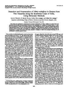
individual properties. These include whole milk powder (WMP), skimmed milk powder (SMP), whey protein concentrate (WPC), whey protein isolate (WPI), milk protein concentrate (MPC), milk protein isolate (MPI), casein and caseinates (Lagrange et al., 2015). Dairy powders can be used in fortification of other dairy products (Karam et al., 2013), as well as an ingredient in a wide array of foods including soups and sauces, confectionary (Sharma et al., 2012), infant formula, sports dietary supplements and in foods for health recovery (Gill et al., 2001; Lagrange et al., 2015). However, the increased production of dairy powders may create safety and economic risks to the dairy sector, specifically when controlling microbial loads in these products. Several key steps are involved in producing dairy powders including pasteurization, separation, evaporation, and spray drying (Figure 1). These thermal and mechanical processes can reduce the microbes present in the milk. However, spore forming bacteria may survive. It has been shown that the spore-forming bacterial composition of raw milk differs considerably from their associated dairy powders (Miller et al., 2015), highlighting that the processing of milk into powder changes the composition of the specific spore-formers present. Post-production, powders can be stored for extended periods and in the absence of water, bacterial metabolic activity and growth is limited (Deng et al., 2012), thus preventing spoilage and product defects. However, under these conditions, bacterial spores can remain dormant until more favorable conditions are encountered, when germination and outgrowth can proceed (Setlow, 2003, 2014).
BACTERIAL CONTAMINANTS OF DAIRY POWDERS Sources of Bacterial Contamination of Dairy Powders Spore-forming bacteria can contaminate dairy powders through a variety of means. Bacteria can originate from the soil (Heyndrickx, 2011), feces, bedding, feed, or milking equipment (Gleeson et al., 2013), or can enter the raw milk via contaminated teats, milking cups and bulk tanks. Additionally, contamination can occur during transport from the farm to the processing plant (Pantoja et al., 2011), and also within the processing facility itself from poor handling and contaminated equipment (Burgess et al., 2010; Faille et al., 2014). The formation of homogeneous or heterogeneous multicellular bacterial communities on the surface of processing equipment in the form of biofilms is a particular concern for the dairy processing sector and, when present, can lead to recurring problems of microbial contamination. The biofilms, which are themselves resistant to cleaning, can serve as a reservoir for bacterial spores which can slough off and contaminate dairy powders (Branda et al., 2001; Faille et al., 2014).
Common Bacterial Contaminants Common contaminants identified in dairy powders include species of the class Bacilli (Table 1), many of which are capable of forming endospores (Checinska et al., 2015). Taxa
Frontiers in Microbiology | www.frontiersin.org
2
January 2017 | Volume 8 | Article 109
McHugh et al.
Spore-Forming Bacteria in Dairy Powder
FIGURE 1 | Sample dairy powder production pipelines.
from within this group. B. cereus strains can contain many enterotoxins which are associated with diarrheal food poisoning including non-hemolytic enterotoxin (Nhe; Lund and Granum, 1996; Lindback et al., 2004), hemolysin BL (Hbl; Beecher and Wong, 1997), and cytotoxin K (CytK; Lund et al., 2000). It should be noted that the description of CytK as a viable enterotoxin has been called into question as, in isolation, the presence of the corresponding gene has not been linked to virulence in diarrheal pathogenesis (Castiaux et al., 2015). Other molecules previously thought to be enterotoxins associated with food poisoning but which have since been reclassified include EntFM (Tran et al., 2010) and BcET (Choma and Granum, 2002). Some strains of B. cereus also produce an emetic toxin, cereulide (Ces), a product of non-ribosomal peptide synthesis, which can cause emetic food poisoning (Horwood et al., 2004; Toh et al., 2004).
the fermentation of indigestible by-products of soya bean oil production to yield a suitable food source for monogastric animals (Wongputtisin et al., 2014). B. cereus sensu lato is the most important group of species identified from a pathogenic perspective (Bottone, 2010). This group, containing up to 11 individual, highly related species (Okstad and Kolsto, 2011; Liu et al., 2015), includes species that are regarded as nonpathogenic (Okstad and Kolsto, 2011). Other species include B. thuringiensis which is used as pesticides (Schnepf et al., 1998; Bravo et al., 2013); B. cereus, a class 2 pathogen capable of food poisoning which gave this species group its name (Bottone, 2010) and even a class 3 human pathogenic species, B. anthracis (Rasko et al., 2005). All of these are notoriously difficult to classify and differentiate from each other (Helgason et al., 2000; Radnedge et al., 2003; Rasko et al., 2005; Liu et al., 2015). B. cereus is the main cause of food poisoning
Frontiers in Microbiology | www.frontiersin.org
3
January 2017 | Volume 8 | Article 109
McHugh et al.
Spore-Forming Bacteria in Dairy Powder
a cascade of phosphorylation including five autokinases and two phosphorelay proteins (Molle et al., 2003). Spo0A binds to DNA and influences the expression of over 500 genes (Molle et al., 2003). It does so directly, for example it can control efficient replication of a single chromosome for both the mother cell and fore spore by binding to the origin of replication in the mother cell (Boonstra et al., 2013). But it can also work indirectly, through regulation of other transcription factors (Molle et al., 2003). There are over 100 genes known to be required for spore formation, with more being identified as research in the field develops (Meeske et al., 2016). Steps involved in spore formation include segregation of DNA, formation of a septum, engulfment and formation of a fore spore, formation of spore protein layers, cortex, membranes and spore coat and maturation of the spore before lysing the mother cell and being released. This process has previously been comprehensively reviewed elsewhere (Sella et al., 2014; Pompeo et al., 2016). Following its formation, an endospore can remain dormant and can persist in unfavorable environmental conditions without moisture or nutrients due to the protective structure and properties of the endospore.
TABLE 1 | Contaminants of the class Bacilli identified in powdered dairy products. Bacilli contaminants
Reference
Bacillus lichenformis
Ronimus et al., 2003; Ruckert et al., 2004; Rueckert et al., 2005; Reginensi et al., 2011; Buehner et al., 2015; Miller et al., 2015; Sadiq et al., 2016; VanderKelen et al., 2016
Bacillus subtilis sensu lato
Ronimus et al., 2003; Ruckert et al., 2004; Rueckert et al., 2005; Reginensi et al., 2011; Miller et al., 2015; Sadiq et al., 2016
Bacillus pumilus
Ruckert et al., 2004; Reginensi et al., 2011; Buehner et al., 2015; Miller et al., 2015; Sadiq et al., 2016; VanderKelen et al., 2016
Bacillus circulans
Ruckert et al., 2004; Sadiq et al., 2016
Bacillus coagulans
Ruckert et al., 2004; Sadiq et al., 2016
Bacillus cereus sensu lato
Reyes et al., 2007; Buehner et al., 2015; Miller et al., 2015; Sadiq et al., 2016; Zhang et al., 2016
Bacillus megaterium
Reginensi et al., 2011; Buehner et al., 2015
Bacillus sonorensis
Buehner et al., 2015; Sadiq et al., 2016
Bacillus altitudinis
Buehner et al., 2015
Oceanobacillus spp.
Buehner et al., 2015
Bacillus clausii
Miller et al., 2015; Sadiq et al., 2016
Bacillus thermoamylovorans
Miller et al., 2015; Sadiq et al., 2016
Anoxybacillus spp.
Miller et al., 2015; Trmcic et al., 2015; Sadiq et al., 2016
Anoxybacillus flavithermus
Ronimus et al., 2003; Ruckert et al., 2004; Rueckert et al., 2005; Reginensi et al., 2011; Sadiq et al., 2016; VanderKelen et al., 2016
Geobacillus spp.
Miller et al., 2015; Trmcic et al., 2015
Geobacillus stearothermophilus
Ronimus et al., 2003; Ruckert et al., 2004; Rueckert et al., 2005; Buehner et al., 2015; Sadiq et al., 2016
Geobacillus thermoleovorans group
Sadiq et al., 2016; VanderKelen et al., 2016
Ureibacillus spp.
Miller et al., 2015
Urebacillus thermosphaericus
Ruckert et al., 2004
Aeribacillus pallidus
Miller et al., 2015; Sadiq et al., 2016
Lysinibacillus spp.
Miller et al., 2015
Lysinibacillus sphaericus
Sadiq et al., 2016
Paenibacillus spp.
Miller et al., 2015
Paenibacillus cookii
Sadiq et al., 2016
Paenibacillus macerans
Sadiq et al., 2016
Bacillus aerophilus sensu lato
Sadiq et al., 2016
Brevibacillus brevis
Sadiq et al., 2016
Brevibacillus parabrevis
Sadiq et al., 2016
Virgibacillus proomi
Sadiq et al., 2016
Bacillus shackletonii
Sadiq et al., 2016
Sporosarcina contaminans
Sadiq et al., 2016
Laceyella sacchari
Sadiq et al., 2016
Bacillus amyloliquefaciens
VanderKelen et al., 2016
Spore Structure Endospores contain several thick layers. The outer coat, or exosporium, is a thick layer only found in some species, usually those of B. cereus sensu lato (Matz et al., 1970; Lai et al., 2003). The exosporium contains two layers, a basal layer surrounded by an external layer with hair like projections consisting mainly of the glycoprotein Bacillus collagen-like protein A (BclA; Sylvestre et al., 2002; Stewart, 2015). The exosporium, and especially BclA, contributes to hydrophobicity and aids the binding of spores to their substrates, including food preparation surfaces and stainless steel. This, along with its ability to assist spores in their avoidance of innate immune cells (Stewart, 2015), and also aids the spores’ survival, spread and pathogenicity potential in the food chain. The exosporium, if present, surrounds the spore coat. The spore coat is a complex, semipermeable, proteinaceous layer found on all endospores. It is the outermost layer of B. subtilis spores (Setlow, 2006) and gives resistance to chemicals and enzymes, as well as structurally holding the spore together. It excludes large molecules, while allowing nutrients pass through and interact with germination receptors deeper in the spore structure (Driks, 2002; Lai et al., 2003). The spore coat surrounds an outer membrane, which encapsulates the cortex. The cortex is made of specific peptidoglycan (Popham, 2002) that is assembled into rod shaped structures, located perpendicularly to the spore surface (Li et al., 2016). It confers resistance to wet heat and is essential in the dormancy of the spore as well as reducing the water content of the core (Setlow, 2006). The cortex surrounds the germ cell wall, which becomes the bacterial cell wall following germination (Setlow, 2006; Wells-Bennik et al., 2016). The germ cell wall surrounds an inner membrane. This too protects the bacterial spore against chemicals, and contains the proteins required for germination back to active cells (Setlow, 2003). Proteins including transporters (some of which are associated with efflux processes and unique to the spore inner membrane), proteases (essential for sporulation and germination), DNA repair and replication enzymes (including
Spore Formation Endospores are formed in Bacillus and Clostridium species in response to environmental stress, by the activation of the master transcriptional regulator Spo0A (Hoch, 1993) following
Frontiers in Microbiology | www.frontiersin.org
4
January 2017 | Volume 8 | Article 109
McHugh et al.
Spore-Forming Bacteria in Dairy Powder
in dairy powders should ideally be