Development of a Low Cost Spectrometer for Studies of Diffuse Reflectance with. Dermatological Science and Applications. S. DomÃnguez-DomÃnguez1, R.
Development of a Low Cost Spectrometer for Studies of Diffuse Reflectance with Dermatological Science and Applications. 1
S. Domínguez-Domínguez1, R. Domínguez-Dominguez1, G. Romo-Cárdenas1 School of Engineering and Technology, Montemorelos University, Montemorelos Mexico
Abstract— From an optical perspective, skin with all its layers and surfaces are made from different tissues that have different or unique pattern of reflectance, which could help to differentiate normal or healthy tissues from those with a presence of any type of injury or pathology. The application of optical tool for the characterization of bio- tissues have gained importance due to its noninvasive nature. In this work it is proposed to conduct a study to develop an inexpensive spectrometer and a light source of broad spectrum; as well as the protocol that would allow the exploration of diffuse reflectance of colloids with the intention of develop a tool that could be used to identify any type of pathology. In this paper, the preliminary results of the study, involving the development of the light source, a low-cost spectrometer and the measurement and analysis protocol to explore the feasibility of implementation in areas of biomedical science areas are discussed. Beside the development of the system, agar and dyed gelatin tissue phantoms were used in order to study the performance of the system. Results show a good consistency in the performance of the developed spectrometer, in its capability to acquire the diffuse reflectance from the tissue samples. As well, as in accordance to the irradiated wavelength and the amount of dispersion induced in the tissue phantom. Still, results cannot be conclusive in order to determine significate differences that could lead to the application of this technology in health applications, it is expected to obtain a better specificity with future work. Keywords— Dermatology, biomedical optics, diffuse reflectance.
invasive and therefore, patients are not going to be exposed to painful procedures during diagnosis or treatment. In order to increase the accuracy of such systems needed more accurate models of the interaction of light with skin tissues are. Although reports of progress, much remains to be done in this area [3]. From studies in Cuba, it was shown from the statistical point of view that after the general practicing and pediatrics, dermatology follows in order of frequency in consultations offered, with a rate of 8.7 per 100 inhabitants. The number of patients seeking medical care dermatology is high: the skin lesions affecting between one third and one quarter of the population and 10-15% of visits to general practitioners are related to this body [4]. What makes important the constant generation of technology for this types of pathologies. By studying the processes involved in the interaction of light with skin, it is possible to develop better techniques to diagnose medical conditions such as moles, erythema and skin lesions. In this case, absorption coefficient, scattering coefficient, and the concentrations of chromophores in the skin are the fundamental properties which can provide information essential for many diagnostic applications. Furthermore, by studying the processes involved in the interaction of light with skin, can be developed parameters and protocols to find better techniques to diagnose medical conditions. II.
I. INTRODUCTION
The study and optical analysis of human skin is relevant in a variety of fields such as medicine and cosmetology [1], [2]. Where color and appearance of skin is important in the field of medicine. During the diagnosis of skin diseases such as pigmented lesions, careful observation and visual assessment of the area sick is always the first step and the most important [3]. The optical imaging is used to detect and treat the diseased area on the skin. Optical techniques are non-
METHODOLOGY
For this project, a prototype light source of broad spectrum and a spectrometer with light integrating systems was developed, which can irradiate and analyze the spectrum of light reflected on artificial tissue phantoms to set a precedent in prototypes developed and protocols in the near future, diffuse reflectance studies in human skin models. A commercial bulb Neodymium 100W (Sunglo, Exoterra) was used, as it is known to emit a spectrum
1313
D.A. Jaffray (ed.), World Congress on Medical Physics and Biomedical Engineering, June 7-12, 2015, Toronto, Canada, IFMBE Proceedings 51, DOI: 10.1007/978-3-319-19387-8_319
1314
S. Domínguez-Domínguez, R. Domínguez-Dominguez, and G. Romo-Cárdenas
ranging from 350 to 2500nm [5] for the construction of the light source, as it can be seen in figure 1.
Fig.1 Emission spectrum of incandescent source Neodymium [5]. Such light source was mounted in a cylindrical structure with a reflective film that would collimate the beam, to control both the direction of emission and radiation heating in the support cylinder (Figure 2).
Fig. 3. Schematic spectrometer. a) Side view b) azimuthal view. For the acquisition of diffuse reflectance light, an integrative structure that focuses was constructed. The reflected light directed in multiple directions by a colloid sample used as tissue phantom is captured and focused at the spectrometer’s integrating cylinder (Figure 4).
Fig. 2. Schematic structure of collimating broad spectrum light. For the construction of the spectrometer, a LB connector was used as the housing for the spectrometer, completely sealed to prevent light contamination by other light sources. In the front of the connector, an integrating cylinder was attached parallel to the position of the input cavity, through which light enters into the sensor. Inside, a CMOS sensor (Steren, COM-111) placed with a front bend angle along with a 0.8 micron diffraction grating placed in the front of the sensor, as shown in figure 3.
Fig. 4. Experimental setup. In order to make a first exploration of this device, a study was performed on samples based on gelatin and a dispersive agent at different concentrations. The samples were made with the same base of gelatin with 40ml, 80ml, 120ml and 160ml of the dispersive agent. Spectra were acquired via software AmpCap and processed for analysis by means of programs: Cellphone Spectrometer (University of Illinois-Urbana) and Physics Sensor (National University of ColombiaMedellin). This technique generates a spectral resolution of 5.6nm.
IFMBE Proceedings Vol. 51
Development of a Low Cost Spectrometer for Studies of Diffuse Reflectance
III. RESULTS
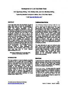
To make the initial validation of the device, a direct measurement of the spectrum of the light source was analyzed, which was compared with that reported by a manufacturer of such products. The results are shown in Figure 5.
1315
a consistency in the reflectance spectra of all samples and on the other hand, is notable difference in the intensity of reflected light (UA) in the sample especially between 580 and 640 nm.
Fig. 6 diffuse reflectance spectrum colloid samples acquired with dispersant fluid. A validation experiment was made acquiring the emission spectrum of an a 50W bulb infrared HAGEN using a commercial spectrometer (OceanOptics, USB2000 + UV-VIS-EN). The results of the emission spectrum acquired by both devices from the IR bulb are shown in figure 8. Prototype
48
Prototype
42 36 30 24 18
100
200
300
As shown in the graphs of Figure 5, the spectra are very similar with valleys approximately at 500nm and 600nm, just as the subsequent increase can notice the latter wavelength. Furthermore, with respect to the experiments performed with compounds colloid dispersing agent. It can be seen in Figure 6, where it is to call attention to two important results. On the one hand it can be noticed
O.C. Vis-UV
500
600
700
800
900
700
800
900
O.C. Vis-UV
2000
Fig.5. Above, spectral distribution of neodymium bulbs manufacturers [6]. Below, spectrum measured by prototype spectrometer.
400
Wavelenght (nm)
2500
1500 1000 500 0 -500
100
200
300
400
500
600
Wavelenght (nm)
Fig. 8 In the picture below a spectrum of infrared bulb 50 Watts acquired with a commercial spectrometer is observed. On top, a similar spectrum obtained with the spectrometer developed. This is a device that requires special specifications for the realization of this project.
IFMBE Proceedings Vol. 51
S. Domínguez-Domínguez, R. Domínguez-Dominguez, and G. Romo-Cárdenas
1316
IV. DISCUSSION The results presented are considered very significant progress, it is important to note that despite the traditional nature of the experiments, it is possible to obtain consistent results, both in the development of the experiments, as compared to data reported in the literature. However, it is clear that is required to make a deeper study of both prototypes and devices used, as well as the experimental protocols followed. These results invite them to compare performance with business in order to meet the operational differences and limitations of the prototype developed spectrometers.
Significant progress is considered in the project which aims to study in human skin samples.
ACKNOWLEDGMENT We thank the University of Montemorelos for their support in conducting this project and also to CONACYT for funding this research.
CONFLICT OF INTEREST The authors declare that they have no conflict of interest.
REFERENCES
V. CONCLUSIONS
The realization of this project shows the feasibility of developing a spectrometer dedicated to the study of diffuse reflectance. Have shown significant progress, however, it is necessary to perform more studies and tests to understand in more detail the performance of the device developed as well as developing a specific software for the analysis of acquired spectra. Meeting these goals will allow us to explore the feasibility of applying these devices to the medical area. The operation of low-cost spectrometer is limited for analyzing spectra of various samples, initially as its sensitivity is limited to the visible range. Although similarity of the graphs of the emission spectra observed in some spectral graphs lamp manufacturer neodymium. The methodology used to measure samples and the working properly proposed for diffuse reflectance studies, considering the fact generate in the colloidal dispersion resembling skin samples human. The developed device design protocol ensures capture reflected light. However, it does not have a standard that allows a calibration of this device and then analyze in detail the performance of the same. The hypothesis of using light sources broad spectrum of diffuse reflectance studies for dermatological studies and to develop a low-cost spectrometer, could be seen as valid, nevertheless require further work to obtain more specific results and conclusive.
[1] E. Angelopoulou, "Understanding the color of human skin," in Photonics West 2001-Electronic Imaging, 2001, pp. 243-251. [2] M. Yamaguchi, M. Mitsui, Y. Murakami, H. Fukuda, N. Ohyama, and Y. Kubota, "Multispectral color imaging for dermatology: application in inflammatory and immunologic diseases," in Proceedings of 13th Color Imaging Conference (Society for Imaging Science and Technology/Society for Information Display, 2005), 2005, pp. 52-58. [3] T. Igarashi, K. Nishino, and S. K. Nayar, "The appearance of human skin," 2005. [4] A. S. Rodríguez, C. R. Cerdeira, R. V. Garrido, and J. L. R. Peralto, "DERMATOSIS ERITEMATOESCAMOSAS," Manual y atlas de las enfermedades de los genitales del varón, p. 193, 2008. [5] C. D. Elvidge, D. M. Keith, B. T. Tuttle, and K. E. Baugh, "Spectral identification of lighting type and character," Sensors, vol. 10, pp. 3961-3988, 2010. [6] R. Sekuler. (2013). Throwing some (bluish) light on the subject. Available: http://people.brandeis.edu/~sekuler/SensoryProce ssesMaterial/colorRenderingLight.html
IFMBE Proceedings Vol. 51