Bio., Univ. of Texas, Austin, TX, 8Dept. ... development in Ceratopteris fern spores (Chatterjee et al., ... main modules in the software to facilitae experimental.
DEVELOPMENT OF A MICROFLUIDIC ION SENSOR ARRAY (MISA) TO MONITOR GRAVITYDEPENDENT CALCIUM FLUXES IN CERATOPTERIS SPORES A.R. De Carlo1, M. Rokkam2, A. ul Haque3, S.T. Wereley3, P.P. Irazoqui4, H.W. Wells5, W.T. McLamb6, S.J. Roux7, D.M. Porterfield1,8 1 Dept. of Ag. & Bio. Eng., 2Dept. of Elec. & Comp. Eng., 3Dept. of Mech. Eng., 4Weldon School of Biomed. Eng., Purdue University, West Lafayette, IN, 5Bionetics Corporation, Kennedy Space Center, FL, 6Dynamac Corporation, Kennedy Space Center, FL., 7Molecular, Cellular & Devel. Bio., Univ. of Texas, Austin, TX, 8Dept. of Hort. & Landscape Arch., Purdue Univ., West Lafayette, IN. Early development of the germinating Ceratopteris fern spore involves the establishment of cellular polarity based on gravity sensing in the single cell system (Edwards and Roux; 1994). This process of cell polarization culminates in directional nuclear migration which determines the plane of the first cell division. The product of this first mitotic division is a prothalial cell (top) and a rhizoid (bottom). Previous experiments have shown that polar Ca2+ currents are involved with gravimorphogenic development in Ceratopteris fern spores (Chatterjee et al., 2000). In a series of experiments that covered the time period of polarity development that leads up to spore germination, it was possible to measure calcium flux at the top, bottom, and sides of the spore using a selfreferencing Ca2+ electrode (Porterfield, 2002). Using the single sensor we were able to determine that there is a transcellular Ca2+ current (into the bottom and out of the top) that correlates with responsiveness of the cell to gravity. More recently we have attempted to identify the differential role of ion channels and pumps in this process of cell polarity development using both molecular and electrophysiological methods. We used a microsystem to rotate individual cells 180° and measure changes in Ca2+ ion currents after reorientation using a single Ca2+selective microelectrode operated as a self-referencing sensor. This approach was limited as we could only effectively measure before and after signals from a single position, and was only able to do these experiments in a 40-second time frame (Stout et al., 2003). These experiments were not able to measure how fast the transcellular calcium current reorients, because of the basic limitations in the sensor technology used. In order to overcome the technical limitations to studying this system associated with the current sensor technology we have been working towards developing new sensor systems based on adaptation of MEMS fabrication techniques. The goal is to fabricate and test dynamic sensing systems for advanced-throughput physiological measurements of calcium signaling events at the cellular level. Using silicon micromachining we have constructed and tested prototype versions of the Microfluidic Ionic Sensor Array (MISA) lab-on-a-chip device for conducting real-time multidimensional calcium flux measurements around 16 fern spores arranged in a vertically positioned 4x4 matrix. A microfluidic pore sustains each fern spore in basic growth media while four solid-state calcium-selective electrodes measure the calcium concentration differential pairwise between four positions around the cell (1 top, 1 bottom, and 2 sides).
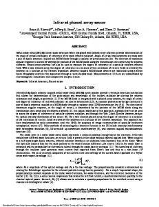
The microfluidic system and sensor array are fabricated on a silicon substrate. The pyramidal pores which hold the fern spores in place are etched at an angle of 54° using KOH etching, and the electrodes and bonding pads are defined by depositing silver and gold on top of photoresist and lifting the photoresist off. Chloride plating of the electrodes is accomplished through immersion in 6% (w/w) NaClO. SU-8 photoresist (MicroChem, USA)is applied to the chip as a structural and insulating layer, and Ca++-selective PVC membrane made with ETH-5234 ionophore (Sigma-Aldrich, St. Louis MO) is spin coated onto the electrode surfaces. The dimensions of the whole chip are 9.7mm x 11mm, with 250µm x 1000µm silver/gold composite bonding pads on all sides and 20µm x 20µm electrodes. For a single wafer there are 41 MISA chips (Figure 1), of which the current process yields approximately 80-90%.
Figure 1. Picture of the 9x11 mm silicon fabricated MISA chip.
The MISA chips are mounted and wire bonded in the center of a printed circuit board (PCB) that contains an array of 64 non-inverting amplifiers, using low-noise (~3nV/√Hz), low-drift (~0.8µV/°C) operational amplifiers, corresponding to the 64 electrodes. Each of the amplifiers provides a signal gain of 30 and filters out noise above 50Hz. The PCB is connected to four data cables through 16-pin connectors with 2mm spacing. The total size of the board is 30.25in2. The output from the PCB is then multiplexed by a 512-point crosspoint matrix, then processed and stored using a 32-input 18-bit data acquisition (DAQ) card in differential mode. MISAPlot 2.0 software was written in LabVIEW (National Instruments, Austin TX) for the DAQ. There are four main modules in the software to facilitae experimental functionality. The software also provides accesss tomoothing and compression functions are written in, as well as functions for logging acquired data, switching data and notes from the user. Data extraction and rapid calibration (Figure 2) are also possible. Gravitational and Space Biology Bulletin 19(2) August 2006
123
AR DeCarlo et al. – Microfluidic Ion Sensor Array We have also developed a novel coupling method, dual-electrode differential coupling (DEDC), so that the differentials between two working electrodes could be amplified and digitized directly without the use of reference electrode. This method was validated in tests (Figure 3) that involved placing the two ion-selective probes in separate Ca++ solutions, connected by a 3M KCl salt bridge. One probe was calibrated in 100µM, 1mM and 10mM solution while the second electrode remained in a standard solution. Each calibration was done three times in each of the standard solutions.
Raw Electrode Output (mV)
-60
-80
-100
-120
REFERENCES
-140
-160 0.1
1
10
Calcium Concentration (mM)
Figure 2. Calibration (Average +/- SD) of a planar array of 64 separate calcium sensors using the ETH-5234 calcium-selective membrane (Anker et al. 1981, Bühlmann et al. 1998). After spin coating to form the membrane, the sensor array was left to sit overnight in a dust-free container, then soaked in a solution of 10µM CaCl2 + 10mM NaNO3 (Konopka et al., 2004). Potentials for the electrodes were measured in calibration solutions of 100µM, 1mM and 10mM CaCl2. The potentials were normally distributed around a slope of 27mV which is in good agreement with the theoretical slope predicted by the Nernst equation. 60
Raw Electrode output (mV)
40
Anker P, Wieland E, Ammann D, Dohner R, Asper R, Simon W. 1981. Neutral carrier based ion-selective electrode for the determination of total calcium in blood serum. Analytical Chemistry 53: 1970-1974. Bühlmann, P., Pretsch, E., Bakker, E. 1998. Carrier based ion-selective electrodes and bulk optodes. 2. ionophores for potentiometric and optical sensors. Chemical Reviews. 98: 1593-1687. Chatterjee, A., Porterfield, D.M., Smith, P.J.S., Roux, S.J. 2000. Gravity-directed calcium current in germinating spores of Ceratopteris richardii. Planta 210: 607-610. Edwards, E.S., Roux, S.J. 1994. Limited period of graviresponsiveness in germinating spores of Ceratopteris richardii. Planta 195: 150-152.
20
0
-20 10.0mM 1.0mM 0.1mM
-40
-60 0.01
0.1
1
10
100
Calcium Concentration (mM)
Figure 3. The DEDC (dual-electrode differential coupling) method was developed so that transcellular ions concentration differentials could be measured without need for system offset settings. Two glass-bodied probes were placed in separate Ca++ solutions, connected by a salt bridge. Each series was replicated using each of three standard solutions as a base solution (figure legend). For each series the Nernst slope was approximately 27mV, with the potential equal to zero when both electrodes are in the same solution.
These basic tests have validated the functionality of the technology. The MISA chip will be able to measure the calcium concentration differentials associated with transcellular currents from individual fern spores in real time. The array of 64 electrodes is organized in quads within 16 fluidic pores, enabling simultaneous
124
measurement of calcium concentrations differentials around sixteen spores. The MISA system will be used in parabolic flight/microgravity based physiological experimentation, to study the role of polar calcium currents in gravity-dependent cellular development. Future uses of this in silico cell physiology lab-on-a-chip technology include biomedical applications such as advanced through-put cell viability and cancer detection. Possible pharmacological applications include testing the effectiveness and dose-response curves of drugs. We anticipate adapting this base system by development and adaptation of ionophores for other ions. We are also developing different versions of this system for amperometric electroanalytical chemistry, which will be used for analytes like oxygen, nitric oxide, and various neurotransmitters to name a few. This will also provide the technical foundation for adapting enzyme based biosensors for use on the system.
Gravitational and Space Biology Bulletin 19(2) August 2006
Konopka, A., Sokalski, T., Michalska, A., Lewenstam, A., Maj-Zurawska, M. 2004. Factors affecting the potentiometric response of all-solid-state solvent polymeric membrane calcium-selective electrode for lowlevel measurements. Analytical Chemistry 76: 6410-6418. Porterfield, D.M. 2002. The use of microsensors for studying the physiological activity of plant roots. In Waisel, Y., Eshel, A., Kafkafi, U. (eds) Plant roots the hidden half, 3rd Edition. Marcel Decker, New York. 333347. Puglisi, J., Bers, D. 2001. LabHEART: An interactive computer model of rabbit ventricular myocyte ion channels and Ca transport. American Journal of Physiology – Cell Physiology 281: C2049-C2060. Stout, S.C., Porterfield, D.M., Roux, S.J. 2003. Calcium signaling during polarity development. Annual Meeting of the American Society for Gravitational and Space Biology Abstract #39.