J Med Syst (2011) 35:559–569 DOI 10.1007/s10916-009-9392-4
ORIGINAL PAPER
Development of a Portable Linux-Based ECG Measurement and Monitoring System Tan-Hsu Tan & Ching-Su Chang & Yung-Fa Huang & Yung-Fu Chen & Cheng Lee
Received: 8 September 2009 / Accepted: 15 October 2009 / Published online: 14 November 2009 # Springer Science + Business Media, LLC 2009
Abstract This work presents a portable Linux-based electrocardiogram (ECG) signals measurement and monitoring system. The proposed system consists of an ECG front end and an embedded Linux platform (ELP). The ECG front end digitizes 12-lead ECG signals acquired from electrodes and then delivers them to the ELP via a universal serial bus (USB) interface for storage, signal processing, and graphic display. The proposed system can be installed anywhere (e.g., offices, homes, healthcare centers and ambulances) to allow people to self-monitor their health conditions at any time. The proposed system also enables remote diagnosis via Internet. Additionally, the system has a 7-in. interactive TFT-LCD touch screen that enables users to execute various functions, such as scaling a single-lead or multiple-lead ECG waveforms. The effectiveness of the proposed system was verified by using a commercial 12-lead ECG signal simulator and in vivo experiments. In addition to its portability, the proposed system is license-free as Linux, an open-source code, is utilized during software development. The cost-effectiveness of the system significantly enhances its practical application for personal healthcare. Keywords Portable Linux-based ECG measurement and monitoring system . Twelve-lead ECG signals . Embedded Linux platform . Open-source codes T.-H. Tan (*) : C.-S. Chang : C. Lee Department of Electrical Engineering, National Taipei University of Technology, Taipei, Taiwan e-mail:
[email protected] Y.-F. Huang Department of Information and Communication Engineering, Chaoyang University of Technology, Taichung, Taiwan Y.-F. Chen Department of Health Services Administration, China Medical University, Taichung, Taiwan
Introduction A significant social problem is that most individuals suffer great stress due to working and life pressures. Although the relationship between work-related stress and death remains unclear, it was found that those with considerable workrelated stress have a 5–20% probability higher than those with little work stress in suffering from cardiovascular diseases (CVDs) [1]. A number of studies demonstrated a strong correlation between stress and CVDs in Japan, North America and Western Europe [2]. Several studies examined the relationship between psychosocial work-related stress and CVDs [3–5]. Of these studies, 24 investigated the association between job stress and CVDs for men and six investigated this relationship for women. Most studies demonstrated a strong positive correlation between job stress and CVDs; additionally, psychosocial work stressors were identified as risk factors for hypertension and CVDs [6]. In 2003, the World Health Organization (WHO) claimed that CVD alone was responsible for 29.2% of total deaths globally, and this was still increasing annually [7]. The WHO also reported that over 50% of these deaths can be prevented by the use of cost-effective monitoring devices for accurate diagnosis. However, people typically ignore their health status due to a lack of an appropriate monitoring device; they are unable to check their health condition anywhere at any time, greatly increasing the probability of sudden death. One solution to this problem is to develop a portable system for monitoring health status ubiquitously. Since the procedures for measuring heart rate (HR) and heart rate variability (HRV) have been standardized [8], using an electrocardiogram (ECG) to identity CVDs has received much attention [9–10]. Several systems have been developed for monitoring CVDs; however, most are only available in medical institutions due to high costs. Consequently, several personal computer (PC)-based ECG monitoring systems have been developed [11–13] to greatly
560
reduce device cost and increase accessibility as PCs are now ubiquitous. A Windows-based client-server architecture for measuring and monitoring ECG signals was presented in [11]. In the system the client (the ECG unit) extracts ECG signals, and the server imports or exports the signals. A PC-based system that captures a patient’s ECG signals via an acquisition card and displays them on a monitor was proposed in [12]. In that system, three electrodes are utilized as sensors that detect ECG signals; one electrode is placed on the left and one on the right wrist, and the third electrode, the reference (ground), is placed on an ankle. A graphical user interface (GUI) for ECG monitoring was designed using a LabVIEW software module developed by National Instruments to allow users to monitor signal waveforms. Additionally, digitized ECG data can be saved as a text file and then retrieved and displayed. Furthermore, visual alert for the abnormal heartbeat is provided. A PC-based ECG measurement and monitoring system employing Visual Basic 6.0 to perform HRV analysis in the time and frequency domains was recently presented in [13]. That system uses the modified Tompkins QRS detection algorithm to determine HRV via ECG signals. A universal serial bus (USB) controller employing Windows API was used to capture and deal with USB data. Furthermore, this system determines the HRV of users in real-time and provides the frequency spectrum of ECG signals for diagnosis. Although these PC-based systems are useful for monitoring heart function based on ECG signals, they are not portable and cannot monitor heart function anywhere at any time. Several Windows-based portable systems have been developed in recent years [14–16]. For example, Kara [14] developed a compact ECG with graphic LCD screen and phonocardiogram unit, enabling physicians and patients to hear heart sounds simultaneously while examining ECG signals. The system allows patients to understand their personal health status directly by hearing heart sounds and listening to the explanation provided by physician in a realtime manner. It implemented a receiver program by using Microsoft Visual C++ programming language. In [15], a portable ECG recorder with USB storage was used to acquire ECG signals. In the system, ECG signals are collected from four electrodes via standard lead placement. A six-lead EGG can be selected via the lead-selection circuit and the acquired EGG signals are then stored in the USB storage for subsequent processing by a PC. In [16], a portable ECG recording system integrating ECG signal acquisition, Bluetooth modules and a LabVIEW software module was proposed. In addition, ECG signals are analyzed to determine HR, HRV and ECG spectrum. However, the price of such monitoring devices is high due to licensing fees for the Microsoft Windows operating system and may incur a high economic burden for many people.
J Med Syst (2011) 35:559–569
Isolation amplifiers can be used to break ground loops, eliminate source ground connections, and provide isolation protection to patient and electronic equipment [17]. In a bio-potential amplifier, the main purpose of the isolation amplifier is to protect the patient by eliminating the hazard of electric shock resulting from the interaction among patient, amplifier and other electric devices in the patient’s environment, specifically defibrillators and electrosurgical equipment. It also has the capability to prevent line frequency interferences. In this work, we present a license-free portable Linuxbased system for monitoring heart functions based on ECG signals. High usability, portability, ease-of-use and low cost are the design objectives of the proposed system. It can be installed virtually anywhere (e.g., offices, homes, healthcare centers and ambulances). The system allows people to self-monitor their heart conditions. Additionally, the data can be sent to a server at a distant hospital via Internet for remote diagnosis. Furthermore, the proposed system has a 7-in. interactive TFT-LCD touch screen that allows users to execute various functions, such as scaling single-lead or multiple-lead ECG waveforms. Also, an isolation circuit is added to prevent electrical shocks. The effectiveness of the proposed system was verified by employing a commercial 12-lead ECG signals simulator [18] and in vivo experiments. The remainder of this paper is organized as follows. Section “System implementation” describes the implementation of the proposed portable Linux-based ECG measurement and monitoring system. Section “Experimental results” then illustrates experimental results and accuracy of the measured clinical ECG signals. Conclusions are finally drawn in Section “Conclusions”.
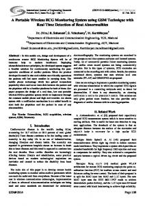
System implementation Hardware architecture Figure 1 presents the hardware architecture of the proposed system, which consists of an ECG front end and an embedded Linux platform (ELP). The ECG front end consists of signal amplifiers, an analog-to-digital (A/D) circuit and control circuit module, and the ELP is a modular platform that composed of signal storage, signal processing and graphic display modules for a 12-lead ECG signals measurement and monitoring system. The key features of the proposed system are as follows: 1. Low cost due to the utilization of license-free Linux open-source code. 2. Easy to use due to the fully interactive TFT-LCD touch screen.
J Med Syst (2011) 35:559–569
561
Fig. 1 a Block diagram and b hardware architecture of the proposed system USB Interface
Signal storage
Signal processing
Graphic display
embedded Linux platform (a) ECG Connector
ECG Simulator
USB Interface
ECG frond end
embedded Linux platform (b)
3. High portability due to a total weight of only 1.5 kg or so. 4. Allowing people to monitor health status anytime and anywhere. 5. Enhancing safety by adding an isolation circuit to prevent electrical shocks. 6. Enabling remote diagnosis via wireless network interface. The function of each part is described as follows: 1. ECG front end: This subsystem acquires ECG signals from electrodes and then delivers them to the ELP via a USB interface. 2. Signal storage: The 12-lead ECG signals obtained are stored in the RAM disk and SD (Secure Disk) card of the embedded Linux platform. 3. Signal processing: This module performs time and frequency domains analyses of the 12-lead ECG signals using the equations and the Discrete Fourier transform (DFT) suggested by [19]. Table 1 lists these equations. 4. Graphic display: This module demonstrates the measured values and graphs of ECG signals in time and frequency domains on the ELP. 5. ECG simulator: This module is used for testing the proposed system. In this study, a scheme proposed in [19] for detecting 12lead ECG signals and diagnosing CVDs was adopted. The functions of this scheme are summarized as follows. 1. Acquisition of ECG signals from a patient using 10 electrodes. 2. Amplification and digitalization of acquired ECG signals.
3. Derivation of the 12-lead ECG signals in the timedomain from signals acquired using 10 electrodes. 4. Derivation of frequency-domain spectrums of the 12-lead ECG signals. 5. Establishment of the base value of ECG signals using the determined HR by multiplying HR in beats/second by a scaling quantity ranging from 3 to 7 (preferably 5). 6. Health evaluation of the heart status by dividing an area obtained by conducting integration from 0 Hz to the base value in the frequency domain, by another area obtained by performing integration from the base value to infinity. 7. Peaks analysis of the power spectrum in the frequency domain for diagnosing CVDs. Table 1 Twelve-lead time-domain ECG signals and their evaluation equations Lead I II III aVR aVL aVF V1 V2 V3 V4 V5 V6
ECG signals = = = = = = = = = = = =
LA-RA LL-RA LL-LA RA-(RA + LA + LL)/3 LA-(RA + LA + LL)/3 LL-(RA + LA + LL)/3 C1-(RA + LA + LL)/3 C2-(RA + LA + LL)/3 C3-(RA + LA + LL)/3 C4-(RA + LA + LL)/3 C5-(RA + LA + LL)/3 C6-(RA + LA + LL)/3
562
Acquisition of ECG signals All ECG signals are obtained by sensors placed at 10 sampling points (Fig. 2) which are located on the right arm (RA), left arm (LA), right leg (RL), left leg (LL) and the chest (C1–C6) close to the heart. A 12-lead ECG signals connector (DB15 connector) is employed to transmit ECG signals to the ECG front end. Figure 3 shows the pin assignment of DB15 connector. Design of ECG front end The circuits proposed in [20] and [21] were adopted and improved in designing the ECG front end circuit. Since the circuit layout presented in [20] requires a larger space than the proposed system can accommodate, a significant space reduction is achieved by the proposed layout. Additionally, the A/D converter (AD7888) in [21] does not fit the proposed system as the resolution of the AD7888 is not compatible with the ELP; thus, the circuit layout was redesigned. Figure 4 presents the block diagram of the improved 12-lead ECG front end circuit. In addition to designing a highly portable and low-cost system, another study goal is to achieve long-term ECG monitoring. Therefore, the circuit should be simplified as
Fig. 2 Sampling points of ECG signals
J Med Syst (2011) 35:559–569
much as possible. To this end, the power of all amplifiers is supplied by the ELP, thereby decreasing device size and avoiding the additional cost of any power supply circuits. Design of amplifiers circuits Skin contact may result in disturbance during ECG signal acquisition [22]. To suppress such disturbance meanwhile amplify the ECG signals extracted by eight ECG sensors, two precision instrumentation amplifier (AD8221) chips and one quad input and output amplifiers (AD8604) chip [23] are integrated to produce V1, V2, V3, V4, V5 and V6 signals, as illustrated in the amplifiers block Fig. 4. Moreover, three amplifiers depicted in blocks P10-RALL, P11-LALL and P12-RALL (Fig. 4) are used to generate RA-LL, LA-LL and RA-LA signals, respectively, such that the 12-lead ECG signals can be monitored and analyzed. Design of analog-to-digital (A/D) circuit The A/D circuit block is depicted as P06-MPU in Fig. 4. To achieve the design objectives, an A/D converter, the ADuC841 [23], which integrates a high-performance self-calibrating multi-channel analog-to-digital converter (ADC), a dual digital-to-analog converter (DAC) and an optimized single-cycle 20-MHz 8-bit MCU on a single chip, is utilized as the main chip in the A/D circuit. The output signals from nine amplifier circuits are applied to the ADuC841 chip for digitization. Notably, each digitized signal is represented by 12 bits. Moreover, a program was designed on an 8051 built-in ADuC841 chip (speed = 20 MIPS) to extend a 12-bit signal to a 16-bit signal by filling the four most significant bits with 0s, and storing them in the flash ROM embedded in the ADuC841 chip. Via this program, the ADuC841 chip can output 16-bit data that comply with the data format to be processed by the selected USB controller chip. Therefore, the design complexity of the USB circuit is significantly reduced. This ADuC841 chip is also powered by the ELP via a USB. Connection of A/D circuit and ELP via a USB interface A common USB port is adopted for connecting the A/D circuit and ELP by taking advantage of the USB 2.0 interface, which supports a maximum data rate of 480 Mbps and low-voltage peripherals. Additionally, the USB hubs and power-efficient storage drives can directly use power from the ELP. The PL2303 USB-to-serial bridge controller [24] is adopted as the USB controller in the proposed system. The design and development of the device driver are according to [23] and chip data sheets described in [24]. Furthermore, the power of the PL2303 controller and laptop is supplied by the ELP, details of which can be found in blocks P02-USB and P03-Laptop (Fig. 4). Notably, the usability of the proposed system can be extended by employing the Laptop block, such as execution of remote medical diagnosis via wireless network.
J Med Syst (2011) 35:559–569
563
1
1
Fig. 3 Pin assignment of DB15 connector
P06-MPU
P02-USB
P04-ISOLATION
USB_5V USB_GND
5V1 GND1
USB_RST USB_UPG
RESET UPG
Viso GND2
P05-POWER Viso
P03-Laptop NB_5V NB_GND
5VD DGND
5VD DGND
RSTN
RST
RST
V3L V4L
TXD# RXD#
5VA AGND UPG
P13-12LEAD IN0+ IN1D+ DIN-
D1 D2 LL
OUT0 IN0+ OUT1 IN1REF D+ 5VA DINAGND
D3 D4 LL
V1 V2 LL
V3 V4
UPG
TXDN RXDN
RXDN TXDN
P09-VIEW56 P07-AFE V5L V6L 5VA AGND
5VD DGND
OUT0 OUT1 REF 5VA AGND P08-VIEW34 P07-AFE
Viso
NB_RST USB_UPG NB_TRX4 NB_RX4
REF 5VA AGND
5VD DGND
Viso RSTN
USB_TXD# USB_RXD#
Viso GND2
P07-VIEW12 P07-AFE V1L V2L REF 5VA AGND
5VD DGND
A/D converter
Isolation circuit
OUT0 OUT1 REF 5VA AGND
IN0+ IN1D+ DIN-
V5 V6
D5 D6 LL
P10-RALL P10-AFE1
RAL VCC29 5VA AGND
IN+ OUT INREF VCC29 COM 5VA AGNDSHIELD
RA LL
RA
COM0 SHD0
COM0 SHD0
LA LL
LA
P11-LALL P10-AFE1 LAL VCC29 5VA AGND
IN+ OUT REF INVCC29 5VA COM AGNDSHIELD
COM1 SHD1
COM1 SHD1
P12-RALA P10-AFE1 RALA VCC29
VCC29 5VA AGND
IN+ OUT INREF VCC29 5VA COM AGNDSHIELD
RA LA COM2 SHD2
COM2 SHD2 VCC29 AGND
Amplifiers
Fig. 4 Block diagram of the improved circuit of the 12-lead ECG front end
VCC29 AGND
1
J Med Syst (2011) 35:559–569
1
564
1
+OUT -OUT
S
D
1
+IN -IN
Fig. 5 The proposed isolation circuit
Design of isolation circuit The power for the amplifier chips (AD8221 and AD8604) and A/D converter chip (ADuC841) are supplied by the ELP via the USB port. Notably, users of the proposed ECG monitoring system may have CVD. Therefore, one must account for risk of electrical shock caused by a static charge, a requirement of certified medical facilities. Thus, an isolation circuit block, the P04-isolation (Fig. 4), was added to prevent electrical shocks. Figure 5 shows details of the isolation circuit, in which the ADuM1401C digital isolator chip [23] functions as a DC/DC converter which transforms the input DC voltage into a lower DC value. The feature of the DC/DC converter is adopted in the design to supply DC power to other chips in addition to achieving the purpose of isolation. Notably, it has important features as follows: 1. 5 kV rms isolation rating with IEC-60601–1 [25] medical safety approval. 2. Short-circuit protection for xD+ and xD− lines (USB lines must automatically switch between actively driving D+/D−, receiving data, and allowing external resistors to set the idle state of the bus). 3. Class 3A (4,000 to